Note: Descriptions are shown in the official language in which they were submitted.
CA 02158479 2004-07-06
WO 94/23638 PCT/US94/03426
-1-
TITLE OF THE INVENTION
DIAGNOSIS AND TREATMENT OF SUPRAVALVULAR
AORTIC STENOSIS AND WILLIAMS SYNDROME
BACKGROUND OF THE INVENTION
The present invention is directed to a process for
the diagnosis and prevention of supravalvular aortic
stenosis (SVAS) and Williams syndrome. SVAS and
Williams syndrome is diagnosed in accordance with the
present invention by analyzing the DNA sequence of the
elastin gene of an individual to be tested and comparing
the DNA sequence to the known DNA sequence of a normal
elastin gene. Alternatively, the elastin gene of an
individual to be tested can be screened for mutations
associated with SVAS or Williams syndrome. Prediction of
SVAS and Williams syndrome will enable practitioners to
prevent these disdorders using existing medical therapy.
The publications and other materials used herein to
illuminate the background of the invention or provide
additional details respecting the practice
and for convenience are
respectively grouped in the appended List of References.
Supravalvular aortic stenosis (SVAS) is an
inherited vascular disorder (1). As its name implies,
narrowing of the ascending aorta is a dominant feature
of this disease, but other arteries, including the
pulmonary arteries, may be affected. If uncorrected,
SVAS may lead to increased intracardiac pressure,
myocardial hypertrophy, heart failure and death. The
incidence.of SVAS is estimated to be 1 in 25,000 live
births. The vascular abnormalities typical of SVAS can
be inherited as an isolated, autosomal dominant trait
WO 94/23638 21594 7 9 PCT/US94/03426 ~
-2-
(1-3) or as part of a second disease, Williams syndrome,
a developmental disorder that affects multiple organ
systems (2-4). In addition to vascular disease, manifes-
tations of Williams syndrome include hypertension,
mental retardation, an unuqually gregarious personality,
premature greying of the hair, premature aging of the
skin, joint laxity early in life followed by joint
contractures, dysmorphic facial features and infantile
hypercalcemia (4). The relationship between SVAS and
Williams syndrome was previously undefined. Occasionally
patients with Williams syndrome have been noted in
families with SVAS.
SVAS was first described in 1842 (5), but the
pathogenesis of this disorder was unknown until now.
Mechanistic hypotheses have been based on clinical and
pathological studies, but these data are conflicting.
It is not clear, for example, whether hypertrophy or
hyperplasia of medial smooth muscle is the more
prominent feature of this disorder. O'Conner et al. (6)
examined tissue from six individuals with SVAS; two of
these cases were familial, one was a sporadic case of
SVAS, and three had Williams syndrome with SVAS. These
investigators did not discover any significant patho-
logic differences between the individuals with different
SVAS inheritance patterns. They noted that the medial
layer of the aorta in all patients showed a haphazard
arrangement of thick elastic fibers, excessive collagen,
hypertrophied smooth-muscle cells, and scant ground
substance. This contrasts with normal medial tissue,
which is highly organized and arranged in parallel
layers of connective tissue and smooth muscle. They
also observed that smooth muscle cells formed clumps or
bundles and were the major component of the medial
layer. In a study of a single individual with SVAS,
Perou also showed that the diseased media contained
excessive smooth muscle (7). The resulting pattern was
WO 94/23638 215" T 79 PCT/US94/03426
-3-
that of irregular fascicles of smooth muscle surrounded
by fibrous and collagenous tissue. In contrast to these
studies, Pober et al. recently reported a study of seven
individuals with SVAS and Williams syndrome, noting that
the medial layer of affected aortas contained an
increased number of smooth muscle cells and normal to
decreased collagen. These investigators also observed
an elevated level of platelet-derived growth factor
(PDGF) and concluded that increased quantities of PDGF
stimulate smooth muscle proliferation and cause the
cardiovascular abnormalities of SVAS (8).
The pathogenic mechanisms underlying Williams
syndrome are unknown, but many hypotheses have invoked
a mechanism of abnormal calcium metabolism (9). These
hypotheses are based on the intermittent observation of
infantile hypercalcemia in Williams syndrome as well as
studies showing that excessive vitamin D administration
can produce pathologic changes in the supravalvular
aortic wall of rabbits during development (10). Recent
attempts to repeat this work, however, have failed and
the hypothesis involving vitamin D has been questioned
(11). Researchers have tried to identify a causal
relationship between calcitonin-gene-related peptide
(CGRP) and Williams syndrome. Hitman et al. investigated
13 families with Williams syndrome and infantile hyper-
calcemia for germ line mutations in the CGRP gene, and
found no mutations (12). Using linkage analysis,
investigators have also excluded the CGRP gene, which is
located on chromosome 11, in a family with autosomal
dominant SVAS (13).
Diagnosis of SVAS has been based on family history,
physical examination and echocardiography. Unfortu-
nately, these tests may be ambiguous, making early
detection of this disorder difficult. The recent advent
of high quality two-dimensional and color flow Doppler
echocardiography have improved non-invasive screening
WO 94/23638 PCT/US94i03426
-4-
for SVAS (14), but invasive tests such as cardiac
catheterization and angiography are more sensitive.
Currently, vascular surgery irz? 4the only treatment option for SVAS.
Therefore, it 1s- an object of the present
invention to provide better diagnostic methods for
screening for SVAS and Williams syndrome which will
ultimately lead to new medical therapies for their
prevention.
SUMMARY OF THE INVENTION
The present invention demonstrates the molecular
basis of SVAS and Williams syndrome. More specifically,
the present invention has determined that molecular
variants of the elastin gene cause or are involved in
the pathogenesis of SVAS and Williams syndrome. Analysis
of the elastin gene will provide an early diagnosis of
subjects with SVAS and Williams syndrome. The diagnostic
method comprises analyzing the DNA sequence of the
elastin gene of an individual to be tested and comparing
it with the DNA sequence of the native, non-variant
elastin gene. In a second embodiment, the elastin gene
of an individual to be tested is screened for mutations
associated with SVAS or Williams syndrome. The ability
to predict SVAS will enable physicians to prevent the
disease with medical therapy like beta blocking agents.
Finally, this invention has implications for the cause
and treatment of common vascular disease, like
atherosclerosis.
BRIEF DESCRIPTION OF THE FIGURES .
Figure 1 shows the pedigree structure and elastin
genotypes for SVAS families K1773 (Fig. lA), K1779 (Fig.
1B), K1861 (Fig. 1C) and K2049 (Fig. 1D). Affected
individuals having the characteristic pattern of
WO 94/23638 215 8479 PCT/US94/03426
-5-
elevated Doppler velocity and narrowing of the ascending
aorta or pulmonary arteries on echocardiograms are
represented by filled circles (females) and squares
(males). Unaffected individuals are represented by open
squares or circles. Family members who had an equivocal
phenotype or for whom no phenotypic data were available
are represented by stippled squares and circles. Above
each symbol, individual alleles are listed for the
elastin polymorphic PCR marker (15). At this marker
locus the restriction enzyme BstNI revealed two distinct
alleles within the families. The disease gene cosegre-
gates with the 244-bp allele (allele 2) in families
K1773 and K1779. Alleles shown in parentheses were
inferred. Individuals carrying a translocation are
indicated by T and those not carrying the translocation
are indicated by N in family K1861.
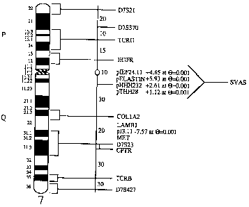
Figure 2 shows the approximate map location of the
SVAS disease gene. Genes and polymorphic loci mapped to
chromosome 7 are shown at right. The approximate genetic
distance between these loci in cM is shown on the
diagram at center (16). The approximate subsegmental
location of these loci is shown on the idiogram at left.
In K1773 and K1779, SVAS is linked to pHHH232,
pIEF24.11, pTHH28 and elastin, which are mapped
approximately to 7q11 between the centromere and COL1A2
(collagen). Pairwise lod scores between a DNA marker and
the disease phenotype are indicated.
Figure 3A shows that hybridization of a full-length
elastin cDNA probe to Not I digests of DNA from SVAS
patients II-1 (affected with SVAS) and 111-2 (affected
with SVAS and phenotypic features of Williams syndrome)
reveals aberrant restriction fragments of 450 and 1000
kb. The 700 kb fragment is seen in both unaffected
(individual I-i) and affected members of kindred 1861
and represents the normal allele.
WO 94/23638 2158479 PCT/US94/03426
-6-
Figure 3B shows that hybridization of a 2.8 kb
elastin genomic probe (N-2.8) to Bam HI digests of DNA
from members of K1861 reveals 12 kb aberrant fragments
in SVAS patients. Pedigree"'information is identical to
panel A. The probe N-2.8 also hybridizes with two Bam HI
fragments of 8 kb and 9 kb in both affected and unaffec-
ted individuals of K1861. These fragments represent the
normal elastin allele. The aberrant fragment was not
seen in DNA samples from 100 controls.
Figure 4 shows PFGE analyses of DNA from kindred
1861. (A) Ideogram of chromosome 7 showing location of
the elastin locus with the intron and exon structure,
and probes used to define the translocation. Numbering
of exons was first described for the bovine elastin
gene; the human elastin gene lacks exons 34-35 (39).
(B) An elastin subclone (N-2.0) proximal of the
translocation breakpoint detects a 450 kb anomalous Not
I fragment in DNA from SVAS patients including the
individual with features of Williams syndrome. By
contrast, a subclone (N-2.8) distal of the breakpoint
detects a 1000 kb anomalous Not I fragment in DNA from
affected members of kindred 1861. Both probes detect
the normal 700 kb Not I fragment which is also seen in
unaffected family members. These data prove that the
SVAS-associated translocation disrupts the elatin gene.
Figure 5 shows restriction maps of the T1 trans-
location allele. (A) Restriction map of N, the
nontranslocated elastin allele (chromosome 7) cloned
from SVAS patient III-1. Elastin exons are indicated by
filled bars and numbered. Restriction enzymes (B, Bam
HI; H, Hind III; E, Eco RI; K, Kpn I; P, Pst I; S, Sma =
I) are indicated. Expanded restriction map for a 3.0 kb
subclone (N-3.0) is shown below. (B) Restriction map of
Tl, a genomic clone from SVAS-patient III-i containing
a transiocation breakpoint. The site of the breakpoint
region is indicated. Chromosome 7 sequences containing
w 94'23638 215 8 4 7 9 PCT/US94103426
-7-
elastin exons are at left. Translocated sequences from
chromosome 6 are at right. Expanded restriction map for
2.2 kb of T1-14 (a subclone of T1) is shown below.
Figure 6 shows nucleotide sequence of the T1
translocation breakpoint. Nucleotide sequence of the Ti
(translocation allele, top) and N (nontranslocated
elastin allele, bottom) showing complete identity until
the translocation breakpoint indicated by an arrow.
Nucleotide identity is indicated as a dash. This
rearrangement disrupts elastin exon 28, resulting in a
new stop codon (TGA) as indicated by a small box.
Sequences encoding elastin exon 28 in the normal clone
are underlined. Alu sequences within intron 27 of the
normal elastin clone were identified near the transloca-
tion breakpoint and are enclosed in a large box.
Figure 7 shows PCR analyses indicating that T1
represents a translocation breakpoint. (A) Map of the
Ti clone showing the translocation breakpoint and the
location of oligonucleotide primers. (B) Oligonucleo-
tide primers directed across the translocation
breakpoint (TBF and TBR) yield a PCR product of the
predicted size (370 bp) in affected family members of
kindred 1861 and in a subclone of T1 (T1-14), but not in
unaffected family members or in a normal elastin
subclone (N-3.0).
Figure 8 shows that hybridization of elastin probe
to Bcl I digests revealed 8.5 kb abberent fragments in
DNA from affected members of K2049. The 12.5 and 11.5
kb fragments were observed in affected and unaffected
members of this kindred. These data indicated that 3'
elastin sequences are deleted in affected members of
this SVAS family.
Figure 9A shows the restriction map of the elastin
locus showing the intron and exon structure and the
location of probes used to define the SVAS-associated
mutation. Restriction sites are indicated (Bc, Bcl I;
WO 94/23638 PCT/US94/03426 ~
-8-
S, Sac I; H, Hing III; RV, Eco RV; Pv, Pvu II). The
predicted location of the mutation is shown.
Figure 9B shows that several 5' elastin probes detect a 550 kb anomalous;Not I
fragment in DNA from
affected members of K2049.,- By contrast, 3' probes fail
to detect a Not I anomy. All probes detect the normal
700 kb Not I fragmentwhich is also seen in unaffected
members of this kindred. These data indicated that 3'
elastin sequences are deleted in affected members of
this SVAS family.
Fig. 10 shows hybridization of an elastin genomic
probe to Pst I digests of DNA from Williams syndrome
patient 11-2 which reveals a restriction fragment of 3.5
kb but does not show the common 2.0 and 1.6 kb frag-
ments. Williams syndrome patient III-1 did not inherit
the 3.5 kb Pst I fragment from his father; he only
inherited the 2.0 and 1.6 kb fragments from his mother.
These data show that affected father and son both carry
null mutations of the elastin gene. Identical results
have been found in a second family with Williams
syndrome.
Fig. 11 shows pedigree structure for Williams
syndrome kindreds. Individuals with the characteristic
features of Williams syndrome are indicated by filled
circles (females) or squares (males). Empty circles or
squares indicate unaffected individuals. In kindred
1806 (Fig. 11A) and Kindred 2042 (Fig. 11B), the
Williams phenotype is transmitted from parent to child.
Kindreds 1998 (Fig. 11C) and 2016 (Fig. 11D), 2767 (Fig.
11E), 1866 (Fig. 11F), 1868 (Fig. 11G) and 1888 (Fig.
11H) represent sporadic cases of Williams syndrome.
Hemizygosity at the elastin locus was demonstrated in
all affected members of these kindreds by Southern and
PCR analyses, proving that mutations in the elastin gene
cause Williams syndrome.
WO 94/23638 2158479 pCT/US94/03426
-9-
Fig. 12 shows predicted amino acid structure of the
elastin protein showing potential sites for desmosine
crosslinking (K) and disulfide bridging (C).
DETAILED DESCRIPTION OF THE INVENTION
The present invention is directed to the determina-
tion that SVAS and Williams syndrome map to the elastin
gene and that molecular variants of the elastin gene
cause or are involved in the pathogenesis of SVAS and
Williams syndrome. The present invention is further
directed to methods of screening humans for the presence
of elastin gene variants associated with SVAS or
Williams syndrome. Since SVAS or Williams syndrome can
now be detected earlier (i.e., before symptoms appear)
and more definitively, better treatment options will be
available in those individuals identified as having SVAS
or Williams syndrome. Finally, the present invention
has implications for the cause and treatment of ommon
vascular disease, such as athersclerosis.
Supravalvular aortic stenosis (SVAS) is an inheri-
ted disorder that causes hemodynamically significant
narrowing of the ascending aorta and other arteries.
SVAS can be inherited as an isolated trait or as part of
the Williams syndrome, a developmental disorder that
also results in hypertension, mental retardation, a
personality disorder and premature aging of the skin.
Two approaches have been utilized herein to
identify the elastin gene as the cause of SVAS and
Williams syndrome, namely linkage analysis and the
identification and characterization of disease-
associated gene abnormalities. In linkage analysis, one
attempts to identify co-inheritance of a phenotype
(e.g., SVAS) and a genotype (DNA polymorphism).
Disease-associated gene abnormalities include
chromosomal rearrangements, gross and microscopic
WO 94/23638 2158479 PCl'/US94/03426 ~
-10-
deletions or additions, and sequence differences. These
two approaches led to the identification of the elastin
gene as the cause of SVAS and Williams syndrome. SVAS was completely linked to
the elastin locus on
chromosome 7qll by phenotypic and linkage analyses
(details provided in the Examples) by studying two
multigenerational families with SVAS. Kindred 1773 is
a Kentucky family of Irish decent and consists of 47
family members who are at risk for SVAS (Fig. lA). The
second family, K1779, is of German decent and contains
9 family members (Fig. 1B) including spouses, most of
whom live in Indiana. The clinical features of these
families are similar and typical of familial SVAS. In
both families, several affected members required sur-
gical correction of SVAS, and in one family (K1773),
three family members died of this disorder. The linkage
analysis was performed using highly polymorphic DNA
markers spanning the genome. Proof that the elastin
gene causes or is involved in the pathogenesis of SVAS
was performed by characterizing the disease-associated
gene abnormality of a 6p21/7q11 translocation in a SVAS
family. The 6p2l/7qll translocation consegregates with
the SVAS phenotype in the unique family K1861 (Fig. 1C).
It was found that this translocation disrupts the
elastin gene. In a second SVAS family, K2049, a
deletion of the 3' end of the elastin gene was
identified. Thus, molecular variants of the elastin
gene have been found to cause or be involved in the
pathogenesis of SVAS.
Proof that the elastin gene causes or is involved
in the pathogenesis of Williams syndrome was
accomplished by analyzing the disease-associated gene
abnormality in two familial and six sporadic cases of
Wiliams syndrome. It was found that hemizygosity at the
elastin locus, i.e., one allele of the elastin gene was
2158479
WO 94/23638 PCT/US94/03426
-11-
absent, causes or is involved in the pathoigenesis of
Williams syndrome.
The identification of the association between
elastin gene mutations, SVAS and Williams syndrome
permits the early presymptomatic screening of
individuals to identify those at risk for developing
SVAS or Williams syndrome. To identify such
individuals, the elastin alleles are screened for
mutations. The elastin alleles are screened for
mutations either directly or after cloning the alleles.
The alleles are tested for the presence of nucleic acid
sequence differences from the normal allele using any
suitable technique, including but not limited to, one of
the following methods: fluorescent in situ hybridization
(FISH), direct DNA sequencing, PFGE analysis, Southern
blot analysis, SSCP analysis, linkage analysis, RNase
protection assay and allele specific oligonucleotide
(ASO) dot blot anlaysis. For example, either (1) the
nucleotide sequence of both the cloned alleles and
normal elastin gene or appropriate fragment (coding
sequence or genomic sequence) are determined and then
compared, or (2) the RNA transcriptions of the elastin
gene or gene fragment are hybridized to single stranded
whole genomic DNA from an individual to be tested, and
the resulting heteroduplex is treated with Ribonuclease
A (RNase A) and run on a denaturing gel to detect the
location of any mismatches. Two of these methods can be
carried out according to the following procedures.
The alleles of the elastin gene in an individual to
be tested are cloned using conventional techniques. For
example, a blood sample is obtained from the individual.
The genomic DNA isolated from the cells in this sample
is partially digested to an average fragment size of
approximately 20 kb. Fragments in the range from 18-21
kb are isolated. The resulting fragments are ligated
into an appropriate vector. The sequences of the clones
WO 94/23638 ~ 1 C Q~~ 9 PCT/US94/03426
JO -12-
are then determined and compared to the normal elastin
gene.
Alternatively, polymerase chain reactions (PCRs) are performed with primer
pairs for the 5' region or the
exons of the elastin gene. PCRs can also be performed
with primer pairs based~on any sequence of the normal
elastin gene. For example, primer pairs for one of the
introns can be prepared and utilized. Finally, PCR can
also be performed on the mRNA. The amplified products
are then analyzed by single stranded conformation
polymorphisms (SSCP) using conventional techniques to
identify any differences and these are then sequenced
and compared to the normal elastin gene sequence.
Individuals can be quickly screened for common
elastin gene variants by amplifying the individual's DNA
using suitable primer pairs and analyzing the amplified
product, e.g., by dot-blot hybridization using allele-
specific oligonucleotide probes.
The second method employs RNase A to assist in the
detection of differences between the normal elastin gene
and defective genes. This comparison is performed in
steps using small (-500 bp) restriction fragments of the
elastin gene as the probe. First, the elastin gene is
digested with a restriction enzyme(s) that cuts the
elastin gene sequence into fragments of approximately
500 bp. These fragments are separated on an electro-
phoresis gel, purified from the gel and cloned
individually, in both orientations, into an SP6 vector
(e.g., pSP64 or pSP65). The SP6-based plasmids
containing inserts of the elastin gene fragments are
transcribed in vitro using the SP6 transcription system,
well known in the art, in the presence of [a-32P]GTP,
generating radiolabeled RNA transcripts of both strands
of the elastin gene.
Individually, these RNA transcripts are used to
form heteroduplexes with the allelic DNA using conven-
~~~8479
WO 94/23638 PCT/US94/03426
-13-
tional techniques. Mismatches that occur in the RNA:DNA
heteroduplex, owing to sequence differences between the
elastin fragment and the elastin allele subclone from
the individual, result in cleavage in the RNA strand
when treated with RNase A. Such mismatches can be the
result of point mutations or small deletions in the
individual's elastin allele. Cleavage of the RNA strand
yields two or more small RNA fragments, which run faster
on the denaturing gel than the RNA probe itself.
Any differences which are found, will identify an
individual as having a molecular variant of the elastin
gene and the consequent presence of SVAS, Williams
syndrome or predisposition to common vascular disease.
Genetic testing will enable practitioners to
identify individuals at risk for SVAS and Williams
syndrome at, or4 even before, birth. Presymptomatic
diagnosis of SVAS and Williams syndrome will enable
prevention of these disorders. Existing medical
therapies, including beta adrenergic blocking agents,
will prevent and delay the onset of severe vascular
disease in SVAS and Williams syndrome (currently the
only therapy for these disorders is open-chest surgery).
Finally, this invention changes our understanding of the
cause and treatment of common vascular disease like
athersclerosis, a disease that kills hundreds of
thoussands of individuals. Existing art has focused on
cholesterol and high blood pressure in the cause and
treatment of vascular disease. This invention
demonstrates that inelasticity of blood vessels can
cause vascular obstruction. This finding will lead to
a new avenue for medical therapy of vascular disease.
Therefore, individuals with molecular variants in the
elastin gene may be predisposed to common vascular
disease.
The present invention is further detailed in the
following Examples, which are offered by way of illus-
WO 94/23638 PCT/US94/03426 ~
t+ -14-
tration and are not intended to limit the invention in
any manner. Standard techniques well known in the art
or the techniques specifically described below are
utilized.
A 5 '~= EXAMPLE 1
Methods for Phenotypic Evaluation
Two multigenerational families with SVAS (Figs. 1A
and 1B) were studied. Kindred 1773 was of Irish descent
and Kindred 1779 was of German descent. Informed consent
was obtained from all study participants or their
guardians, in accordance with standards established by
local institutional review boards. To determine if
family members and spouses had signs of SVAS or Williams
syndrome, physical examinations were performed. The
method of identification of SVAS was similar to that
described previously (14). Echocardiograms were
performed with the use of Hewlett-Packard Sonos 500 and
VingMed CFM 700 machines with 2.5 to 5.0 MHz duplex
imaging and Doppler probes as determined by patient
size. A 2.5 MHz continuous-wave offset imaging pencil
probe was used for suprasternal continuous-wave Doppler
sampling. Color flow Doppler was used in all of the
patients to assess blood flow acceleration in the aortic
root and pulmonary arteries. Each examination was
recorded on 0.5 in. (1.27 cm) VHS videotape.
Standard parasternal long axis, short axis, apical
four chamber, subcostal and suprasternal views were
recorded in all patients when technically feasible. In
addition, the proximal ascending aorta was imaged in the
long axis from high left or right parasternal views.
Two-dimensional echocardiographic measurements of the
left ventricular outflow tract, aortic annulus, aortic
root at the sinus of Valsalva, ascending aorta at the
sinotubular junction (or its narrowest point), descend-
WO 94123638 215S4r~ 9 PCT/US94/03426
-15- [
ing aorta below the origin of the left subclavian
artery, main pulmonary artery, and the narrowest
portions of the proximal right and left pulmonary
arteries were determined for each patient when tech-
nically feasible. Peak blood flow velocities were
measured by Doppler from the ascending aorta at the apex
and suprasternal notch (continuous wave), main pulmonary
artery (pulsed wave), right pulmonary artery (pulsed or
continuous wave), and left pulmonary artery (pulsed or
continuous wave). The velocity time integral and
ejection time were recorded from the ascending aorta,
left ventricular outflow tract and pulmonary artery.
Peak aortic and pulmonary artery flow velocities were
compared with the normal range of values (aortic: adult
1.0-1.7 m/s, children 1.2-1.8 m/s; pulmonary: adult 0.6-
0.9 m/s, children 0.7-1.1 m/s; ref. 19). M-mode measure-
ments were recorded from the left and right ventricular
cavities, septum, posterior wall of the left ventricle,
aorta, aortic valve opening, and left atrium (14).
To determine the phenotype of individuals, all
Doppler echocardiographic data were independently
reviewed without knowledge of genotypic data. Indivi-
duals were classified as affected, uncertain, and
unaffected based on catheterization, angiography and
surgical findings. If catheterization data were
unavailable, phenotype was determined based on
echocardiographic impression of narrowing of the aorta
at the sinotubular junction and the supravalvular
pulmonary region, increased Doppler blood flow velocity
in the ascending aorta, increased flow velocity in the
main pulmonary artery, and/or increased blood flow
velocity in the peripheral pulmonary arteries. Family
members were scored on a scale from -6 (no evidence of
SVAS) to +6 (strong evidence of SVAS). For linkage
analysis, individuals with impression scores of -2 and
lower were classified as unaffected, +2 and greater as
CA 02158479 2004-07-06
WO 94123638 PCT/US94/03426
-16-
affected, and -1, 0 and +1 as uncertain. Phenotypic
criteria were identical for females and males.
EXAMPLE 2
Methods for DNA Analysis
Approximately 40 mis of blood were obtained from
each family member for genetic analyses. Human genomic
DNA was purified from leukocytes and from Epstein-Barr
virus-transformed cell lines (20, 21). Five mg of DNA
from each individual was digested with restriction
endonucleases (Molecular Biology Resources, Milwaukee,
WI) overnight under conditions recommended by the
manufacturer. Digestion reactions included 4 mM
spermidine. DNA fragments were separated by agarose gel
electrophoresis, soaked in 0.4 N NaOH for 30 min and
transferred overnight (22) to nylon membranes (Hybond
N+, Amersham, Inc.). After transfer, filters were
washed once in 0.5 M Tris (pH 7.5) and once in 0.1 X
SSC/0.1% SDS before hybridization. Prehybridization of
membranes was carried out in a hybridization solution
containing 10% polyethylene glycol, 7% SDS, 1.5 X SSPE
and 250 mg/ml human placental DNA at 65 C for 24 hours.
Plasmids were denatured and labeled with [32P]dCTP (New
England Nuclear) by random primer synthesis (23) to high
specific activity (typically 1-5 X 10 cpm/mg DNA).
Radiolabeled probe DNAs were hybridized overnight to the
human genomic DNA transfers at 65 C in fresh hybridiza-
tion solution. After hybridization, membranes were
washed twice for 15 min. each at room temperature in 0.1
X SSC and 0.1% SDS and then washed for 30 min. at 65 C.
Membranes were exposed to X-ray films backed by
intensifying screens at -70 C overnight. Included in the
140 polymorphic probes used were: pTHH28 (24), pIEF24.11
(25), pHHH232 (26), pJ3.11 (26).
* Trademark ,
WO 94/23638 2158479 PCTIUS94/03426
-17-
Polymorphic genomic sequences at the elastin locus
(15) were amplified by polymerase chain reaction (PCR)
with a final volume of 25 ml containing 200 ng genomic
DNA template. Reactions contained 0.4 mM of each
unlabeled oligonucleotide primers:
HEIG15: 5'-CGCTCTAGACAAGGCCTGGGGGAAATTTACATCC-3'
(SEQ. ID NO:1) and
HEIG16: 5'-CGCAAGCTTCTGGAGGCCTGGGAGCCAGTTTG-3'
(SEQ. ID NO:2) (15).
The reactions further contained 200 mM each of dNTPs
(Pharmacia), 1 x PCR buffer (10 mM Tris pH 8.3, 50 mM
KC1, 1.5 mM MgCl), and 1.25 U Taq DNA polymerase
(Perkin-Elmer-Cetus). Samples were overlaid with mineral
oil and processed through 30 PCR cycles: 1.5 min. at 94
C, 1 min. at 65 C, 1 min. at 72 degrees C and a final
extension step of 7 min. at 72 C. Amplified products
were incubated with pstNI according to the manufac-
turer's recommendations. Digestion products were run
for 3 hours on a 4%:0.5% Nusieve/LE agarose gel and
stained with ethidium bromide.
EXAMPLE 3
Methods for Linkage Analysis
Polymorphic patterns were determined for each
individual without knowledge of phenotype. Genotypic
data were entered into a computer relational data base,
and the output listings were checked against the
autoradiograms to avoid clerical errors. Linkage
analyses were performed using the programs MLINK and
LINKMAP of the LINKAGE package (27). Lod scores were
calculated at various recombination fractions for each
probe. Based on results from segregation analysis, an
autosomal dominant inheritance of a single gene with a
penetrance of approximately 0.90 was assumed. Allele
frequencies for markers were from previous calculations
WO 94/23638 2158479 PCT/US94/03426
-18-
(15, 24-27). Male and female recombination fractions
were assumed to be equal.
EX.AMP E 4
Analysis of Phenotypic Evaluation
Kindred 1773 was of Irish descent and included 47
family members at risk for SVAS. The second family,
Kindred 1779, was of German descent and had seven family
members at risk for this disorder. Seven affected
members of these kindreds required surgical correction
of SVAS, and at least three died of this disorder; two
individuals died in early childhood (18 months and 3
years) during catheterization and surgery, respectively,
and one died at age 39 of heart failure after refusing
surgery. There was no evidence that these families were
related. The clinical features of affected family
members, including variability of cardiac expression,
were typical of familial SVAS with one exception. In
addition to severe SVAS, one affected member of Kindred
1779 (III-1) had learning disability (IQ of 76),
gregarious personality, hoarse voice, joint contrac-
tures, and mild dysmorphic facial features. These
characteristics satisfied the arbitrary diagnostic index
for Williams syndrome (28).
Segregation analyses indicated an autosomal
dominant pattern of SVAS gene inheritance with incom-
plete penetrance,. This analysis suggested that some SVAS
gene carriers appeared unaffected by the disease. To
avoid misclassifying individuals, a conservative
approach to phenotypic assignment was taken. Each
individual was given an impression score based on the
extent of observed SVAS, supravalvular pulmonic stenosis
or peripheral pulmonary artery stenosis. Impression
scores, coupled with catheterization, angiographic and
surgical data, were used to classify family members as
WO 94/23638 21 58¾ 7 9 pCT/US94/03426
-19-
affected, unaffected. or uncertain. Forty-seven
individuals from K1773 and 7 individuals from K1779 were
examined, and the results are shown in Table 1. As a
result, 17 family members were classified as affected,
23 as unaffected and 14 as uncertain. As history and
physical examination for spouses were normal in all but
one instance, it was assumed that spouses were not
affected by this rare disorder; one spouse had a
click-murmur, and echocardiogram confirmed mitral valve
prolapse.
WO 94/23638 2158 479 -20- PCT/US94/03426 ~
-
~o ;D::)zzz:D:D4 4zzz4zz4z44oz
U
(', (f~ x -
a)
T
44 = ~;, ~ ~ N ~ -
'~ UI 1 1 1 1 1 I 1cn I+ ~ 1 I 1~ I> I 1~ I 1
+
1 . .. ..
b + + +
U
0
tA $4
M O I I i I tO tO N N 0 1 1 ch d' N I tO crf tO er cM 1 I sM
tU I I I 1 1+ 1 1 1+ 1+ I
H H
co
0 O
,p -H .
00 %O N N CO [l- 00 01
+J U U 1 1 I I N r-1 O% O- %p 1 I I tn r 1 r1 0o r r-I CO I 1 ch
. . . . . . . . . . N . .
4,4 ~~ - NNe-ir-Ie-1 Nr-1 NNe-1r-I N
~-I 0 -f-i 17
o
-=-~
4j
E tv H iA 1 00 e-I d' fd CO e-1 l- N O 01 C1 01
0 44 I 1 I I = = = I = I I O = = I = = = = = I I =
H
(d 0 O O H H O r-1 O ri .--I O O O
W
a~
0
4-) rl (A N01C'l~10 C'f NO C~11nIf1G000 U')
w I I I 1 ====.
O e-I O i-i N r-i i I
e-1 r-i r-i I r-I r-1 r-1 O i--1 I I r-i
ai a~i ai ai a~i
~ Rf Rf to OO%OOOd'H !o to C1e-iODI~C1N01tONl- e-ItG
U C) C~OOP10t0%0%0 U UV e0 t7clMMNlnlfl'd'd'f7
'Cf TJ 'C3 'O TJ
H
'.~ =
H ch v tn [- OA ri
'J m e-I c'n r-i C7 d' l- 00 01 r-i W--1 C--1 e-I e-1 e-1 N
-r1 t- r-i d' u1 %O l- ON H C-t I I I I I 1 1 1 I I I 1 I
T% t- e-IN 1 1 1 1 1 1 1 1 HHHHHHHHHHHHH
.j", e-i I I HHHHHHHH HHHHHHHHHHHHH
H -Yi H H H H H H H H H H H H H H H H H H H H H H H
WO 94/23638 21 584-y 9 PCTIUS94/03426
-21- (
ZZ,44z::)zZ044 xoZoxD:D4z4z44xz4
wrn
w
~~1 1 1 - 1 - ~ I>=~{- I -F ~ i> 1 1 1 B 1 1 I -F- -h
i 1 1 0 1
-r4 UI iA I
T~t1 I
.. ..
to +
~-
U
0
=ri O
rnlN
tA O tf1 tn cn tO d1 e-1 I I I I I N N M r-1 t0 r-1 .4 tn eM 1 1n N tO to r)
IRN
O 1 I + -1- 1 I I - } - I+ I-i- -1- -F I 1 1-F 1 1+
14
b
r-1 C
~=
0 0
~-1 = t~ t~ rl OD tD t~ l~ G1 I- e-I 01 N
}I O CO CO O r-1 10 OO tO f- d' Ol 00 C) N 00 t- 00 O!- t0 l~
4-) U U = = = = = = 1 I I 1 I = . . . . . . = = 1 = = = = = =
~~ N CV r-i e-I N r - - 1 e- N N rl e~ r-i r-i N r-i i-I r-i i-1 O
O '1 hn
U
....
r-i
0 .
-I qI 01 CO %O 00 0 l, 00 V--f OV--I 0 O- t- O O N CO O 1-
r.
~ F*~ = = 1 = I I I I I i r-i.O = = = = = = o= = 1 = = = = =
o
O~ o 0 O o r-1 r-1 .-1 ~--I o i-I i-1 N o~-I ~-1
a
0
r-i N d'd'O %00 srCOf~1DOtOOlf1~- Nd'c7sPHt-
Gy . . . 1 = . I I 1 I 1 . . . . . . . . . I = . . . . .
Oi e-1 ~--I N e- N ~-I ~-i ~-I i--1 ~-1 ~--i .-~ N~--1 r-1 r-i cV1 r-I ~--I ~-
1
d
0
I~NOOMOI- tGv r-i01 tf1MN00d'%CNG1[- fo r-100C+1e-I 0
NMCn 10 f`'1C'fMCn C7MN NNe-le-le-Ir-I r-i r-Ir-I
~
_ 'CS
~--i
lo
-~-1 NCMl%;r 1C00010i--INMsM
> NNNNNN(") f'1c''f c")C=! 0 e-INc')10 1n%0
-rl I 1 I I 1 I 1 I I 1 I H N c7 d~ tn ~O I~ 00 G1 f-1 i-1 r-I r-1 H e-1 V-1
'CJ H H H H H H H H H H H I 1 1 1 I I I I 1 I 1 1 I I I i
O H H H H H H H H H H H 'J'J'J "J'J'J > 'J'J'J'J'J'J'J'`/'J
H H H H H H H H H H H H H H H H H H H H H H H H H H H H
WO 94/23638 2158479
-22- PCT/US94/03426
~~ ~ ~ z m ~ N = ~ b
r1 0 0 to - i-N Aa
wrn 0 b+J C 0
W t!! tOd O> Gl 1.)-U
Gl = A1
U~ I + 1~ r -
O~~
0 C
=~U tn tn ~'33s~~~y
.. ..
+ + d Mwbr-1'0
Ur-I
V W it ~-~ >
U'dA*~ -
0 U4 ~ t~A-U
O = O -'-i=ri ~ S~d
Ul O N I I I I I d~ ~O -V b
~ UU] + 1 ~ =11 ed
d w
H O
J-I+idOro
~s~U33~,d
W >1 Rf~
ed 't: (1) 44
-rl
0 0- y 't3~ to
Tl +J U U I 1 1 1 1 I~ . ~~ N O b
= U r-1 e-i
iJ 0 ~ O G -~ 'IZ',~ U
,~ 17 ~'~~~.~t~-~
U y 9.~.~ 0~ d t-~
UaW~
~N 1A.rO`~tllr~
w~ 1 1 1 e 1 1= ~a' a'xtc' `o
E3 0 rq b . 1 b i-1
a 0 p
0 r4
M C n ~
o, ~'b=N
't3
ttf.,j to)
O M ~lUH
r-i
w~ 1 1 1 1 1 1~ tV [~H
O ~ O U d H tp
'4 a=.'UU
RS 0 :3 O 3 N
a-,j
'o
cnMt~l
ror,l~H
ai o ~ ,~1-1
1G d' ~) fh N ~--1 e-i R7-~ ~'~T"~ v rl
41 omNO% ~
N
~4J ~MH-> ~
'"'~ -O ~' ~~". V H -I C3 r>
-1
44 o W H O H ~
dR44
f~-~ =U
> oN cq r-I r-i = as > -=H
-ri t- r-I r1 V tn 1 1 1040 Q O =e1 cA 4j
'CS I- e-I I I I I H H 104 (0 p,, er b'J $4
ri I HHHH HH 0 r-1 O- 1 .t".a' O
H X H HHHH HH 0 (0004p-iH-o
~ WO 94/23638 2158479 PCT/US94/03426
-23-
Fig. 1 shows pedigree structure and elastin geno-
types for SVAS families K1773 (Fig. 1A) and K1779 (Fig.
1B). Individuals having the characteristic pattern of
elevated Doppler velocity and narrowing of the ascending
aorta or pulmonary arteries on echocardiogram are
represented by blackened circles (females) and squares
(males). Unaffected individuals are represented by open
squares or circles. Family members who have an equivocal
phenotype or for whom no phenotypic data are available
are represented by stippled squares and circles. Above
each symbol, individual alleles are listed for the
elastin polymorphic PCR marker. The disease gene
cosegregates with the 244 bp allele (allele 2). Alleles
shown in parentheses are inferred.
EXAMPLE 5
Linkage Analysis
A. Marker Linkage Data
To determine the chromosomal location of an SVAS
gene, linkage analysis was performed using highly
polymorphic DNA markers that span the genome. One
hundred and forty markers were successfully scored and
more than 28% of the genome was excluded (lod score -2
or lower) before linkage was identified.
Evidence for linkage was first identified using the
marker pHHH232 (D7S395) (Table 2) (26). In K1773, the
logarithm of the likelihood for linkage (lod score) was
+2.47 at a recombination fraction (9) of 0.001. For
K1779, the pairwise lod score at this locus was +0.14,
again at 9=0.001. The combined lod score for both
families was +2.61. As pHHH232 had previously been
mapped to the long arm of chromosome 7(7q11), these
data suggested that a gene for SVAS was located in that
chromosomal region.
WO 94/23638 2158479 PCT/US94/03426
-24-
To improve the statistical support for these
findings, linkage studies with two polymorphic markers
known to be located near 7qll, pTHH28 (D7S371) and
pIEF24.11 (D7S448) we`re performed (24, 25). A signifi-
cant lod score of +4.78 (6=0.001) was identified in
K1773 with pIEF24.11. Combined lod scores were +1.12
for pTHH28 and +4.65 for pIEF24.11, strongly supporting
the assignment of a SVAS gene to chromosome 7q.
The elastin gene is located near these polymorphic
markers. To test whether elastin (ELN) could cause or
be involved in the pathogenesis of SVAS, linkage
analysis using a PCR-based polymorphic marker at the
elastin locus (15) was performed. A lod score of +5.43
was obtained for K1773 and +0.50 for K1779, both at
8=0.001. These data confirm the localization of an SVAS
gene to the long arm of chromosome 7 and support the
involvment of elastin in the etiology of SVAS.
i= WO 94/23638 21584( 9 PCT/US94/03426
-25-
MNIn lAOtf1 0 ~-i.-1 tnOtn
` = O O O M O M N O N 01 O O1
O . . . . =
0 . . . . . .
r-IOe-I 00 000 000
+++ ++-f +++ + +
00 01 t- d' N 10 OO LO M =-1 r4 0
= e!'Oln 01001 MOll;r riOrl
O = = = = = = = . = = = =
N O N O O O O O O CO N O N
OD + -1- + tn . + + + + -t- le -H 1 +
CN r-I U~
r A ~ M r r
rn M
t~ 0 t- m
e-i =r'1 fV l- rl co Ll N -W %O [- d' e-i LO LO cM e-I
-ti4 dJ = t- N C1 tf1 O LO Ll ln e-1 tO r-1 rl O e-I
U 0 .Ci . = = = = = = = = e-1 = = =
~ ri MOC ) N e-IOri 000 = MOM
74 +J -h + + M + + -F oo + + i- -F 0 -F
RS W ~ xx x W
cn O W E H
t~ =r-1
e-i 4-1 e-i tnU)01 MCO~-I 01001 d'l- I-
~i ~ = COMi--I OOe-i tONOO 0 0 G1
Fy O = s = = = = = = = = = =
N ~I =rl st'O!n NON 000 sNOM
440 .~ +++ +++ +++ + i +
GU N U
H ~ a
O ln %D N co LO r-1 10 t0 ln e-1 d' 0 st'
U O N d' tC N9--i M I- N 0 d= .--i M
U] . . . = . . . .
O InOlf) NON OOr-1 d~Od'
~ + + + + + + + + + + i +
O
N
=i-I r-I M 0 M I~ sr e-1 CV 0 N CO M tf)
f$~ O d= LO 01 d' ri %G CO M.-i f- Ci tC
O = . . . = .
=ri = If)Oln NON OOe-I 48 d'
a O +++ +++ +++ +s+
o Ep o 0
E H H P
M ON M 01 M O1 M O1
~n n~ nn nn
n~ nn n~ n~
~ ~ ~ ~ ~ ~ ~ ~
~ ~ ~ ~ ~ v ro 10
=,i =~ =~ -.~ =,i =~ -,~ =,-~
WO 94/23638 2158479 PCT/US94/03426 ~
-26-
. 0.0 ~4 LO 34 M 2f
to0.1.10t00l4 G)
C%J 4) .t2 N + 3
ch
Aa> ~ m r ~ ~+
W O O r-i O N
O o Z N p ~ w+ =' O
[ j
.4 4,O
cn oo O C ¾+3
U '"i'4''i j0pG1
44 M t0 ~''d O ~ ~-I ~
W
= v r.{ +
In
O
14
p 0
O k= O Om O
m N
,sL O ', 44 W O
oo ~4 ~ ~
0 N I~ ~G ~=t.P O OD
Q4 RS O
4J = A i-1 dj p + it RS
U O = c~ 0, it . 4-1
$-I --I d G.C; 4J O c 1U1
44 rl
= N
UI43 30N 4-)
~ 0 O ~ ~ ~ W
~- r-I p V p~ a U 4) O 44
. M 14
41 0
'"~ p N ~~NS W 01~~U
U 4)~=~WwO~
N W O~~~ N 44
tn l~ 10 ''-I S-i ~+'d W
E i o 1
go~ t1l ~~Q~
~ ~ -~ 3 0 0 .N
cn.C O O~N 41TJ
e-I n 4J~ ~+ d W
C
o ~ H ~~ ~ 3
O
O 1 ~ N4) 44 .C
Gl I 0 d,~ Cf 4J
- 4-) a0r~='~44
--C') ~
r a)N
Ul ~ ,N ~y0
r-I
.r{ 0W
~H
~
~ ,'3Uw~1Sa
N l-044)
~ ~ ~ ~ =
~ ~W ~4
eYi P,W
~~
4 -) r. N~ =~
O R! fo Rf 00 .~ p
O U cn r-i co ln
'C3 A
Unn+x 1
=~ ~p'x-T~' r~d~~-ir~-I OeN-i O
w t~i0 X W +) w~~~
WO 94123638 2158 4 7 9 PCT/US94/03426
-27-
B. Mult ipo int Linkage Data
Although all four markers used in this study have
been localized to the same region of chromosome 7 (15,
25, 26), the marker order is unknown. Determination of
the order of these loci using the CEPH database was
attempted, but marker order could not be determined with
certainty as CEPH mapping data were either incomplete
(pTHH28, pHHH232, pIEF24.11) or not done (elastin).
Next, a determination of marker order was attempted,
using data from SVAS families as these families were
typed for elastin. Again, marker order could not be
determined with certainty because the families were too
small to yield significant marker-marker lod scores
(greater than +3). Nevertheless, the best estimate of
recombination distance between markers was consistent
with linkage. The highest lod score was +2.7 at = 0.06
between elastin and pIEF24.11. A multi-point analysis
using these two markers was completed, and yielded a
maximum lod score of +8.4 at the elastin locus. This
substantial increase in lod score supports the assign-
ment of an SVAS gene to the long arm of chromosome 7.
EXAMPLE 6
Methods for Translocation Analysis
A. Cell lines
Epstein-Barr virus transformed cell lines were
established for each member of K1861 (20, Fig. 1C).
Cells for isolation of total genomic DNA or for
preparation of plugs for PFGE analysis were cultured in
RPMI 1640 medium (Cellgrow/Mediatech) supplemented with
15% fetal calf serum (Hyclone). Human/rodent somatic
cell DNAs for NIGMS mapping panel 1 were obtained from
the Coriell Institute for Medical Research.
CA 02158479 2004-07-06
WO 94/23638 PCTIUS94/03426
-28-
B. Southern Analysis
DNA restriction enzyme digestions were carried out
as recommended by the manufacturer (New England Biolabs)
with the exception that 2-5 fold excess enzyme was used.
5 ug of each digested DNA was separated on 0.7-1.0%
agarose gels in 2X Tris-acetate buffer. Gels were
depurinated for 10 min. in 0.25M HC1, rinsed briefly in
H20, and soaked in 0.4N NaOH for 30 min. Transfer to
nylon membranes (Hybond N+, Amersham) was carried out
overnight in 0.4N NaOH (22). Following transfer the
membranes were neutralized in 0.5M Tris-HC1 (pH 7.0) for
10 min. and allowed to dry.
DNA for PFGE analysis was prepared in agarose plugs
(30) and incubated with restriction enzymes in situ as
recommended by the manufacturer (New England Biolabs).
The resultant fragments were separated using contour
clamped homogeneous electric field electrophoresis
(CHEF) in a CHEF-DRII apparatus (Bio-Rad). Switch times
were 13 to 150 sec for 27.3 hours. PFGE took place in
1. 0% agarose gels using 0.5X TBE buffer. PFGE gels were
transferred to nylon membranes as described above.
Radioactive DNA probes were prepared to high
specific activity, >2x10⁹ cpm/ug DNA, by random hexamer
priming as described by Feinberg and Vogelstein (23).
Membranes were prehybridized for >2 hours in a solution
containing 10% polyethylene glycol, 7% SDS, 1.5X SSPE
and 500 ug/ml total human DNA. Hybridization was carried
out in fresh solution following the addition of radio-
labelled probe to >1x10⁶ cpm/ml. All hybridizations were
performed at 65iC for >8 hours. Filters were washed in
2X SSC/0.1% SDS for 10 min. at 25 C followed by two
washes in 0.1X SSC/0.1% SDS for 10 min. at 25 C and a
final wash in 0.1X SSC/ 0.1% SDS for 15 min. at 65 C.
Filters were air dried and exposed to X-ray film (Kodak
X-OMAT AR) overnight at -70 C with two intensifying
screens (Lightning Plus, Dupont).
* Trademark
CA 02158479 2004-07-06
WO 94/23638 PCT/US94/03426
-29-
C. PCR Amnlification
DNA clone inserts, somatic cell hybrid DNA and
total human DNA samples were amplified by the polymerase
chain reaction (31). One hundred ng of genomic DNA or
10 ng of plasmid DNA was amplified in a 25 ul reaction
containing 20 pmol of each oligonucleotide primer, 200
mM each of dCTP, dGTP, dTTP, and dATP, 1.5 mM MgC12, 10
mM Tris (pH 8-.3 at 20 C), 50 mM KC1 and 2 units of Taq
polymerase (Boehringer Mannheim). Amplification condi-
tions were 940 C/10 min followed by 30 cycles of 64
C/60sec, 72 C/60sec and 94 C/60sec. Three PCR primer
sets were synthesized for this study. Chromosome 6
specific primer sequences from the translocation
breakpoint region were:
T6F: 5'-GGAGAGAGCCAGGCAATGC-3' (SEQ ID NO:3);
T6R: 5'-AAAATGCGCAGGGCATTGCCAA-3' (SEQ ID NO:4).
Chromosome 7 specific primer sequences were:
T7.F: 5'-CCTGGACTTGGAGTTGGTGCTGG-3' (SEQ ID NO:5);
T7R: 5'-CCGAGCCCTCCAAGGACC-3' (SEQ ID NO:6).
Primers for amplification across the translocation
breakpoint were:
TBF: 5'-ATCGTTCAGAAATGGAACACTCA-3' (SEQ ID NO:7);
TBR: 5'-ACCTGGACCCGCGGTTAACTTA-3' (SEQ ID NO:8).
D. Genomic Library Construction and Screening
Genomic phage libraries of translocation patient
III-1 were constructed in lambda FIX II (Stratagene)
according to the manufacturers' recommendations.
Approximately 2x10⁵ primary recombinants were incubated
with Fd._ goli strain LE392 and plated at low density.
Duplicate plaque lifts were made with 0.2 um Biotrans*
filters (ICN) by the method of Benton and Davis (32).
Prehybridization of library filters was carried out in
an aqueous solution consisting of 5X SSPE, 5X Denhardt's
solution, 0.5% SDS and 500 ug/mi sheared, denatured
salmon sperm DNA for >2 hr. Hybridization was carried
* Trademark
CA 02158479 2004-07-06
WO 94/23638 PCT/US94/03426
-30-
out in fresh hybridization solution following the
addition of radiolabelled probe DNA to >2x10⁶ cpm/ml.
Hybridization was performed overnight at 65 C. Filter
washes consisted of one 25 C wash in 2X SSC/0.1$ SDS for
15 min. followed by one 25 C washes in 0.1X SSC/0.1% SDS
for 15 min. and a final 65 C wash in 0.1X SSC/0.1% SDS
for 5 min.
E. DNA Constructs and Sequencincq
Elastin cDNA probes were cloned from human cDNA
using PCR. The oligonucleotide primers used in these
cloning experiments were derived from published sequence
data (33, 34). These primers were:
ELN1F: 5'-AGATGGCGGGTCTGACGG-3' (SEQ ID NO:9);
ELN2F: 5'-TCCCAGGAGCTCGGTTCCCCG-3' (SEQ ID NO:10);
ELN3R: 5'-CACCTGGGATCCCAGCAGGTG-3' (SEQ ID NO:11);
ELN4R: 5'-GGCCACAAGCTTTCCCCAGGCA-3'(SEQ ID NO:12).
Clones generated by PCR spanned bases 513-2229 of the
mature cDNA.
DNA fragments from genomic phage were isolated and
subcloned into pBluescript* II SK(-) (Stratagene) as
described (35). Clones N-3.0 and N-2.0 were constructed
by digesting genomic N-type phage with Hind III,
followed by gel purification of the appropriate frag-
ments and ligation into Hind III digested pBluescript II
SK(-). Clones N-2.8 and T1-14 were prepared by
digesting N or Ti genomic phage with Hind III (genomic
site) and Not I (phage vector site), gel purification of
correct size fragments and ligation into pBluescript II
SK(-). Plasmid DNAs were isolated from E. coli cultures
by the alkaline lysis method and purified by centrifuga-
tion through CsCl gradients (35). Sequencing of double
stranded DNA templates was carried out using the dideoxy
chain termination method (36) employing the Sequenase
2.0 kit (US Biochemicals). Sequence alignment was done
using the Intelligenetics*program suite running on a Sun
* Trademark
WO 94/23638 215 8 4'7 9 PCTIUS94/03426
-31-
workstation. Sequence analysis was performed using the
FastDB algorithm of the IG Suite and through the BLAST
server at NCBI (37).
EXAMPLE 7
Translocation Analysis
A. Identification of anomalous restriction
fragments in DNA from SVAS patients
To test whether a t(6:7)(p21.1;q11.23) balanced
translocation identified in a patient with SVAS disrupts
the elastin locus, elastin cDNA and genomic probes were
generated to screen for anomalous restriction fragments
in DNA from members of SVAS kindred 1861 (Fig. iC).
High molecular weight DNA extracted from lymphoblastoid
cells of affected and unaffected family members was
incubated with the infrequently cutting restriction
enzyme Not I. The resultant restriction fragments were
separated by pulsed field gel electrophoresis (PFGE) and
transferred to nylon membranes. Hybridization with an
elastin cDNA probe revealed Not I fragments of 1000 kb,
700 kb and 450 kb in affected members of kindred 1861
(Fig. 3A). By contrast, in unaffected members of this
family and in control individuals, only the 700 kb Not
I fragment was observed. These additional fragments are
thus unlikely to be neutral polymorphisms or the result
of variable methylation. These data suggest that the
SVAS-associated translocation disrupts the elastin gene,
producing novel Not I restriction fragments. Alterna-
tively, the translocation may disrupt sequences near the
elastin locus, but not disrupt the elastin gene itself.
It is unlikely that these aberrant fragments are caused
solely by a deletion or an insertion since the elastin
cDNA identified two anomalous fragments, one larger
(1000 kb), and one smaller (450 kb) than the fragment
observed in controls.
WO 94/23638 PCT/US94/03426
-32-
To confirm these findings and to determine how
close the SVAS-associated translocation breakpoint is to
the elastin gene, these experiments were repeated using
restriction enzymes that cut human genomic DNA more
frequently. Elastin cDNA probes identified anomalous
Bam HI, Hind III and Eco RV restriction fragments of 12
kb, 2 kb and 5 kb respectively, in affected family
members but not in unaffected members. A 2.8 kb genomic
probe derived from the 3' end of the elastin gene (N-
2.8) also identified the anomalous Bam HI restriction
fragments of 12 kb in affected family members but not in
unaffected members (Fig. 3B). The 12 kb anomalous Bam
HI fragment was not observed in DNA samples of more than
100 unrelated control individuals, suggesting that it is
not a neutral polymorphism. These findings confirm PFGE
data and indicate that the SVAS-associated translocation
disrupts sequences very near the elastin locus.
B. The SVAS-associated translocation
disrupts the elastin gene
If the germline translocation identified in SVAS
kindred 1861 disrupts the elastin gene, probes from
either end of that locus should detect different
anomalous restriction fragments, each derived from one
of the two translocation chromosomes. To test this
hypothesis, genomic subclones from different regions of
the elastin gene were used to probe filters of DNA from
affected and unaffected family members after digestion
with Not I (Fig. 4A). In affected members of this
family, a 2.0 kb elastin genomic probe encompassing
exons 24-27 (N-2.0) detected the 450 kb anomalous Not I
fragment (Fig. 4B, left). By contrast, an elastin probe
containing exon 36 (N-2.8) defined the 1000 kb anomalous
Not I fragment (Fig. 4B, right). Both probes also
hybridized to the normal 700 kb Not I fragment in
affected family members, unaffected members and controls
WO 94/23638 21584 ~ PCT/US94/03426
9
-33-
(Fig. 4B). As noted above (Fig 3A), a full-length
elastin cDNA probe detected all three Not I fragments in
affected members of this family. These data show that
the SVAS-associated translocation lies within the
elastin gene.
To confirm this finding and refine the location of
the translocation breakpoint, a bacteriophage library
was generated from partially cut, size-selected DNA
obtained from individual III-1 of kindred 1861 (Fig.
1C), an affected child who had undergone surgery for
correction of SVAS. When an elastin cDNA subclone
spanning exons 18-36 was used to screen this library,
two classes of clones were identified, N and Tl.
Restriction maps of class N clones (Fig. 5A) were
consistent with published maps of the elastin locus (38,
39). These clones represent this patient's
nontranslocated elastin allele. Restriction maps from
the T1 clone identified some shared fragments with N-
type clones and published maps, but the Ti map diverged
significantly at the 3' end, resulting in approximately
12.7 kb of DNA which does not correspond to restriction
maps of the elastin locus (Fig. 5B). The likely
explanation for these restriction mapping data is that
Ti represents a translocation allele containing elastin
sequences from chromosome 7 as well as chromosome 6
sequences, resulting in novel restriction sites.
To test whether T1 contains a translocation
breakpoint, Hind III fragments from both N and T1 were
subcloned and sequenced. Directed sequence analyses and
restriction mapping (Fig. 5A and 5B) of shared fragments
showed that N and T1 both contained elastin exons 19-27.
However, in 3' sequences these clones diverge; N clones
contained elastin exons 28-36 (Fig. 5A) whereas PCR and
sequence analysis failed to detect elastin exons in the
12.7 kb of divergent T1 DNA (Fig. 5B). Instead,
sequences derived from this subclone mapped to
WO 94/23638 PCT/US94/03426
215 8 465
-34-
chromosome 6, as determined by PCR analyses of a panel
of DNA samples from chromosome-specific somatic cell
hybrids (Table 3). These data indicate that clone T1 is
one of the germline alleles derived from the
t(6;7)(p21.l;qi1.23) balanced translocation and suggest
that the SVAS-associated translocation disrupts the
elastin gene near exon 28 (Fig. 5B).
TABLE 3
Somatic Cell Hybrid Mapping of
Seguences Flankina the Translocation Breakpoint
(1) Segregation of T7F + T7R with Human Chromosomes
in Human-Rodent Mapping Panel
Concordant Discordant
Hybrids Hybrids
Chromosome f+ -j- +/+ -~- Discordant
1 4 6 0 8 45
2 5 5 1 7 45
3 8 4 2 4 34
4 9 4 2 4 28
5 6 3 3 6 50
6 9 3 3 3 34
7 12 6 0 0 0
8 11 4 2 1 17
9 0 6 0 12 66
10 6 3 3 6 50
11 6 5 1 6 39
12 8 4 2 4 34
13 6 4 2 6 44
14 10 4 2 2 22
15 11 5 1 1 11
16 1 5 1 11 66
17 12 3 3 0 17
18 7 5 1 5 33
19 7 4 2 5 39
20 10 5 1 2 17
21 5 3 1 7 55
22 6 4 2 6 44
X 2 5 1 10 61
Y 4 5 1 8 44
WO 94/23638 215 84 79 PCT/US94/03426
-35-
TABLE 3 (Cont.)
Somatic Cell Hybrid Mapping of
Secruences Flanking the Translocation Breakpoint
(2) Segregation of T6F + T6R with Human Chromosomes
in Human-Rodent Mapping Panel
Concordant Discordant
Hybrids Hybrids t
Chromosome +f+ -~- /+ -1- Discordant
1 4 6 8 0 45
2 5 5 7 1 45
3 8 4 4 2 34
4 10 5 2 1 17
5 8 4 4 2 34
6 12 6 0 0 0
7 9 3 3 3 34
8 11 4 1 2 17
9 0 5 12 1 72
10 8 5 4 1 28
11 6 5 6 1 39
12 9 4 3 2 28
13 7 5 5 1 34
14 11 4 1 2 17
15 9 4 3 2 28
16 3 5 5 1 56
17 12 3 0 3 17
18 7 5 5 1 34
19 9 6 3 0 17
20 9 4 3 2 34
21 7 4 5 2 39
22 8 5 4 1 28
X 2 4 10 2 67
Y 3 4 9 2 61
C. Sequence analysis of the T1
translocation breakooint
To prove that T1 represents a translocation allele,
the divergent restriction fragments were subcloned and
sequenced. Refined restriction mapping of the 3 kb Hind
III fragment from N (subclone N-3) and the 14 kb of T1
DNA containing divergent sequences (subclone T1-14),
demonstrated that these fragments share -1.3 kb of DNA
and then diverge. To characterize this divergence more
completely, sequence analyses of N and T1 subciones near
the breakpoint were performed. The sequences of N-3 and
T1-14 are identical only for 1337 bases (Fig. 6). The
WO 94/23638 2155419 -36- PCT/US94/03426
point of divergence represents the translocation break-
point and lies within elastin exon 28. The rearrangement
encodes a new stop cqdon 6 bp downstream of the
breakpoint (Fig. 6). No significant other open reading
frames were identified within 1 kb of the breakpoint;
moreover, a second in-frame stop codon was identified
129 bp downstream of the first stop codon.
To confirm that clone Ti represents a translocation
allele and is not an artifact of cloning, oligonucleo-
tide primer pairs were constructed for PCR analysis of
the putative breakpoint (Fig. 7A). These primers
produced a product of the predicted size in PCR
reactions performed using template DNA from T1 and with
genomic DNA of members of kindred 1861 who carry the
SVAS-associated translocation (Fig. 7B). By contrast,
identical amplification conditions using DNA from
unaffected family members and controls gave no product
with these primers. As expected, primers generated from
sequences on either side of the breakpoint yielded PCR
products of the predicted size from all subjects.
Analyses of somatic cell hybrid mapping panels showed
that sequences on one side of the breakpoint mapped to
chromosome 7 whereas sequences on the other side of the
breakpoint mapped to chromosome 6 (Table 3). These data
indicate that sequences derived from clone T1 represent
one of the two translocation alleles and prove that the
breakpoint disrupts one elastin allele.
D. Characterization of the translocation in a family
member with SVAS and features of Williams syndrome
All affected members of kindred 1861 had isolated
SVAS with one exception; individual 111-2 had SVAS,
full cheeks, a hoarse voice and bilateral fifth finger
clinodactyly, all features seen in Williams syndrome.
One possible mechanism for this phenotypic variation is
instability of the translocation chromosome, resulting
CA 02158479 2004-07-06
WO 94/23638 PCT/US94/03426
-37-
in a more severe mutation of the elastin gene or
involvement of a second gene. However, PFGE and
Southern analyses indicated that the anomalous
restriction fragments identified in individual 111-2
were identical to the anomalies found in other affected
family members (Fig. 3B and 3C). Furthermore, PCR
analyses of the translocation allele in this patient
showed products of the identical size as other family
members with the translocation (Fig. 7B). To confirm
that these mutations were identical, PCR was used to
clone and sequence the translocation breakpoint from
individual 111-2. The translocation sequences in this
individual were identical to those identified
previously. These data indicate that the additional
features of Williams syndrome seen in individual 111-2
of this SVAS family are not due to instability at the
translocation locus.
EXAMPLE 8
Methods for Mutation Analysis
Phenotypic evaluations were performed as described
above. DNA analysis was performed as described in
Example 2 except that the markers used in probing were
genomic clones for elastin: 5-9, 5-2, 5-3, 5-4, 5-2.6,
a large elastin phage clone 5, and elastin cDNA.
Pulsed field gel electrophoresis were performed as
follows. Plugs were made from 1 x 10⁸ cells from
established lymphoblastoid cell lines. Cells were
resuspended in NET (0.1 M EDTA, 20mM NaCl, 10 mM Tris pH
7.5) and 1% low melt agarose in NET. Plugs were
incubated overnight with 10 mg proteinase K, 0.45 M
EDTA, .9 mM Tris pH 7.5, and 1% sarcosyl. Plugs were
washed over 4 days in TE-3 followed by TE-4. Plugs were
then digested with Not I according to manufacturer's
conditions in 200 ul total volume with spermidine.
* Trademark
CA 02158479 2004-07-06
WO 94/23638 PCT/US94103426
-38-
*
Plugs were run on a Biorad chef field gel apparatus in
1$ LE agarose in 0.5 X TBE (20 X TBE: 0.9 M Trizma*Base,
0.9 M Boric Acid, 20mM EDTA) at 14 C. Running
conditions were initial A time, 13 sec; final A time,
150 sec.; start ration 1; run time, 27.3 hours; 200
volts. Probing conditions were identical to those
described.
Genomic phage and cosmid libraries of high
molecular weight DNA from K2049 individual 11-2 were
constructed in lambda DASH II strategies as described in
Example 6 except that approximately lx105 primary
recombinants were incubated with E. col' strain LE392
and two 25 C washes in 0.1X58C/0.1% SDS performed. DNA
fragments from genomic phage were subcloned into pBSSK+
(Stratagene) and sequenced as described in Example 6.
EXAMPLE 9
Mutation Analysis
A. Identification of PFGE and Southern
anomalies in DNA from SVAS patients
An elastin cDNA probe was used to screen for
anomalous restriction fragments in DNA from members of
SVAS K2049, a two generational family with two affected
individuals from Nevada (Fig. 1D). High molecular
weight DNA extracted from lymphoblastoid cells of
affected and unaffected family members was incubated
with the restriction enzyme, Not I. The resultant
restriction fragments were separated by pulsed field gel
electrophoresis (PFGE) and transferred to nylon
membranes. Hybridization with a full-length elastin
phage clone revealed Not 1 fragments of 700 kb and 550
kb in both affected members of K2049 (Fig. 9B). By
contrast, in unaffected members of this family and in
controls, only the 700 kb Not I fragment was observed.
These anomalous restriction fragments were not
* Trademark
W 94/23638 - 21584( 9 PCT/US94/03426
-39-
identified in DNA from controls, so they are unlikely to
be neutral polymorphisms or the result of variable
methylation. These data suggest that an SVAS-related
mutation is located near the elastin locus.
To confirm these findings and to determine how
close the SVAS-associated DNA anomalies are to the
elastin gene, these experiments were repeated using
Southern analysis. An elastin cDNA probe identified
anomalous Bcl I restriction fragments of 8.5 kb in
affected family members but not in unaffected members
(Fig. 8). The 8.5 kb Bcl I fragment was not observed in
DNA samples of more than 100 unrelated control
individuals, suggesting that it is not a neutral
polymorphism. These findings confirm PFGE data
indicating that the SVAS-associated mutation disrupts
sequences near the elastin locus. Possible mutations
that could explain these data include Bcl I and Not I
site polymorphisms, a deletion, an insertion, an
inversion or a translocation.
B. The SVAS-associated mutation
disrupts the elastin gene
To define the location and character of the SVAS-
associated mutation, genomic subclones that span the 3'
half of the elastin gene (Fig. 9A were used) to probe
Not I filters of DNA from affected and unaffected family
members. In affected members of this family, the 5'
elastin probes (5-9, 5-2, 5-3) detected the 550 kb
anomalous Not I fragment (Fig. 9B) and the 700 kb
fragment which was also detected in unaffected family
members and in controls. By contrast, the 3' elastin
probes (5-4, 5-2.6) identified only the 700 kb Not I
fragment in both affected and unaffected family members
(Fig 9B). These data suggest that the SVAS-associated
mutation is a deletion affecting sequences in the 3'
region of the elastin gene.
WO 94/23638 PCT/US94/03426
-40-
E}~AMPLE 10
1=
Coinheritance otan Elastin Null
Allele and 'Williams syndrome
To determine if mutations in the elastin gene cause
or are involved in the pathogenesis of Williams
syndrome, elastin cDNA and genomic clones were used to
screen for anomalous restriction fragments in DNA from
individuals with Williams syndrome.
DNA extracted from lymphoblastoid cells of affected
and unaffected individuals was incubated with the
restriction enzyme Pst I. The resultant restriction
fragments were separated by agarose gel electrophoresis
and transferred to nylon membranes. Hybridization with
an elastin genomic probe revealed Pst I fragments of
3.5, 1.3, and 1.2 kb in an affected member of kindred
1806 (individual II-1, Fig. 10). By contrast, in DNA
from controls, Pst I fragments of 2.0 and 1.6 kb were
also observed and the 3.5 kb fragment was uncommon
(8/100). Since no additional restriction fragment
anomalies were identified with this probe in DNA from
this patient, the 3.5 kb Pst I fragment represents a
site polymorphism. The absense of 2.0 and 1.6 kb Pst I
fragments in this individual can be explained by
homozygosity for this uncommon polymorphism or by
hemizygosity at the elastin locus.
To determine if Williams syndrome patient II-1 was
hemizygous at the elastin locus, DNA samples from the
rest of kindred 1806 (Fig. 11A) were examined. This
kindred is unusual in that it shows father to son
transmission of the Williams syndrome. Hybridization of
the elastin genomic clone to DNA from these family
members showed that the phenotypically unaffected
grandmother (individual 1--1, Fig. 10) was heterozygous
at this marker locus, showing both the uncommon 3.5 kb
fragment as well as the common 2.0 and 1.6 kb Pst I
WO 94/23638 21C84(ry 9 PCT/US94/03426
J -41-
fragments. Unfortunately, the grandfather (individual
I--2) was not available for phenotypic or genotypic
analyses, so the pattern' of inheritance could not be
determined. However, the affected son (individual III-1)
of the Williams patient (individual II-1) failed to
inherit the 3.5 kb Pst I fragment from his father; he
only inherited the 2.0 and 1.6 kb fragment from his
mother. Since this family showed typical codominant
inheritance of informative polymorhic markers from four
different genomic loci, it is very unlikely (likelihood
<1/1000) that these findings are due to false paternity.
Rather, these data indicate father to son transmission
of a null allele at the elastin locus and suggest that
hemizygosity at the elastin locus may cause Williams
syndrome.
EXAMPLE 11
Hemizyaosity at the Elastin Locus in Williams syndrome
To determine if hemizygosity at the elastin locus
is important in the pathogenesis of Williams syndrome,
polymorphic markers at this locus were used to screen
for null mutations in DNA samples from additional
patients from Kindreds 1998, 2016, 2767, 1866, 1868 and
1888 (Figs. 11C-11H, respectively). These experiments
showed that individuals with sporadic Williams syndrome
failed to inherit an elastin allele from a parent. DNA
analyses with highly informative polymorphic markers
from four different loci showed the expected pattern of
codominant inheritance in all families, indicating that
these findings were not due to misinheritance or DNA
sampling errors. Instead, these observaations can be
explained only by hemizygosity at the elastin locus.
Since all parents were heterozygous at the elastin
locus, hemizygosity in their affected children resulted
from de novo mutations. Finally, parent to child
WO 94/23638 -42- PCT/US94/03426
21~$
"
transmission of a null mutation at the elastin locus was
discovered in a second family with Williams syndrome
(K2042, Fig. 11B). These data indicate that hemi-
zygosity at the elastin locus is responsible for
Williams syndrome in these individuals.
To summarize, the above Examples demonstrate that
a gene for SVAS is located on the long arm of chromosome
7, near elastin. No recombination between elastin and
the disease phenotype was observed suggesting that
elastin is the SVAS gene. The Examples further demon-
strate linkage between SVAS and a polymorphism within
the elastin gene. The Examples also show that a
heritable translocation in a family with SVAS disrupts
the elastin gene, demonstrating that a mutation in
elastin sequences causes or is involved in the patho-
genesis of SVAS in this family. The Examples show that
a deletion in the 3' end of the elastin gene cause SVAS
in another family. Finally, the Examples show that
mutations in the elastin gene also cause or are involved
in the pathogenesis of Williams syndrome.
It is not yet clear whether the pathologic features
of SVAS result from quantitative or qualitative defects
in elastin. This protein is a highly hydrophobic,
nonglycosylated polypeptide of approximately 830 amino
acids and is thought to form a random coil. After
secretion, individual elastin molecules are covalently
cross-linked to one another via lysine residues by the
copper-dependent enzyme, lysyl oxidase, to form a
complex interlocking network of elastic fibers (40).
The heritable translocation described here disrupts the
elastin gene at exon 28, resulting in a new stop codon
6 bp downstream of the breakpoint. It is not known if
this mutant elastin allele is expressed; but, if it is,
the resultant protein would lack two consensus sites for
desmosine cross-linking and two conserved cysteine
WO 94/23638 . 215V 4 ` c7 PCT/US94/03426
-43-
residues near the carboxyl terminus (Fig. 12). These
cysteine residues are thought to be important for
interaction with the cysteine-rich protein fibrillin in
arrays of microfibrils. A truncated protein lacking
domains critical for intermolecular interaction might
have a dominant-negative effect on elastin encoded by
the normal allele, disrupting post-translational
processing and development of elastic fibers.
Alternatively, the mechanism of SVAS may involve a
quantitative loss of normal elastin resulting from
reduced production or stability of mRNA or protein. In
this case, the pathologic changes seen in SVAS would
result from adaptation of an inelastic vessel to
hemodynamic stress from recurrent injury and repair.
The mechanism of vascular stenosis in SVAS involves
increased hemodynamic damage to the endothelium of
inelastic arteries, causing intimal proliferation of
smooth muscle and fibroblasts and eventual fibrosis.
This mechanism is supported by clinical improvement of
pulmonary artery stenosis seen in SVAS patients after
postnatal reduction of pulmonary artery pressure. By
contrast, narrowing of the aorta generally progresses
over time, coincident with sustained increases in
systemic blood pressure after birth.
It is intriguing to note that some of the patho-
logic features of SVAS are also seen in atherosclerotic
vessels. Intimal proliferation of vascular smooth muscle
and fibroblasts with subsequent fibrosis also appears to
be a critical feature of the atherogenic process. With
time, atherogenesis may also affect the architecture of
the media of a vessel, including the elastic elements.
The present data show that abnormalities in structural
proteins, like elastin, that comprise the arterial wall
play a role in vascular disease and indicate that these
proteins are targets for therapy.
WO 94/23638 -44- PCT/US94/03426
Although elastin is found in the extracellular
matrix of many organs,,previously studied SVAS patients
have no obvious abnormalities of skin, lungs and other
distensible tissue. The reasons for this observation
are not known. Differential expression of a family of
elastin genes could account for this finding, but only
one elastin gene has been identified (29). Clinical and
pathologic studies of SVAS patients have focused on the
vascular system, so it is possible that abnormalities of
other tissues have been overlooked (41). Alternatively,
the complex structure of elastic fibers in the vascular
system may be more sensitive to subtle mutations in the
elastin gene.
Studies showing identical vascular pathology in
SVAS and Williams syndrome have suggested that these
disorders are related. Patients have been identified
with Williams syndrome features in autosomal dominant
SVAS families (42). The SVAS genotype cosegregated with
the Williams phenotype in these patients, a finding not
likely due to chance. The present studies show that
hemizygosity at the elastin locus is responsible for
Williams syndrome and that SVAS and Williams syndrome
are allelic disorders.
These studies have practical implications for
treatment of this vascular disorder. Currently, the
only treatment option for SVAS is vascular surgery, a
procedure that has significant morbidity and mortality.
Since increased hemodynamic stress on inelastic arteries
causes vascular obstruction in SVAS, reduction of this
stress will ameliorate the problem. Existing pharmaco-
logic agents, like beta-adrenergic blockers, reduce
heart rate and blood pressure and will prove effective
in SVAS.
Genetic variants in the elastin gene leading to
SVAS or Williams syndrome are identified by various
techniques, including but not limited to, linkage
_2158479
WO 94/23638 PCT/US94/03426
-45-
analysis, fluorescent in situ hybridization (FISH),
allele specific oligonucleotide (ASO) dot blot analysis,
pulsed-field gel electrophoresis, Southern analysis,
single-stranded conformation polymorphisms (SSCP), RNase
protection assay and DNA sequencing of the variant
genes. These techniques are also used for screening
individuals for early identification of SVAS or Williams
syndrome.
Diagnosis of SVAS and Williams syndrome can be made
in some patients by noninvasive color flow Doppler
echocardiography. Unfortunately, these studies are not
completely sensitive or specific, especially in
detecting peripheral pulmonary stenoses, which may be
difficult to assess in larger patients (4, 43). Also,
these techniques are only useful after the disease
process is advanced. Invasive cardiac catheterization
and angiography are more sensitive, especially in
identification of peripheral pulmonary artery stenoses,
but carry a significant risk of serious complications,
and again are only useful after the disease had become
manifest. The data described herein make diagnosis of
SVAS and Williams syndrome definitive at, or even
before, birth. The identification of the SVAS and
Williams syndrome gene as the elastin gene by the
linkage, translocation and mutations data makes genetic
testing possible for additional families, including
sporadic cases. Early, definitive diagnosis will
benefit both affected and unaffected family members.
Finally, the onset and magnitude of vascular obstruction
caused by SVAS and Williams syndrome can be delayed by
reducing the force of cardiac contraction, heart rate
and blood pressure with pharmacologic agents.
Vascular disease is one of the more common causes
of morbidity and mortality in industrialized societies.
Over the last decade, a great deal has been learned
about the environmental and metabolic causes of
WO 94/23638 PCT/US94103426
-46-
hypertension, hyperlipidemia, and diabetes, important
vascular risk factors, but relatively little is known
about additional genetic factors that play a role in
vascular disease. ,,SVAS and Williams syndrome offer
important genetic`"ciues about the pathogenesis of more
common vascular disorders.
While the invention has been disclosed in this
patent application by reference to the details of
preferred embodiments of the invention, it is to be
understood that the disclosure is intended in an
illustrative rather than in a limiting sense, as it is
contemplated that modifications will readily occur to
those skilled in the art, within the spirit of the
invention and the scope of the appended claims.
=,S
WO 94/23638 - 21584( 9 PCT/US94/03426
-47-
LIST OF REFERENCES
1. Eisenberg, R., Young, D., Jacobson, B., Boito, A.,
Familial supravalvular aortic stenosis. Am. J. Dis.
Child. 108, 341-7 (1964).
2. Grimm, T., Wesselhoeft, H., Zur genetik des
Williams-Beuren-syndroms und der isolierten from
der supravalvularen aortenstenose utersuccungen von
128 familien. Zeitsch. Kardiol. 69, 168-72 (1980).
3. Noonan, J.A., Syndromes associated with cardiac
defects. Engle, M.A. (ed). Pediatric Cardiovascular
Disease. Philadelphia: FA Davis Co. 97-116 (1981).
4. Morris, C.A., Demsey, S.A., Leonard, C.O., Dilts,
C., Blackburn, B.L., Natural history of Williams
syndrome: Physical characteristics. J. Pediat. 1~3,
318-26 (1988).
5. Chevers, N., Observations on the diseases of the
orifice and valves of the aorta. Guys. Hosp. Rep.
7, 387-421 (1842).
6. O'Connor, W., Davis, J., Geissler, R., Cottrill,
C., Noonan, J., Todd, E., Supravalvular aortic
stenosis: clinical and pathologic observations in
six patients. Arch. Pathol. Lab. Med. 109, 179-85
(1985).
7. Perou, M., Congenital supravalvular aortic steno-
sis. Arch. Path. 71, 113-26 (1961).
8. Pober, B. et al., Platelet-derived growth factor in
supravalvular aortic stenosis of Williams syndrome.
(Abstract). Williams Syndrome Association Profes-
sional Symposium 1990; Boston, MA.
9. Jones, K.L., Williams syndrome: An historical
perspective of its evolution, nat ural history, and
etiology. Am. J. Med. Genet. A, 89-96 (1990).
10. Friedman, W.F., Roberts, W.C., Vitamin D and the
supravalvular aortic stenosis syndrome. The trans-
placental effects of vitamin D on the aorta of the
rabbit. Circ. 34, 77-86 (1966).
11. Chan, G.M., Buchino, J.J., Mehlhorn, D, Bove, K.E.,
Steichen, J.J., Tsang, R. C. , Effect of vitamin D on
pregnant rabbits and their offspring. Pediat. Res.
33, 121-126 (1979).
WO 94/23638 PCT/US94103426 ~
-48-
LIST OF REFERENCES (Cont'd)
12. Hitman, G.A., Garde, L., Daoud, W., Snodgrass,
G.J., Cohen, R.D., The calcitonin-CGRP gene in the
infantile hypercalcaemia/ Williams-Beuren syndrome.
J. Med. Genet. 26, 609-613 (1989).
13. Pastores, G.M., Michels, V.V., Schaid, D.J., Dris-
coll, D.J., Feldt, R.H., Thibodeau, S.N., Exclusion
of calcitonin/a-CGRP gene defect in a family with
autosomal dominant supravalular aortic stenosis. J.
Med. Genet. 29, 56-57 (1992).
14. Ensing, G.J., Schmidt, M.A., Hagler, D.J., Michels,
V.V., Carter, G.A., Feldt, R.H., Spectrum of find-
ings in a family with nonsyndromic autosomal
dominant supravalvular aortic stenosis: a Doppler
echocardiographic study. JACC 13, 413-9 (1989).
15. Tromp, G., Christiano, A., Indik, Z. et al., A to
G polymorphism in ELN gene. Nucl. Acids Res. 19,
4314 (1991).
16. Tsui, L-C., Farrall, M., Report of the committee on
the genetic constitution of chromosome 7. Cyto-
genet. Cell. Genet. 58, 337-81 (1991).
17. Merritt, A.D., Palmer, C.G., Lurie, P.R., Petry,
E.L., Supravalvular aortic stenosis: genetic and
clinical studies [Abstract]. J. Lab. & Clin. Med.
62, 995 (1963).
18. Kahler, R.L., Braunwald, E., Plauth, W.H., Morrow,
A.G., Familial congenital heart disease. Am. J.
Med. 40, 384-99 (1966).
19. Hatle, L., Angelman, B., Doppler ultrasound in
cardiolocrv: physical princiules and clinical
applications. 1985; 2nd ed. Philadelphia: Lea &
Febiger. 93.
20. Kunkel, L.M., Smith, K.D., Boyer, S.H., et al.,
Analysis of human Y-chromosomes-specific reiterated
DNA in chromosome variants. Proc. Nat. Acad. Sci.
USA 74, 1245-9 (1977).
21. Bell, G., Karem, J.H., & Rutter, W.J., Polymorphic
DNA region adjacent to the 5' end of the human
insulin gene. Proc. Nat. Acad. Sci. USA 78, 5759-63
(1981).
~ WO94/23638 2158479 PCT/US94/03426
-49-
LIST OF REFERENCES (Cont'd)
22. Southern, E.M., Detection of specific sequences
among DNA fragments separated by gel electropho-
resis. J. Mol. Biol. 98, 503-17 (1975).
23. Feinburg, A.P., & Vogeistein, B., Addendum: a
technique for radiolabeling DNA restriction endo-
nuclease fragments to high specific activity. Anal.
Biochem, 137, 266-7 (1984).
24. Holm, T., Nakamura, Y., Ballard, L. et al., Isola-
tion and mapping of a polymorphic DNA sequence
(pTHH28) on chromosome 7p [D7S371]. Nucl. Acids
Res. 16, 9887 (1988).
25. Dean, M., Stewart, C., Perry, A. et al., D7S448
detects a indIII polymorphism located in the
centromere region of chromosome 7. Nucl. Acids Res.
19, 200 (1991).
26. Lathrop, G.M., O'Connell, P., Leppert, M. et al.
Twenty-five loci form a continuous linkage map of
markers for human chromosome 7. Genomics 5, 866-73
(1989).
27. Lathrop, G.M., Lalouel, J.M., Julier, C., Ott, J.,
Strategies for multilocus linkage analysis in
humans. Proc. Nat. Acad. Sci. USA 81, 3443-6
(1984).
28. Preus, M., The Williams syndrome: objective defini-
tion and diagnosis. Clin. Genet. 24, 433-8 (1984).
29. Fazio, M.J., Mattei, M-G., Passage, E. et al.,
Human elastin gene: New evidence for localization
of/to the long arm of chromosome 7. Am. J. Hum.
Genet. 48, 696-703 (1991).
30. Schwartz, D.C., and Cantor, C.R. (1984). Separation
of yeast chromosome-sized DNAs by pulsed field gel
electrophoresis. Cell 37, 67-75.
31. Saiki, R.K., Gelfand, D.H., Stoffel, S., Scharf,
S.J., Higuchi, R., Horn, G.T., Mullins, K.B., and
Erlich, H.A. (1988). Primer-directed enzymatic
amplification of DNA with a thermostable DNA
polymerase. Science 239, 487-491.
32. Benton, W.D., and Davis, R.W. (1977). Screening 1-
gt recombinant clones by hybridization to single
plaques in situ. Science 196, 180-182.
WO 94/23638 PCT/US94/03426
-50-
LIST OF REFERENCES (Cont'd)
. '-,. =
33. Indik, Z., Yoon, K., Morrow, S., Cicila, G.,
Rosenbloom, J., Rosenbloom, J., and Ornstein-
Goldstein, N. (1987b). Structure of the 3' region
of the human elastin gene: great abundance of Alu
repetitive sequences and few coding sequences.
Conn. Tis. Res. 16, 197-211.
34. Fazio, M.J., Olsen, D., Kauh, E., Baldwin, C.,
Indik, Z., Ornstein-Goldstein, N., Yeh, H.,
Rosenbloom, J., and Uitto, J. (1988). Cloning of
full-length elastin cDNAs from a human skin
fibroblast recombinant cDNA library: further
elucidation of alternative splicing utilizing exon-
specific oligonucleotides. J. Invest. Derm. 91,
458-464.
35. Maniatis, T., Fritsch, E.F., and Sambrook, J.
(1982). Molecular Cloning: A Laboratory Manual
(Cold Spring Harbor, New York: Cold Spring Harbor
Laboratory), pp. 86-403.
36. Sanger, F., Nicklen, S., and Coulson, A.R. (1977).
DNA sequencing with chain-terminating inhibitors.
Proc. Nati. Acad. Sci. USA 74, 5463-5467.
37. Altschul, S.F., Gish, W., Miller, W., Myers, E.W.,
and Lipman, D.J. (1990). Basic local alignment
search tool. J. Mol. Biol. 215: 403-410.
38. Indik, Z., Yeh, H., Ornstein-Goldstein, N.,
Sheppard, P., Anderson, N., Rosenbloom, J.,
Peltonen, L., and Rosenbloom, J. (1987).
Alternative splicing of human elastin mRNA
indicated by sequence analysis of cloned genomic
and complementary DNA. Proc. Natl. Acad. Sci. USA
$4, 5680-5684.
39. Bashir, M., Indik, Z., Yeh, H., Ornstein-Goldstein,
N., Rosenbloom, J., Abrams, W., Fazio, M., Uitto,
J.,=Rosenbloom, J. (1991). Characterization of the
complete human elastin gene. J. Biol. Chem. 264:
8887-91.
40. Davidson, J.M. (1987). Elastin: structure and
biology. In Connective Tissue Disease: Molecular
Patholoctv of the Extracellular Matrix, J. Uitto and
A. Perejda, eds. (New York, New York, Marcel
Dekker), pp. 29-54.
WO 94/23638 21584ry 9 PCT/US94/03426
-51-
LIST OF REFERENCES (Cont'd)
41. Grimm, T., and Wesselhoeft, H. (1980). Zur genetik
des Williams-Beuren-syndroms und der isolierten
from der supravalvularen aortenstenose
utersuccungen von 128 familien. Zeitsche Kardiol.
69, 168-172.
42. Morris, C.A. and Moore, C.A. (1991).The inheritance
of Williams syndrome. Proc. Greenwood Genet. Ctr.,
10:81-82.
43. Chiarella, F., Bricarelli, F., Lupi, G., Bellotti,
P., Domenicucci, S., Vecchio, C (1989). Familial
supravalvular aortic stenosis: a genetic study. J.
Med. Genet. 26, 86-92.
WO 94/23638 PCT/US94/03426
215$479 -52-
SEQUENCE LISTING
(1) GENERAL INFORMATION:
(i) APPLICANT: Keating, Mark T.
Leppert, Mark F.
Morris, Colleen A.
(ii) TITLE OF INVENTION: DIAGNOSIS AND TREATMENT OF SUPRAVALVULAR
AORTIC STENOSIS AND WILLIAMS SYNDROME
(iii) NUMBER OF SEQUENCES: 12
(iv) CORRESPONDENCE ADDRESS:
(A) ADDRESSEE: Venable, Baetjer, Howard & Civiletti
(B) STREET: 1201 New York Ave., N.W., Suite 1000
(C) CITY: Washington
(D) STATE: DC
(F) ZIP: 20005
(v) COMPUTER READABLE FORM:
(A) MEDIUM TYPE: Floppy disk
(B) COMPUTER: IBM PC compatible
(C) OPERATING SYSTEM: PC-DOS/MS-DOS
(D) SOFTWARE: WordPerfect 5.1
(vi) CURRENT APPLICATION DATA:
(A) APPLICATION NUMBER: US
(B) FILING DATE:
(C) CLASSIFICATION:
(viii) ATTORNEY/AGENT INFORMATION:
(A) NAME: Ihnen, Jeffrey L.
(B) REGISTRATION NUMBER: 28,957
(C) REFERENCE/DOCKET NUMBER: 19780-105509
(ix) TELECOMMUNICATION INFORMATION:
(A) TELEPHONE: 202-962-4800
(B) TELEFAX: 202-962-8300
(2) INFORMATION FOR SEQ ID N0:1:
(i) SEQUENCE CHARACTERISTICS:
(A) LENGTH: 34 base pairs
(B) TYPE: nucleic acid
(C) STRANDEDNESS: single
(D) TOPOLOGY: linear
(ii) MOLECULE TYPE: DNA (genomic)
(iii) HYPOTHETICAL: NO
(iv) ANTI-SENSE: NO
WO 94/23638 2158479 PCT/1JS94/03426
53-
(vi) ORIGINAL SOURCE:
(A) ORGANISM: Homo sapiens
(xi) SEQUENCE DESCRIPTION: SEQ ID NO:1:
CGCTCTAGAC AAGGCCTGGG GGAAATTTAC ATCC 34
(2) INFORMATION FOR SEQ ID NO:2:
(i) SEQUENCE CHARACTERISTICS:
(A) LENGTH: 32 base pairs
(B) TYPE: nucleic acid
(C) STRANDEDNESS: single
(D) TOPOLOGY: linear
(ii) MOLECULE TYPE: DNA (genomic)
(iii) HYPOTHETICAL: NO
(iv) ANTI-SENSE: NO
(vi) ORIGINAL SOURCE:
(A) ORGANISM: Homo sapiens
(xi) SEQUENCE DESCRIPTION: SEQ ID NO:2:
CGCAAGCTTC TGGAGGCCTG GGAGCCAGTT TG 32
(2) INFORMATION FOR SEQ ID NO:3:
(i) SEQUENCE CHARACTERISTICS:
(A) LENGTH: 19 base pairs
(B) TYPE: nucleic acid
(C) STRANDEDNESS: single
(D) TOPOLOGY: linear
(ii) MOLECULE TYPE: DNA (genomic)
(iii) HYPOTHETICAL: NO
(iv) ANTI-SENSE: NO
(vi) ORIGINAL SOURCE:
(A) ORGANISM: Homo sapiens
(xi) SEQUENCE DESCRIPTION: SEQ ID NO:3:
GGAGAGAGCC AGGCAATGC 19
WO 94/23638 PCT/US94/03426
-54-
(2) INFORMATION FOR SEQ ID NO:4:
(i) SEQUENCE CHARACTERISTICS:
(A) LENGTH: 22 base pairs
(B) TYPE: nucleic acid
(C) STRANDEDNESS: single
(D) TOPOLOGY: linear
(ii) MOLECULE TYPE: DNA (genomic)
(iii) HYPOTHETICAL: NO
(iv) ANTI-SENSE: NO
(vi) ORIGINAL SOURCE:
(A) ORGANISM: Homo sapiens
(xi) SEQUENCE DESCRIPTION: SEQ ID NO:4:
AAAATGCGCA GGGCATTGCC AA 22
(2) INFORMATION FOR SEQ ID NO:5:
(i) SEQUENCE CHARACTERISTICS:
(A) LENGTH: 23 base pairs
(B) TYPE: nucleic acid
(C) STRANDEDNESS: single
(D) TOPOLOGY: linear
(ii) MOLECULE TYPE: DNA (genomic)
(iii) HYPOTHETICAL: NO
(iv) ANTI-SENSE: NO
(vi) ORIGINAL SOURCE:
(A) ORGANISM: Homo sapiens
(xi) SEQUENCE DESCRIPTION: SEQ ID NO:5:
CCTGGACTTG GAGTTGGTGC TGG 23
(2) INFORMATION FOR SEQ ID NO:6:
(i) SEQUENCE CHARACTERISTICS:
(A) LENGTH: 18 base pairs
(B) TYPE: nucleic acid
(C) STRANDEDNESS: single
(D) TOPOLOGY: linear
(ii) MOLECULE TYPE: DNA (genomic)
(iii) HYPOTHETICAL: NO
W 94/23638 215 8 4 7 9 PCT/US94/03426
-55-
(iv) ANTI-SENSE: NO
(vi) ORIGINAL SOURCE:
(A) ORGANISM: Homo sapiens
(xi) SEQUENCE DESCRIPTION: SEQ ID NO:6:
CCGAGCCCTC CAAGGACC 18
(2) INFORMATION FOR SEQ ID NO:7:
(i) SEQUENCE CHARACTERISTICS:
(A) LENGTH: 23 base pairs
(B) TYPE: nucleic acid
(C) STRANDEDNESS: single
(D) TOPOLOGY: linear
(ii) MOLECULE TYPE: DNA (genomic)
(iii) HYPOTHETICAL: NO
(iv) ANTI-SENSE: NO
(vi) ORIGINAL SOURCE:
(A) ORGANISM: Homo sapiens
(xi) SEQUENCE DESCRIPTION: SEQ ID NO:7:
ATCGTTCAGA AATGGAACAC TCA 23
(2) INFORMATION FOR SEQ ID NO:8:
(i) SEQUENCE CHARACTERISTICS:
(A) LENGTH: 22 base pairs
(B) TYPE: nucleic acid
(C) STRANDEDNESS: single
(D) TOPOLOGY: linear
(ii) MOLECULE TYPE: DNA (genomic)
(iii) HYPOTHETICAL: NO
(iv) ANTI-SENSE: NO
(vi) ORIGINAL SOURCE:
(A) ORGANISM: Homo sapiens
(xi) SEQUENCE DESCRIPTION: SEQ ID NO:8:
ACCTGGACCC GCGGTTAACT TA 22
WO 94/23638 PCTIUS94/03426 -56-
(2) INFORMATION FOR SEQ ID NO:9:
. Y ..
(i) SEQUENCE CHARACTERISTICS:
(A) LENGTH: 18 base pairs
(B) TYPE: nucleic acid
(C) STRANDEDNESS: single
(D) TOPOLOGY: linear
(ii) MOLECULE TYPE: DNA (genomic)
(iii) HYPOTHETICAL: NO
(iv) ANTI-SENSE: NO
(vi) ORIGINAL SOURCE:
(A) ORGANISM: Homo sapiens
(xi) SEQUENCE DESCRIPTION: SEQ ID NO:9:
AGATGGCGGG TCTGACGG 18
(2) INFORMATION FOR SEQ ID N0:10:
(i) SEQUENCE CHARACTERISTICS:
(A) LENGTH: 21 base pairs
(B) TYPE: nucleic acid
(C) STRANDEDNESS: single
(D) TOPOLOGY: linear
(ii) MOLECULE TYPE: DNA (genomic)
(iii) HYPOTHETICAL: NO
(iv) ANTI-SENSE: NO
(vi) ORIGINAL SOURCE:
(A) ORGANISM: Homo sapiens
(xi) SEQUENCE DESCRIPTION: SEQ ID N0:10:
TCCCAGGAGC TCGGTTCCCC C 21
(2) INFORMATION FOR SEQ ID NO:11:
(i) SEQUENCE CHARACTERISTICS:
(A) LENGTH: 21 base pairs
(B) TYPE: nucleic acid
(C) STRANDEDNESS: single
(D) TOPOLOGY: linear
(ii) MOLECULE TYPE: DNA (genomic)
(iii) HYPOTHETICAL: NO
WO 94/23638 2158479 PCT/US94/03426
-57-
(iv) ANTI-SENSE: NO
(vi) ORIGINAL SOURCE:
(A) ORGANISM: Homo sapiens
(xi) SEQUENCE DESCRIPTION: SEQ ID NO:11:
CACCTGGGAT CCCACGAGGT G 21
(2) INFORMATION FOR SEQ ID NO:12:
(i) SEQUENCE CHARACTERISTICS:
(A) LENGTH: 22 base pairs
(B) TYPE: nucleic acid
(C) STRANDEDNESS: single
(D) TOPOLOGY: linear
(ii) MOLECULE TYPE: DNA (genomic)
(iii) HYPOTHETICAL: NO
(iv) ANTI-SENSE: NO
(vi) ORIGINAL SOURCE:
(A) ORGANISM: Homo sapiens
(xi) SEQUENCE DESCRIPTION: SEQ ID NO:12:
GGCCACAAGC TTTCCCCAGG CA 22