Note: Descriptions are shown in the official language in which they were submitted.
2 ~ 5~
WOg4/27~3 PCT~S94104269
METHOD FOR SELECTIVE OPENING OF ABNORMAL
BRAIN TISSUE CAPILLARIES
q
BACKGROUND OF THE INVENTION
. Field of the Invention:
The present invention relates generally to methods
for increasing the permeability of the blood brain
barrier in order to introduce neuropharmaceutical agents
into the brain. More particularly, the present
invention is directed to a method which selectively
increases permeability of the blood brain barrier in
abnormal brain tissue.
2. ~escriPtion of Related Art:
The publications and other reference materials
referred to herein to describe the background of the
invention and to provide additional detail regarding its
practice are hereby incorporated by reference. For
convenience, the reference materials are numerically
referenced and ~lou~ed in the appended bibliography.
Capillaries within the brain include a barrier
which prevents the delivery of many pharmaceutical
agents to the brain. This blood-brain barrier (BBB) is
present in both normal and abnormal brain tissue. The
treatment of brain tissue abnormalities, such as tumors,
require that the neuropharmaceutical agent be
preferentially directed to the abnormal tissue.
Accordingly, there has been a great deal of interest in
developing techniques which are capable of opening the
blood-brain barrier to allow transport of
neuropharmaceutical agents to the (l, 2, 3, 4 and 5).
None of these methods, however, are capable of
selectively opening the blood-brain barrier only in the
abnormal brain while leaving the blood-brain barrier in
the normal brain intact.
In previous studies, it was demonstrated that
intracarotid infusion of leukotriene C4(LTC4) selectively
W094l27~3 ~1 6 2 ~ ~ 5 - 2- PCT~S94/04269
increases the permeability in brain tumor capillaries
without affecting the permeability in normal brain
capillaries (6-9). The effect-of LTC4 on brain tumor
capillaries is, however, limited to small molecules and
it can only lightly increase the permeability of those
small molecules in abnormal brain tissue. Accordingly,
LTC4 does not significantly the delivery of~ some water
soluble drugs to brain tumors (10-13).
Bradykinin is a naturally occurring peptide formed
from a plasma protein, high molecular weight kininogen
by the action of kallikarein. Bradykinin is a very
powerful vasodilator that increases capillary
permeability. In addition, bradykinin constricts smooth
muscle and stimulates pain receptors. Bradykinin may
reduce cerebral blood flow (14, 15) and, in high doses
will induce breakdown o~ the normal blood brain barrier
(16). U.S. Patent No. 5,112,596 discloses the
intravenous administration of bradyk; ni n to provide a
general increase of blood-brain barrier permeability
which is not selective with respect to tumors or other
abnormal brain tissue.
In view of the above, there is a continuing need to
develop methods for selectively opening abnormal brain
tissue capillaries in order to allow selective passage
of neuropharmaceutical agents into abnormal brain tissue
without increasing the permeability of the normal blood-
brain barrier.
SUMMARY OF THE INVENTION
In accordance with the present invention, it was
discovered that intracarotid artery infusion of low
doses of bradykinin selectively increases the
permeability of abnormal brain tissue capillaries to
both low and high molec~ r weight neuropharmaceutical
agents. Infusion of bradykinin into the carotid artery
has previously been thought to be a drastic measure
2162~55
W094/27~3 PCT~S94/04269
--3--
which, like cortical superfusion, is not to be used with
powerful drugs such as bradykinin except in extreme
cases.
Col.~.ary to prior thinking, the present invention
involves a method wherein bradyk;n;n, at low dosages, is
infused directly into the carotid artery. It was
discovered that such infusion of low levels of
bradykinin selectively open abnormal brain tissue
capillaries without opening normal brain capillaries.
It was discovered that the abnormal brain capillaries
are opened sufficiently by intracarotid infusion of
bradyk; n; n to allow the passage of a variety of
molecular weight, (i.e. about 100 to about 70,000)
neuropharmaceutical agents into the abnormal brain
tissue.
As a feature of the present invention, neuro-
pharmaceutical agents can be co-administered with the
bradyk; n; n to provide selective delivery of the
neuropharmaceutical agent to abnormal brain tissues such
as tumors and cerebral abscesses.
The above discussed and many other features and
att~n~nt advantages will become better understood by
reference to the following detailed description when
taken in conjunction with the accompanying drawings.
~RIEF DESCRIPTION OF THE DRAWINGS
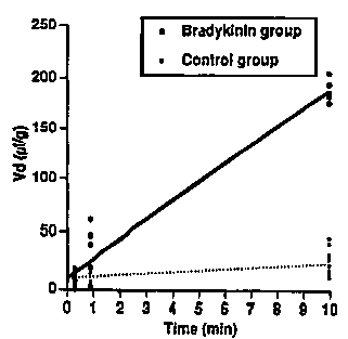
FIG. 1 is a graph showing that the selective
increase in the volume of distribution in brain tumors
is due to an increase in tumor ~ermeability and not
blood volume when treated with bradykinin in accordance
with the present invention.
FIG. 2A is a graph which summarizes test results
showing the selective uptake of dextran by brain tumor
tissue in accordance with the method of the present
invention. Bradykinin selectively incrased permeability
with the tumor 12-fold without increasing permeability
W094/27~3 ~ 5 PCT~S94/04269
--4--
inb rain surrounding tumor (BST), ipsilateral normal
cortex (ipsi carbon) contralateral noraml cortex (contra
cortex) ipsilateral white matter (ipsi wm) contralateral
white matter (contra wm) or ipsilaterial or
contralateral basal ganglia (BGG).
FIG. 2B depicts test results showing selective
uptake of AIB by brain tumor tissue in accordance with
the method of the present invention. In contrast to
dextran, which has a molecular weight of 70,000, AIB has
a molecular weight of 100.
FIG. 3 depicts test results showing the drop in
brain tumor blood barrier permeability during the time
period following infusion of bradykinin into the carotid
artery in accordance with the present invention. The
effect is reversible approximately 20 minutes after
stpp[ing the infusion of bradykinin.
DETAT~ DESCRIPTION OF THE PREFERRED EMBODIMENTS
The present invention is a method for selectively
opening abnormal brain tissue capillaries of a mammal in
order to allow selective passage of both low and high
molecular neuropharmaceutical agent into the abnormal
brain tissues. The present invention is applicable to
treating brain tumors, abnormal tissues resulting from
multiple sclerosis, ischemia and cerebral abscess. The
invention is also applicable to brain tissue which is
inflamed, infected or degenerated due to any number of
different diseases.
The method involves opening the abnormal brain
tissue capillaries by infusing bradykinin or a
bradykinin analog into the carotid artery of the mammal.
The bradykinin or bradykinin analog is infused in an
amount which is sufficient to selectively open the
abnormal brain tissue capillaries to allow passage of
neuropharmaceutical agents, including high molecular
weight agents, into the abnormal brain tissue without
W094/27~3 PCT~S94/04269
~ Q5~ ~
opening the normal brain capillaries to passage of the
neuropharmaceutical agent.
Brady~i ni n i5 a naturally occurring peptide
comprised of nine amino acids. The structure of
bradyk; ni n and methods for isolating and purifying
bradyki n; n are known. Analogs of bradyki~in include
related peptide structures which exhibit the same
properties as bradyki~i~ but have modified amino acids
or peptide extensions on either terminal end of the
peptide. Examples of bradykinin analogs include [phe8
(CH2-NH) Arg9] - bradykinin, N-acetyl [phe8 (CH2-NH - Arg9]
bradykinin and desArg9-bradykinin..
The amount of bradykinin which is infused into the
carotid artery in order to selectively open the abnormal
brain tissue capillaries to allow passage of neuro-
pharmaceutical agents through the BBB may be varied
depending upon the particular abnormal tissue being
treated and the patient weight. The preferred dosage
ranges from between 0.05 ~g/kg body weight/minute to
about 20 ~g/kg body weight/minute. The total amount of
bradykinin which is infused into the carotid artery
during any single treatment is preferably kept below
about 400 ~g/kg body weight. For treating most abnormal
tissues, the rate at which bradykinin is infused into
the carotid artery will be on the order of about 10
~g/kg body weight/minute.
It is preferred that the bradykinin is infused into
the carotid artery over a relatively short time period
on the order of about 5 minutes to about 20 minutes.
The selective opening of the abnormal brain tissue
capillaries resulting from the infusion lasts for
approximately 20 minutes after the bradykinin is
administered. During this time period, a
neuropharmaceutical agent may be introduced
intravenously or also through the carotid artery. The
selectively open abnormal brain tissue capillaries allow
W094/27~3 PCT~S94/04269
~ 0~5 -6-
passage of the neuropharmaceutical agent into the
abnormal brain tissue for treatment.
Any of the well known neuropharmaceutical agents
may be administered in accordance with the present
invention. Low mol~c~llAr weight (100 - 20,000) as well
as high molecular weight (about 20,000 to 70,000)
neuropharmaceutical agents may be used. In addition to
neuropharmaceutical agents, diagnostic agents may be
used including imaging or contrast agents. Exemplary
diagnostic agents include subst~nces that are
radioactively labelled such as 99-Tc glucoheptonate,
gadulium-EDTA, ferrous magnetic or iodinated contrast
agents. Exemplary neuropharmaceutical agents include
antibiotics, adrenergic agents, anticonvulsants,
nucleotide analogs, chemotherapeutic agents, anti-trauma
agents and other classes of agents used to treat or
prevent neurological disorders. Specific
neuropharmaceutical agents which can be ~rin;stered
into abnormal brain tissue in accordance with the
present invention include cisplatin, carboplatin,
methotraxate, 5-FU, amphotercin, immunotoxins, boron
compounds, and monoclonal antibodies.
The bradykinin is a~ri n istered into the carotid
artery by any of the well known infusion ~ec~n;ques.
For example, the bradykin;n may be directly infused into
the carotid artery by the following preferred procedure
used for cerebral angiography where a catheter is
inserted into the femoral artery and directed using
fluoroscopic X-rays into the inter~al cartod artery or
a more distal cerebral artery.
The bradykinin is preferably infused in the form of
a pharmaceutical solution dissolved in 0.9% saline at a
concentration of approximately 10-40 ~g/ml. Any of the
well known pharmaceutical carriers may be used as a
diluent for the bradykinin to provide a solution which
can be infused directly into the carotid artery.
W094/27~3 ~ PCT~S94/04269
Although the present invention is applicable to
selectively treating a wide variety of abnormal brain
tissues, the following examples will be limited to a
demonstration of the invention with respect to brain
tumors with it being understood by those skilled in the
art that the invention is not so limited.
Examples of practice are as follows.
An experimental brain tumor model was made using
female Wyster rats and RG-2 glioma cells. The RG-2
glioma cell line was maintained in a monolayer culture
in F12 medium with 10% calf serum. Female Wyster rats,
each weighing 150 to 250 gm were anesthetized with intra
peritoneal pentobarbital (30 mg/kg). Glial tumors were
implanted into the right hemisphere by intracerebral
injections of 1 x 10⁵ RG-2 glioma cells in five ~l of
1.2% methyl cellulose (F12 medium). One week after
tumor implantation, the rats were used for the brain
tumor model.
The rats were divided into two groups: a bradykinin
group treated with intracarotid infusion of lo
microliters/kg/min of bradykinin or the control group
treated with intracarotid infusion of saline. The
effect of intracarotid infusion of bradykinin was
compared to saline infusions by statistical analysis of
the Ki values using ANOVA and Students T-Tests.
It was determined that the 10 ~l/kg/min dose of
bradykinin dissolved in 0.9% saline did not alter
systemic blood pressure. At infusion rates greater than
20 ~g/kg/min the systemic blood pressure in the rats was
reduced.
The blood volume for the quantitative examination
of permeability was calculated with a graphic method
using [14X] dextran (MW 70,000). The blood volume in
normal brain tissue and tumors were 4.5 and 9.15 ~l/g,
respectively (Fig. 1). The slopes were the
unidirectional transfer constants, the Ki values
W094/27~3 PCT~S94104269
2~ ~2~5 -8-
(~l/g/min), in the two groups. The slope of the line of
the rats treated with bradyk;n; n indicated that the
increased volume of distribution resulted from increased
permeability and not from increased blood volume. The
tumor blood volume was almost twice that of the normal
brain tissue, but the brain and tumor blood volumes were
not altered by intracarotid bradykinin infusion.
[ C] AlB and tl4c] Dextran were used for
quantitative autoradiographic e~A~;nation of regional
permeability. One week after tumor implantation, the
rats were again anesthetized and a polyethylene (PE-10)
catheter was inserted retrograde throughout the external
carotid artery to the common carotid artery bifurcation
ipsilateral to the tumor. The external carotid artery
was then ligated. One femoral artery was cannulated to
monitor systemic blood pressure and the other femoral
artery was cannulated to withdraw arterial blood. Body
temperature was maint~;ne~ at 30OC and arterial blood
gas levels, blood pressure, hematocrit were monitored.
Animals with abnormal physiological parameters were
eliminated . After rat preparation, bradykinin
(10~g/Kg/min in saline) or saline as control was infused
into the right carotid artery at a rate of 53.3 ~l/min
for 15 minutes. Five minutes after the start of the
intracarotid infusion, 100 ~Ci/Kg of the tracer was
injected as an intravenous bolus. A peristaltic
withdrawal pump was used to withdraw femoral arterial
blood at a constant rate of 0.083 ml/min immediately
after injection of tracer for detçrmination of serum
radioactivity. Fifteen minutes after the start of
intracarotid infusions, the animals were killed by
decapitation and the brains were rapidly removed and
frozen. The regional permeability in the brains and
tumor tissues were expressed by the unidirectional
transfer constant, Ki value (~l/g/min). The Ki value of
the tumors for [14C] dextran (MW 70,000) in the
W094/27~3 216 2 Q 5 ~ PCT~S94/04269
bradykinin group was 12-fold higher than that for the
control group (Mean + SD; 17.84 + 1.00 vs. 1.47 + 1.24;
p<0.001) (Fig. 2A). This Ki value corresponded well
with the Ki value derived from the slope in Figure 1.
The Ki values of brain regions without tumor in
either bradykinin treated or control groups were very
low and there was no significant difference between the
two groups. The Ki value of the tumors for tl4c] AIB (MW
103) in the bradykinin group was 1.8-fold higher than
that for the control group (25.91 + 6.78 vs. 13.95 +
4.29; p<0.001 (Fig. 2B). The Ki value of the brain
suLLoullding tumor (BST; areas at 2 mm distance from the
border of the tumor) for tl4c] AIB for the bradykinin
group was also higher than that for the control group
(3.50 + 1.29 vs. 1.83 + 1.78; p<0.05). This result
shows that the effect of bradykinin on brain tumor
capillaries is selective and the effect is more profound
as the size of the tracer molecule increases.
Bradykinin has a short biological half-life because
of its proteolytic inactivation (17). To determine the
duration of the bradykinin effect on tumor capillary
permeability, the Ki at three different time periods was
measured. The rat preparation was the same as described
above. The Ki value was measured in three different
periods by changing the time of t~4c3 dextran injection
as also previously described. The three periods were as
follows: 0 to 10 min during the intracarotid bradykinin
infusion, 0 to 10 min after the infusion, and 20 to 20
min after the infusion. The experiment was terminated
at the end of each period. The Ki value was calculated.
Autoradiography was conducted as follows: The
frozen brains were mounted onto pedestals with M-1
embedding matrix, and 20 ~m coronal sections were cut
with a cryotome. The sections were thawmounted onto
cover slips, and autoradiograms were generated by
coexposing the sections on Kodak XAR-5 film with tissue-
W094/27~3 PCT~S94/04269
--~ 21~ ~ S~ -lo-
calibrated 1~C st~ rds for 2 weeks. The sequential
section was stained with hematoxylin for correlation of
areas of histologically verified tumor with
autoradiograms. The regional radioactivities were
measured in tumor, brain surro~ ; ng the tumor (BST;
areas at around 2 mm distance from the border of the
tumor), ipsilateral cortex to tumor, contralateral
cortex, ipsilateral white matter (WM), contralateral WM,
ipsilateral basal ganglia (BGG), and contralateral BGG.
Quantitative analysis of the regional radioactivity was
performed using a computer (Macintosh II) with a scanner
(UMAX) UC630) and the software, Image 1.45 (NIH).
The effect of bradykinin on tumor permeability was
dim;n; ~h~ 20 minutes after stopping the intracarotid
bradykinin infusion (Fig. 3). The degradation of
bradykinin in rats has been reported to be on the order
of several hours (18). The shorter effect of bradykinin
on tumor capillary permeability is believed to be due to
both the selective intracarotid infusion and the lower
does of bradykinin used. The short effect on tumor
capillaries of intracarotid infusion of bradyk;n;n is,
desirable for the selective delivery of anticancer drug
in the treatment of the brain tumors.
The enzyme that degrades bradykinin is peptidyl
carboxypeptidase kinase II, which is identical to
angiotensin I converging enzyme (ACE) (19). Williams,
et al., using antiserum to the purified pig kidney ACE,
reported that the pig brain capillary contained ACE
(20). Moreover, the ACE inhibitor, captopril, enhanced
the bradykinin effect (14, 21). Whether the rat brain
capillary had ACE was examined using antiserum to the
purified human kidney ACE. Angiotensin converting
enzyme was not recognized in the rat brain capillaries,
whereas this antiserum recognized ACE in the rat kidney
cortex. When an intravenous captopril infusion was used
to enhance the effects of bradykinin on tumor
W094/27~3 ~ 2 0~ PCT~S94/04269
--11--
permeability, hypotension occurred which made it
difficult to maintain normal systemic pressure.
Microscopic analysis was performed using
intravenously injected horseradish peroxidase (HRP) as
described in (22). After rat preparation as previously
described, bradykinin (10 ~g/Kg/min in saline) or saline
as a control was injected into the right carotid artery
for 15 minutes. Five minutes after the start of the
intracarotid infusion, 20 mg/lOOg of horseradish
peroxidase (HRP) was injected by an intravenous bolus.
Ten minutes after the HRP injection rats were perfused
with a mixture of 2% glutaraldehyde and 2% formaldehyde
in 0.1 sodium phosphate buffer solution at pH 7.4
through the heart. After fixation, the brains were
removed and cut at 40 ~m thickness on a vibratome. The
sections were preincubated for 15 min at room
temperature in the medium consisting of 10 ml 0.05 M-
Tris-HCl buffer (pH 7.4), 3,3'-diaminobenzidine
tetrahydrochloride and 0.02% hydrogen peroxide (Sigma).
The sections were trimmed down to the areas of
interests, postfixed for 2 hr in 2% osmium tetroxide
with 0.1 M sodium phosphate, dehydrated, and embedded in
plastic. Plastic sections 1 ~m thick were observed
under light microscopy.
The HRP stain was well recognized in the
extracellular space between tumor cells in the
bradykinin group, whereas the HRP stain was much less in
the control group. In normal brain, bradykinin
increased the HRP staining within t~e cytoplasm of only
a few endothelial cells and there was no extravasation
of HRP between cells. The effect of low dose bradykinin
on endocytosis in endothelial cells in normal brain is,
therefore, small. It has been reported that the
nanomolar concentrations of bradykinin stimulated the
uptake of the fluorescent marker, Lucifer yellow, in the
brain capillary endothelial cells by 40% (23). It also
W094l27~3 ~CT~S94lOn69
~ 2~6~5~ -12-
has been reported that high dose intracarotid infusion
of bradykinin (almost 6 times higher than the dose of
the present invention, caused intravasation of HRP
around the normal brain capillary. Vasodilatation of
microvessels and HRP endocytosis in endothelial cells
was also recognized. The tight junctions of the
endothelium were intact (16). In the above example, the
HRP stain was limited to a few endothelial cells in the
bradykinin group. This demonstrates that in contrast to
other studies using high dose bradykinin, lower doses of
bradykinin in accordance with the present invention
selectively increase the tumor permeability without
increasing the normal brain permeability.
To demonstrate that bradykinin could selectively
deliver other high molecular weight compounds into
tumors. Evans blue (EB) was injected intravenously
instead of radiolabeled tracers as follows: After the
preparation of rats as previously described, 2 ml/kg of
2% Evans Blue (EB) was injected intravenously as also
described above. After the intracarotid bradykinin
infusion, the rat was perfused with 200 ml of phosphate
buffer through the heart to wash out the remaining EB
from the vessels. The brains were removed immediately
and cut as coronal sections.
Since EB binds to serum albumin (MW 67,000) in
blood and distributes with albumin in vivo, EB staining
in the tissue indicates the distribution of albumin (4).
In order to observe the extravasated EB and not the EB
remaining in the vessels in the brain, the blood from
the brain was washed out by perfusing the rats with
phosphate buffer from the heart. The EB staining was
well recognized in the tumor but not in the normal brain
of the bradykinin group. Much less staining was seen in
the control group. This shows that intracarotid
bradykinin infusion selectively increased the delivery
of EB albumin to the tumor.
W094/27133 ~ Q~ r PCT~S94104269
2 ~
-13-
The above examples demonstrate the use of
Jintracarotid bradykinin infusion as a method to
selectively deliver high molecular weight agents to
brain tumors. Intracarotid bradykinin infusion at low
5 doses increases the permeability for the high molec~1lAr
weight tracer dextran by 12-fold, and for low molecular
weight tracer AIB by l.8 fold. Moreover, selective
extravasation of HRP and EB staining in tumors were
caused by intracarotid bradykinin infusion.
lO Accordingly, the method of the present invention is
useful for selectively delivering large molecular weight
compounds to brain tumors.
Having thus described exemplary embodiments of the
present invention, it should be noted by those skilled
15 in the art that the within disclosures are exemplary
only and that various other alternatives, adaptations
and modifications may be made within the scope of the
present invention. Accordingly, the present invention
is not limited to the specific embodiments as
20 illustrated herein, but is only limited by the following
claims.
W094/27~3 PCT~S94/04269
21~2~ 14-
BIBLIOGRAPHY
1. Kumagai AK, Eisenberg JB, Pardridge WM:
Absorptive- ~ ted endocystosis of cationized
albumin and a B-endorphin-cationized albumin
chimeric peptide by isolated brain capillaries.
Model system of blood-brain barrier transport. J
Biol Chem 262: 15214-15219, 1987.
2. Neuwelt EA, Barnett PA, McCormick CI, et al.:
Osmotic blood-brain barrier modification:
monoclonal antibody, albumin, and methotrexate
delivery to cerebrospinal fluid and brain.
Neuro~urgery 17:419-423, 1985.
3. Neuwelt EA, Hill SA, ~renkel EP: Osmotic blood-
brain barrier modification and combination
chemotherapy: concurrent tumor regression in areas
of barrier opening and progression in regions
distant to barrier opening. Neurosurgery 15: 362-
366, 1984.
4. Pardridge WM, Kumagai AK, Eisenberg JB: Ch;mcric
peptides as a vehicle for peptide pharmaceutical
delivery through the blood-brain barrier. 8iochem
Biophys Res Commun 146:307-313, 1987.
5. Iomiwa K, Hazama F., Mikawa H: Neurotoxicity of
vincristine after osmotic opening of the blood-
brain barrier. Neuropathol Appl Neurobiol 9:345-
354, 1983.
6. Black KL, Betz AL, Ar DB: Leukotriene C4 receptors
in isolated brain capillaries. Adv Protagl ~n~; n
Thromboxine Leukotriene Res 17:508-511, 1987.
7. Black KI, Hoff JT: Leukotrienes and blood-brain
barrier permeability: Cereb Flow Metab 5 ~Suppl):
263-264, 1985.
8. Black KL, Hoff JT: Leukotrienes increase blood-
brain barrier permeability following
intraparenchymal injections in rats. Ann Neurol
wog4n7ll3 ~16 2 Q S ~ PCT~594/04269
18:349-351, 1985.
9. Black KL, Hoff JT, McGillicuddy JE, et al:
Increased leukotriene C4 and vasogenic edema
surrounding brain tumors in humans. Ann Neurol
19:592-595, 1986.
10. Black KL, Chio CC: Increased Opening of Blood-
Tumour Barrier by Leukotriene C4 iS Dependent on
Size of Molecules. Neurologic~l Research Vol 7~,
December 1982, pp. 402-404.
11. Black, KL, King WA, Ikezaki K: Selective Opening of
the Blood Tumor Barrier by Intracarotid Infusion of
Leukotriene C4. J. Neurosurg, Vol. 72, June 1990,
pp. 912-916.
12. Baba T, Black KL, Ikezaki K, Chen K, Becker DP:
Intracarotid Infusion of Leukotriene C4 Selectively
Increases Blood-Brain Permeability After Focal
Ischemia in Rats. J Cereb Blood Flow Metab. Vol.
11, No. 4, 1991.
13. Chio CC, Baba T, Black KL: Selective Blood-Tumor
Barrier Description By Leukotrienes. J. Neurosurg.
Vol. 77, September 1992.
14. Yong T, et al., Circ Res 70, 952 (1992).
15. Alvarez AL, et al., Clin Sci 82, 513 (1992).
16. Raymond JJ, Robertson DM, Dinsdale HB, Can ~ Neurol
Sci 13, 214 tl986).
17. Erdos EG, ~ Cardiovasc Pharmacol 15, S20 (1990).;
Vanhoutee PM, et al., Br J Clin Pharmacol 28, 95
(1989).
18. Kumakura S, Kamo I, Tsurufuju S, Br ~ Pharmacol 93,
739 (1988).
19. Ng KKF, Vane JR, Nature 216, 762 (1967).
20. Williams TA, Hooper NM, T.A.J., ~ Neurochem 57, 193
( 199 1 ) .
21. Mombouli JV, Illiano S, Nagao T, Scott BT,
Vanhoutte PM, Circ Res 71, 137 (1992).
22. Nishio S, Ohta M, Abe M, Kitamura K, Acta
WO94/27133 PCT~S94/04269
~ ~2 ~ 16-
Neuropathol (BerlJ 59, 1 (1983).
23. Guillot FL, Audus KL, J Cereb Blood Flow Netab lO,
827 (199O).
24. Dobbin J, Crockard HA, Ross RR, J Cereb Blood Flow
Netab 9, 71 (1989).