Note: Descriptions are shown in the official language in which they were submitted.
2162303
The present invention relates to a novel and useful apparatus
for non-invasively identifying components associated with a liquid
within a container.
Chemical entities are often stored arad transported in a liquid
medium by containers and conduits. For example, medical solutions such
as enteral, parenteral, and other nutrients are compounded or mixed in
an intravenous bag fed by separate tubes leading from pure sources of
material. In particular, 70% dextrose injection U.S.P., 10% Travasol
(amino acid injection), Intralipid 20% I.V. Lipid emulsion, sterile
water, potassium chloride, and the like are combined in this manner.
Many systems have been proposed to determine the identity of components
associated with a liquid passing through a tube' or lying in a container
using electrical characteristics of the particular component within the
tube. For example, such systems are generally invasive, in that the
particular probe or electrode of a probe contacts the fluid within the
container or tube. Tnvasive measurements of this type are not
acceptable in certain fields such as medical fluids which are
intravenously delivered to a patient.
For example, United States patent 3,774,238 describes an
1
21 b23~3
invasive measurement of pipeline material utilizing three capacitors to
determine dielectric properties of the material.
United States patent 4,074,184 employs invasive capacitance
measurements of non-conductive fluids in a tube to determine the vapor-
7.iquid phase ratio.
United States patent 4,227,151 shows an invasive measuring
cell for use in determining the electrical conductivity of fluids in a
container.
United States patent 4,924,702 describes invasive capacitance
sensors that determine the liquid level of a container.
United States patent 4,928,065 shows an invasive measuring
apparatus which provides an electrical fif:ld from electrodes to
determine the characteristics of non-aqueous low conductivity
suspensions.
United States patent 4,935,207 employs an invasive capacitive
sensor which detects analyte ions in a liquid container.
United States patent 5,068,617 teaches an invasive capacitive
device which measures the mixing ratios of composite liquids within a
container.
United States patent 5,208,544 describes an invasive sensor
which produces dielectric measurements on high temperature molten
polymer compositions flowing in a conduit.
United States patent 5,255,656 delineates an electronic sensor
2
2162303
which invasively measures the methanol-gasoline mixture in a fuel line
by the use of capacitive elements.
United States patent 5,266,899 discloses an invasive salt
analyzer which measures the conductivity of saline solutions by
inductive non-contact.
United States patent 5,296,843 shows a fluid or vapor
diagnostic device employing an invasive probe which generates a light
beam passed through a container to a detector to determine the vapor-
liquid ratio within that container.
Several systems have been proposed which non-invasively
determine the characteristics of fluid in a conduit or container. For
example, United States patent 5,239,860 utili2:es a light beam which is
sent through a tube and detected after interaction with a gasoline
alcohol mixture. The wavelength transmission characteristics then
determine the actual alcohol-gasoline mixture within the tube.
United States patent 5,260,665 utilizes a non-invasive
resonance cell with a pair of probes located therewithin to determine
the presence of bubbles within the fluid lane passing through the
resonance cavity.
An apparatus which is capable of non-invasively identifying
components in a container using the electrical conductivity
characteristics of the fluid therewithin would. be a notable advance in
the medical field.
3
2i6~303
SUMMARY OF THE INVENTION
A novel and useful apparatus and method for non-invasively
identifying components in a container are herein provided.
The apparatus and process of the present invention are
particularly useful in identifying components associated with a liquid
in an electrically non-conductive container such as a tube. The
apparatus includes at least a pair of electrodes which are placed
adjacent the exterior of the container and positioned in spaced
configuration from one another along a dimension of the container. In
t:he case of an elongated flow conduit, such dimension would be the
length of such elongated conduit. The first electrode connects to
signal means for generating an input waveforrn. The second electrode
spaced along the dimension of the container would serve as a receiving
or acquiring electrode which out couples the electrical signal after
interaction with the components associated with the liquid in the
container. In certain cases, multiple receiving electrodes may be
employed along the container to ascertain the waveform after interaction
with the components associated with the liquid in the container, each
being positioned at greater and greater distances from the first or
signal waveform propagating electrode. In essence, the propagating and
receiving electrodes farm a capacitive resistive circuit with the
dielectric container walls and the liquid in the container. The
generated signal is modified by the electrical properties of the fluid
4
2i~2303
within the container including conductance, polarizability, and
dielectric constant. The conductance of the intervening solution in the
cell or tube is found in the resulting signal at the receiving
electrodes spaced from the propagating electrodes along the fluid
<:ontainer. "Conductance" is used herein to mean "electrical
conductance". Of course, the resulting signal acquired by the receiving
electrodes is also dependant on the specific cell configuration, i.e.,
t:he cell constant, which is a function of cell and electrode dimensions.
Further, polar or ionic species of fluid contain a charge concentration
which may lead to signal variations that permits distinguishing of polar
from non-polar compounds, which manifest themselves in different signal
profiles on time-voltage plots.
The signal means may further be described as an electrical
waveform generator which is employed to drive t:he propagating electrode.
Analyzing means is also used for receiving an output waveform from the
receiving electrode or electrodes after interaction of the input
waveform with the components within the container. The analyzing means
also synchronizes the input and output wavefo:rms by the use of timing
means which synchronizes the received signal by the receiving electrodes
to the waveform generator. Such timing means is also employed to
activate the analyzing means. The analyzing means acquires data which
may be transformed into voltage quantities representing particular
fluids in the container or to construct a time-voltage profile of the
2162303
particular fluid in the container. Consequently, the peak voltages or
time voltage profiles positively identify and quantify the fluid within
the container.
The signal means for generating an input waveform may be of a
type of exciting voltage known as a step function, i.e., a square wave.
Thus, the use of a step function easily transforms into characteristics
of the fluid within the container being characterized as a function of
voltage with time. Moreover, since the response voltages acquired by
t:he receiving electrodes increase upon excitation, peak, and decay over
time, various compounds may be distinguished on this basis. For
example, different aqueous solutions were observed to reach a peak
voltage at different times after excitation and to possess different
decay characteristics.
In addition, separation of the receiving or signal acquiring
electrodes from the propagating electrode to predetermined distances
generates response waveforms that are furthesr determinative of the
identification of the components within the' liquid medium in the
container. Maximum peak signals acquired with various electrode
separation facilitate such distinguishing of various components within
the container. Moreover, this arrangement is favorable in high
conductivity solutions where the propagated signal is detectable over a
greater distance than in solutions of low conductivity.
It may be apparent that a novel and useful apparatus and
6
21 6 23 3
method for identifying components associated
with a
liquid in a dielectrical container has bE=_en
described.
It is therefore an object of an a spect of the
present invention to provide an apparatus an d method for
identifying components associated with. a liquid in
a
dielectrical container that is capable of operation
while
such components are flowing within the container.
Another object of an aspect of the present
invention is to provide an apparatus for identifying
.LO components associated with a liquid in a dielectric
container that is simple to use and accurate.
Another object of an aspect of the present
invention is to provide an apparatus for identifying
components associated with a liquid in a dielectrical
.L5 container that is non-invasive and may be easily
employed
with medical solutions such as parenteral, and enteral
components.
A further object of an aspect of the present
invention is to provide an apparatus for identifying
:?0 components associated with a liquid in a dielectrical
container that e:Liminates the danger of erroneously
compounding medical solutions.
Yet another object of an aspect of the present
invention is to provide an apparatus for identifying
~?5 components associated with a liquid i:n a dielectrical
container that is useful in the filling of intravenous
containers and may be employed to determine the maximum
content of electrolytes therein.
The invention possesses other objects and
.30 advantages especially as concerns particular
characteristics and features thereof which will become
apparent as the specification continues.
A
CA 02162303 2004-07-29
According to an aspect of the invention, an apparatus for identifying
components associated with a liquid in a dielectric container comprising:
a. a first electrode placed apart from contact with the liquid
and at the dielectric container;
b. a second electrode placed apart from contact with the
liquid and at the dielectric container, said second electrode positioned in
spaced
configuration from said first electrode along the outer surface of the
container;
c. signal means for generating an input waveform, said
signal means being electrically connected to said first electrode; and
d. analyzing means for receiving an output waveform from
said second electrode after interaction of said input waveform with the
components within the container, said analyzing means further including
indicating means for correlating said output waveform to the presence of
particular components in the container, said analyzing means further including
timing means linked to said signal means for synchronizing said signal means
with said analyzing means.
According to another aspect of the invention, a method of determining the
presence of a component associated with a liquid in a dielectric container
comprising:
a. placing a first electrode adjacent the exterior of the
container;
b. placing at least a second electrode adjacent the exterior
of the container at a position in spaced configuration from said first
electrode
along the outer surface of the container;
c. generating an input waveform and feeding said input
waveform to said first electrode; and
d. analyzing an output waveform from the second electrode
after interaction of the input waveform with the component within the
container to
determine an electrical characteristic indicating the presence of a particular
component in the container, said step of analyzing an output waveform
including
the step of providing timing means linked to signal means for synchronizing
said
signal means with analyzing means.
8
CA 02162303 2004-07-29
According to another aspect of the invention, an apparatus for identifying
parenteral and enteral nutrients comprising:
a. a first electrode placed adjacent the exterior of the
container;
b. a second electrode placed adjacent the exterior of the
container and positioned in spaced configuration from said first electrode
along
the outer surface of the container;
c. signal means for generating an input waveform, said
signal means electrically connected to said first electrode; and
d. analyzing means for receiving an output waveform from
said second electrode after interaction of said input waveform with the
components within the container, and comparing said input and output
waveforms, said analyzing means further including indicating means for
correlating said output waveform to the presence of particular components in
the
container, said analyzing means further including timing means linked to said
signal means for synchronizing said signal means with said analyzing means.
According to a further aspect of the invention, an apparatus for identifying
the presence of a liquid in a dielectric transfer tube comprising:
a. a first electrode placed adjacent the exterior of the
container,
b. a second electrode placed adjacent the exterior of the
container and positioned in spaced configuration from said first electrode
along
the outer surface of the container;
c. signal means for generating an input waveform, said
signal means being electrically connected to said first electrode; and
d. analyzing means for receiving an output waveform from
said second electrode after interaction of said input waveform with the
components
within the container, and comparing said input and output waveforms, said
analyzing
means further including indicating means for correlating said output waveform
to the
presence of particular components in the containers, said analyzing means
further
including timing means linked to said signal means for synchronizing said
signal
means with said analyzing means.
8a
CA 02162303 2003-12-12
BRIEF DESCRIPTION OF THE DRAWINGS
FIG. 1 is a side schematic view of a prior art
dielectric cell measuring system.
FIG. 2 is a top plan schematic view of the theoretical system of
the present invention, depicted mechanically and electrically.
FIG. 3 is a block diagram representative of the overall apparatus
and method of the present invention.
FIG. 4 is an isometric view of a partial mechanical embodiment
of the present invention using a single waveform propagating electrode and
multiple receiving electrodes on a dielectric conduit.
FIG. 5 is an electrical schematic view indicating exemplary non-
invasive conductivity values obtained with the apparatus and method of the
present invention depicted in Figs. 2, 3, 4, 6, and 7.
FIG. 6 is an electrical schematic diagram of the circuitry
employed in the present invention.
FIG. 7 is a sectional view of a modified conductivity probe to
obtain data described in detail in Example 1 hereinafter.
FIG. 8 is a sectional view taken along line 8-8 of Fig. 7.
FIGS. 9-11 are graphical representations of quantitative
analyses conducted utilizing the probe of Figs. 7 and 8 and described in
detail
in Example 1.
8b
21b23~3
FIGS. 12 and 13 represent analytical. results delineated in
Example 3.
For a better understanding of the invention reference is made
to the following detailed description of the preferred embodiments
thereof which should be referenced to the prior described drawings.
DETAILED DESCRIPTION OF THE PREFERRED EMBODIMENTS
Various aspects of the present invention will evolve from the
following detailed description of the preferred embodiments thereof
which should be taken in conjunction to the prior described drawings.
The invention as a whole is shown in the drawings by reference
character 10 and is depicted schematically in Fig. 3. The apparatus 10
is employed to identify liquid components in a d_lelectric container such
as a tube, vat, and the like. In particular the apparatus and process
of the present invention are especially applicable to the detection of
parenteral, and enteral components which are emp:Loyed in the composition
of appropriate medical solutions.
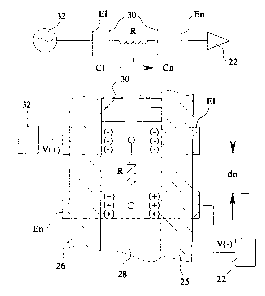
With respect to Fig. 1, a prior art dielectric cell 12 is
depicted and is normally employed to measure the dielectric constant of
solution 14 within cell 12. Electrodes 16 and 18 are normally placed
outside cell 12. Electrode 16 and electrode 18 form the capacitor in a
resonance circuit 21. The resonant frequency of circuit 21 is
proportional to the capacitance of opposing electrodes 16 and 18. The
frequency of resonance is used to determine the electrical
9
2 i 62303
cr~aracteristics of solution 14, namely the dielectric constant. Phantom
electrodes 16 and 18 have also been placed immediately inside the walls
24 of cell 12 to indicate prior invasive measuring techniques for highly
dielectric liquids. Cell 12 characteristics have been determined by
measuring the average voltage developed in an R.-C circuit with a fixed
driving frequency. Cell 12 fails in measuring the dielectric constant
of solutions 14 having a high conductivity. This deficiency is believed
to be due to the dominance of the conductivity of solution 14. The
electrodes 16 and 18 are simply positioned in opposition to one another
across cell 12 as depicted in Fig. 1 to form a ~>arallel plate capacitor
separated by a distance dl.
Fig. 2 is a theoretical mechanical a:nd electronic schematic
depicting workability of apparatus 10 of t:he present invention.
Electrode E1 propagates a waveform (V+) received from a waveform
generator 32. The term "electrode" is employed herein in its broadest
sense as an item or element that emits, col:Lects, or controls the
movements of electrons. Electrode EN represents one or more receiving
electrodes found on the outside wall 25 of dielectric container 26
depicted schematically in Fig. 2. Electrodes E1, En couple (primarily
by an electric field) or induce (primarily by a magnetic field) the
waveform (V+) through the wall 25 of dielectric container 26, and into
or from the material or fluid 28 being sensed within dielectric
container 26. By placing any of the receiving electrodes En along the
CA 02162303 2003-12-12
outside wall 25 of container 26 (distance d1), rather than across or opposite
from one another, cell 12 of Fig. 1, capacitive effects are minimized.
Receiving electrodes En may be positioned non-invasively to lie against the
outside wall 25 of dielectric container 26 or be embedded within wall 25 of
dielectric container 26. In contrast, in the dielectric cell 12 of Fig. 1, the
capacitive effects are maximized. Multiple electrodes, En, may be employed
in apparatus 10 of the present invention at various distances measured along
outside wall 25 of dielectric container 26. In addition, the primary
capacitance
is related to the electric field from each electrode E1, En to the solution
across
the outside cell wall 25, depicted schematically in Fig. 2. The charge
concentration of sample fluid 28 between inner walls 30 is depicted by the (_)
and (-) symbols along the inner wall 30 of dielectric container 26. The
resistance of the sample is indicated by "R". In turn, conductance of the
intervening sample 28 is represented by the formula: 1/R
Fig. 2 represents a resistive capacitive circuit with the electrical
characteristics modified by the conductance, polarizability, ion mobility,
dielectric constant, and other electrical properties of the fluid sample 28
between the dielectric wall portion 30. It should be noted that the resultant
signal on. electrode EN, indicated as V(-) at terminal 22, is dependant on
specific cell configuration formed by container 26, as well as the
conductance,
oolarizabilitv. and dielectric constant ~f the
11
21b2303
intervening fluid. In essence, Fig. 2 indicates that different signal
profiles may be obtained in time-voltage plots using electrodes on the
exterior wall 25 of container 26.
Apparatus 10, shown as a functional block diagram in Fig. 3
includes electrical waveform generator 32 which generates a periodic
signal that may be sinusoidal, a square wave, a saw tooth, or any
modification of the same. Waveform generator 32 passes the waveform
signal to propagating electrode E1 around the cell 34. Cell 34 may take
the form of a container such as a tube, vat, and the like. Fluid may be
flowing through cell 34 or be static therewi.thin. Although E1 is
depicted in singular form, additional propagating electrodes may be
employed (not shown) to provide additional electrical shielding or field
shaping of the waveform in order to modify the cell constant of cell 34.
Collecting, acquiring, or receiving electrodes E2 and E3 are also
depicted in Fig. 3. It should be noted that a plurality of such
receiving electrodes may be employed in the present apparatus 10.
Collecting or receiving electrodes E2 and E3 are spaced from propagating
electrode E1 and positioned along a dimension of cell 34. Where cell 34
is a tube, such particular dimension would be the length of that tube,
which will be shown in detail hereinafter. After modification by the
components associated with a liquid within cell 34, the output waveform
(current (I) or voltage (V)) received by electrc>des E2 and E3 is passed
to first and second amplifying means 36 and 38. The output of
12
2162303
amplifying means 36 and 38 are passed to analyzing means 40 in the form
of a signal which may be a voltage. Timing means 42 synchronizes the
output of waveform generator 32 and the input from first and second
amplifying means 36 and 38 to analyzing means 40. Timing means 42 also
activates analyzing means 40. The timing means signal is indicated by
"t" in Fig. 3. Analyzing means 40 processes the timing signal and the
voltage outputs of first and second amplifying means 36 and 38 are
representative of the fluid in capacitive cell 34 between propagating
electrode E1 and E2 or E3. Thus, a voltage-time profile of the fluid in
the cell may be plotted to positively identify and quantify a particular
component of fluid sample 28. Such identification is especially
important with medical solutions.
Referring to Fig. 4, it may be observed that a dielectric tube
44 is employed to conduct fluid therethrough according to directional
arrows 46 and 48, although fluid may flow in t=he opposite direction.
Propagating electrode E1 and receiving electrodes E2 and E3 are shown
adjacent outer wall 50 of tube 44. Of course, other receiving
electrodes may be used in addition to electrc>des E2 and E3 in this
regard. Electrodes E1, E2, and E3 are generally metallic material such
as stainless steel and include a plurality of electrical conductors 52
shown schematically in Fig. 4. It should be realized that electrodes E1
and E2 are separated from one another along tube 44 at a distance dl.
Also, electrode E1 and E3 are separated by a distance d2. The
13
2162303
significance of this separation will be discussE:d in detail hereinafter
and illustrated in the subsequent examples.
The form of the exciting voltage from waveform generator 32 is
preferably a periodic wave and may be a "step" function, such as a
square wave. By utilizing a step function as 'the source, it has been
found that the characteristics of the intervening fluid within cell 34
can be described above as a function of voltage and time. With
reference to Fig. 5, this phenomena is exemplified where an input square
wave 54 is sent to electrode E1 of Fig. 4. The response signals for 10%
amino acid injection, dextrose, and water are depicted in graph 56, Fig.
5. Response voltages shown in graph 56 rapidly increase upon excitation
by input signal 54, while peak and decay vary ovE~r time depending on the
particular component being present in capacitive cell 34, Fig. 3. For
example, the values for amino acid injection, dextrose, and water, are
quite different at time point "a" and time point "b". In other words,
the analyzing means detection electronics looka at the amplitude of a
response voltage versus time at a particular time or the average
response voltage over an interval of time. In either case, compounds
present in cell 34 are easily distinguishable. Moreover, in graph 56 of
Fig. 5, amino acid injection reached a maximum peak intensity at
approximately (100) nanoseconds prior to the dextrose solution.
However, dextrose exhibited a much slower signal increase and decay than
10% amino acid injection. Thus, a different response signal result is
14
2162303
attained at time instants "a" and "b". It has been found that a square
wave input signal 54 at about 200 kilohertz possesses sufficient speed
to characterize differences in the response of components of Fig. 5 (5-
microsecond response time). In other words, input signal rise times
of between (10) nanoseconds and several microseconds are sufficient to
distinguish most responses in common parenteral nutrients, trace element
solutions, and various electrolytes, depending on cell configuration.
In addition, a capacitively coupled periodic signal such as square wave
input 54 is also helpful in minimizing long term ion migration and
concentration gradients at or near electrodes E1, E2, and E3. Thus,
instability or drift of the response signal over time is greatly
minimized. It should be understood that other periodic waveforms
generated by waveform generator 32 may be employed to enhance the
results depicted in Fig. 5. For example, a ramp waveform, an integral
of a ramp waveform and the like may be employed in this regard.
Fig. 6 represents the electrical schematic of the signal
acquiring circuit used in the apparatus 10 of they present invention. An
IC timer U1, generates an input square wave. C1, C2, and C3 operate as
power supply filters to bypass noise. Integrated circuit (IC) timer U1
is configurated for stable operation as oscillator 66. R1, R2, and C4
are selected to generate a near 50% duty cycle square wave on pin 3 of
U1. R3 provides additional source drive capability for U1. Cell or
tube 34 containing a component to be analyzed, is surrounded in the
2 ~ ~~30~
configuration depicted in Fig. 4 by electrodes E1, E2, and E3. Signal
propagating electrode E1 sends the waveform from oscillator 66 through
th.e components found in cell 34. Acquiring electrodes E2 and E3 pass
the output signal to amplifiers U2 and U3, respectively. U2 is a
transimpedance amplifier (current to voltage). Gain and response times
are determined by R6 and C5. The RC constant for these components is
.44 micro seconds. R4 is a high impedance: ground bias for the
capacitively coupled E2 input to U2. The output of U2 is sent to
oscilloscope 68 and from there to computer 70 via an RS232 link.
Computer 70 may be an IBM PC/486 DX2-66 with 8 megabytes RAM and a
grams/386, version 3.018, level 12 program, from Galactic Industries of
Salem, New Hampshire. Transimpedance amplifier i:T3, receiving the output
from electrode E3, performs a similar function with respect to U2 by
inputing oscilloscope 68. R5, C6, and R7 are analogous to the R4, C5,
and R6 components with respect to amplifier U2.
The following list represents components employed in Fig. 6.
Item Designation Source
C1 0.1 Micro F M.l. Std. CK05BX 104K 50V
C2 0.1 Micro F " "
C3 0.1 Micro F - Series " "
C4 100PF - C114 Kemet " "
C5 2pF Kemet Electronics Corp.
Greenville, SC
C6 2pF "
R1 15 kOhm RN55D Series Dale Electronics (VISHAY)
Columbus, NE
R2 1 kOhm " "
R3 1 kOhm " "
R4 10 MOhm " "
16
212303
R5 10 MOhm " "
R6 220 kOhm " "
RT 220 kOhm " "
U-1 (66) 555 Timer Texas Instruments, Dallas, TX
U-2 (36) OP Amp 843 Analog Device, Norwood MA
U-3 (38) OP Amp 843 " "
Oscilloscope 68 Model 97 Fluke, Inc., Everette, WA
Computer 70 PC/386 w/RS232
In operation, apparatus 10 determines the presence of a
component flowing or static within a container such as dielectric
tube 44 by placing a first electrode E1 adjacent the exterior
surface 50 of tube 44. Additionally, electrodes E2 and E3 are also
fastened adjacent the exterior 50 of tube 44, but are spaced from
ore another and spaced from electrode E1 along tube 44. Such
spacing occurs along a particular dimension such as the length of
tube 44. For example, electrodes E1, E2, and E3 are typically (10-
13) mm long for a (4) mm OD tube and spaced at 10-13 mm intervals.
With respect to the schematic shown in Fig. 6, a waveform is
generated by oscillator 32, 66 and fed, using the=_ circuitry of Fig.
6, to propagating electrode E1. After interaction with the
components within tube 44, electrodes E2 and E3 acquire the signal
arid pass the same to analyzing means 40 in the form of oscilloscope
68 and personal computer 70 via RS232 communications. Oscilloscope
6~~ indicates a characteristic output signal pe:r unit time. Time
voltage points are queried by computer 70 to oscillator 66 via the
RS232 of computer 70. A software program in computer 70 determines
a characteristic, such as conductivity of a component within tube
17
2162303
44. Timing means 42 such as timer U1, Fig. 6, generates the signal
to be used to trigger (synchronize) signals t:o produce further
distinguishing characteristics of the acquired signal along a
certain time span. Components or materials within tube 44 are
easily identified by comparison or signal characteristics
determined by both time and distance signals measured, as modified
by the material s electrical characteristics.
While in the foregoing, embodiments of the present
invention have been set forth in considerable detail for the
purposes of making a complete disclosure of the invention, it may
be apparent to those of skill in the art that numerous changes may
be made in such detail without departing from the spirit and
principles of the invention.
The following examples are included for the purposes of
illustration, but are not intended to limit the scope of the
invention unless otherwise indicated.
EXAMPLE I ~,NVA~,~~O~UCTIVI~
Invasive conductivity measurements using the following
procedure were conducted in order to ascertain whether direct
cc>ntact conductivity measurements could be used to identify
different parenteral nutrients normally availab7.e in a compounding
process. A conductivity meter, Orion Model 126, with a modified
Onion probe 012210, manufactured by Orion, Inc., Boston,
18
CA 02162303 2004-12-21
Massachusetts, was employed to measure the conductivity and to identify
different parenteral nutrients during compounding using a commercial
compounder. The probe employed had a thermocouple for accurate
temperature compensation and a four graphite electrode design to eliminate
any polarization effects with a cell constant of 0.69/cm. With reference to
Figs. 7 and 8, probe 59 was modified by inserting into its interior 62 PVC
tube
55 to form container 61. PVC tubes 57 and 58 were inserted within the
cylindrical cavity 60 of container 61. The solution to be analyzed was passed
into and held within the cavity or chamber 60, allowing fluid contact with the
concentric Orion probe electrodes 64. This procedure decreased the volume
of container 61 and reduced the mixing time of the sample within chamber 60
in contact with the electrodes 64. Container 61 possessed a cell constant of
0.964. In such configuration, the thermocouple was not in contact with the
liquid being tested.
Table I represents the data obtained in this example.
TABLE I
ITEM CONDUCTIVITY, mS/cm
@ 25°C (cell constant
= 0.94/cm
10% Travasol (Amino Acid) Injection 6.63
70% Dextrose Injection USP 0.02
Sterile Water 0.00
Intralipid 20% I.V. Fat Emulsion 0.43
8.5% Travasol (Amino Acid) Injection 5.02
10% FreAmine III** Amino Acid Injection5.32
10% Aminocyn Amino Acid Injection 5.67
6% TrophAmine Amino Acid Injection 4.21
Potassium Phosphates Injection USP;
19
~1~~303
3 mM/ml P, 4.4. mEq/ml K 150.80
MVI Pediatric Vitamins 0.20
Heparin Sodium Injection USP; 1000
USP Units/ml 7.42
Zinc Sulfate Injection USP; lmg Zn/ml 3.07
MTE-5 Trace Element Mixture 8.92
Potassium Chloride Injection
Concentrate USP; 2mEq/ml 210.00
Calcium Gluconate Injection USP 5.53
Potassium Acetate Injection USP;
4 mEq.ml 146.40
Lypholyte Multi-Electrolyte Concentrate 143.10
Sodium Acetate Injection USP; 4 mEq/ml 74.60
A dynamic test was performed with a commercial compounder where
individual tubes were connected to multiple starting solution
bottles and then combined into one tube at a junction or manifold.
Peristaltic pumps forced the starting solutions through the
manifold and into container 61 via tube 57. The solutions tested
included sterile water, 70% Dextrose injection, 10% amino acid
injection solution, and 20% lipid emulsion. Normally the outlet
PV'C tube 58 would have led to an intravenou:a bag, but in the
present example, probes 64 were inserted within cylindrical cavity
60 between the junction or manifold of the compounder and the place
wraere the PVC tube 58 connected to the final container (IV bag).
The conductivity meter was set to manual scaling and the analog
voltage output was connected to an analog-to-digital board in a
personal computer. The resulting counts were recorded as a
function of compounding time, transferred to a computer spreadsheet
program, and plotted.
With reference to Fig. 9, a distinction was detected
2162303
between amino acid and lipid solutions. Water and dextrose, having
much lower conductivities were not identifiable on the plot of Fig.
9. The compounding time plotted against measured conductivity on
Fig. 9 also included the volume of each solution pumped through the
compounder. With reference to Fig. 10, a finer conductivity scale
was employed and illustrated a measurable distinction between
sterile water and 70% dextrose solution. It is believed that the
large spike in conductivity at the beginning of t:he plot of Fig. 10
was due to the expulsion of previously existing amino acid in the
compounder tubing 58. Referring to Fig. 11, different amino acid
injection solutions were shown to be distinguishable. It is also
noted that gas bubbles transferring through the tubing are plotted
as a sharp decrease in conductivity. It was concluded that direct
contact measurements of electrical conductivity of compounds can be
used to identify them during the compounding process.
EXAMPLE 2 NON-INVASIVE CAPACITIVE MEASUREMENTS
Solutions were contained in polyvinyl chloride/PV Acetate
tubing of the type typically used in commercial compounders having
an outer diameter of about 6.35 millimeters and a wall thickness of
about 0.9 millimeters. Static measurements were made on these
tubes and used to positively identify several major nutrients using
a commercial compounder under flow conditions. Fig. 4 represents
a portion of the apparatus employed in this example. The
21
21623x3
propagating electrode E1 and acquiring electrodes E2 and E3 were
formed of conductive tapes, available from 3M Co., St. Paul,
Minnesota. The conductive tapes had a width of 12.7 millimeters
and a thickness of about 0.075 millimeters. Th.e conductive tapes
were placed completely around the tubing such as tubing 44 of Fig.
4, and were spaced 13 millimeters apart. These tapes rested in
aluminum holders machined in a shape that conformed to the exterior
surface of the tubing. A square wave signal was generated with a
frequency of about 220kHz for the following measurements. This
frequency was chosen because good distinction among compounds
tested was obtained, although other frequencies were used as well.
Means for sampling and holding and means for averaging was employed
in. the electronic circuit, i.e., analyzing means 40 of Fig. 6. A
response signal received by one acquiring electrode formed of the
conductive tapes was sent to an analog-to-digital (A/D) converter,
which in turn was connected to the parallel port of a personal
computer. A computer program was written to acquire the signal
from the A/D converter, to display its value, and to identify a
particular chemical entity found in the exit tube from the
compounder. Such computer program is included hereto as an
appendix hereto. Table II represents the values for certain
parenteral nutrients which was determined by th.e above method and
apparatus. It should be noted that there were 2.44 millivolts per
22
2 i 62303
(A./D) count in Table II. As may be seen, the values obtained
indicate that individual compounds may be easily distinguished.
Furthermore, it was found that the absence of a liquid compound in
a tube could also be detected relative to an empty tube. 70%
dextrose is associated with a test count of 260, while sterile
water was associated with 130 counts. This difference indicates
that other dextrose solutions such as 35% dextrose should be
readily distinguishable also from sterile water. It is also
determined that errors of mistakenly switching water and dextrose
tubes on a compounder should immediately be identified with the
invention shown. Figs. 2-4 represent the mechanical, electronic,
and schematic definition of the device employed in the present
example.
NON-INVASIVE CAPACITIVE TEST VALUES
Compound Counts
No tube present 0
Empty tube present 10
Sterile Water 130
70% Dextrose Injection 260
10% Amino Acid Injection 350
20% Lipid Emulsion 680
EXAMPLE 3 NON-INVASIVE CAPAC:j,,~E MEASUREI~ENT_S
TJSING FULL PEAR PROFILE AND MULTIPLE
The electrodes such as those depicted .in Fig. 4, with the
addition of electrodes E4 and E5 (not shown) identical to
23
electrode E2 were employed with the circuit shown in Fig. 6. A
modified square wave input signal was used as the waveform sent to
electrode E1 through timer integrated circuit U1. The input signal
had a period near 220 kHz. An electrodes E2. was successively
placed at 0.5 (12.7mm) inch intervals along a (3) millimeter inner
diameter polyvinyl chloride (PVC) tube connected to an intravenous
bag. Such placements are depicted as E2, E3, E4, and E5. The
electrodes were fitted snugly over the exterior surface of the
polyvinyl chloride tube. Each electrode, E1 and E2, was 0.5 inches
wide and were connected to the circuit depicted in Fig. 6. The
square waveform input was generated and transmitted from the 555
oscillator, through the tube and solution contained in the tube to
electrode E2-E5, the acquiring electrodes. The response waveforms
were then amplified, acquired and measured by an oscilloscope, a
Scopemeter 97 manufactured by Fluke, Inc. of Everett, Washington,
i.e.', triggered to synchronize to the input waveform. The acquired
signal was then fed to the serial port of a computer and collected
for analysis using the Grams/386 Level II software program,
manufactured by Galactic Industries of Salem, Maine. The input and
output or response waveforms were collected from the oscilloscope.
A commercial compounder for parenteral nutrients, i.e., a
cempounder, manufactured by Clintec Nutrition Company under the
trademark Automix 3-3, of Deerfield, Illinois, was used to transfer
24
z ~ ~~~o~
approximately 60 milliliters of solution from sc>urce containers to
th.e intravenous bag via the polyvinyl chloride' tube. Waveforms
were collected during the pumping cycle. Fig. 12 represents the
characteristic response waveforms for 10% Travasol, Lypholyte, and
30% dextrose, three common parenteral nutrients. Response
waveforms shown in Fig. 12 for each nutrient. spans about 2.2
microseconds. The vertical axis represents signal voltage in
volts. Each of the three nutrients was determined to have a
characteristic response profile reaching maximum signal quickly and
decaying over time. The distances shown on the graph of Fig. 12
represents electrode separation between the waveform propagating
electrode E1 and a single electrode E2. The 30% dextrose injection
solution exhibited a broader maximum voltage, reaches such maximum
voltage later than 10% Travasol or Lypholyte pea)c measurements, and
decays more slowly over time. The 10% Travasol injection solution
exhibited a narrower peak at an earlier time than the peaks reached
by the dextrose solution. In addition, 10% Travasol decayed more
quickly than dextrose. Moreover, 10% Travasol exhibited less
variation of peak voltage for changes in E1-E2 distances than
dextrose. Lypholyte, a mixture of electrolytes, showed a similar
response to 10% Travasol, but with a lower maximum signal and very
little peak voltage variation with changes in the E1-E2 electrode
distance. Fig. 13 depicts a change in maximum peak signals of the
216203
nutrients plotted in Fig. 12 using propagating electrodes and
acquiring electrodes El, E2, and the designations E3, E4, and E5
representing different distance intervals of E2 :From E1. Thus, the
10% Travasol and Lypholyte may be distinguished by combining the
results of Figs. 12 and 13. It was concluded that adding a second
or several additional receiving or signal acquiring electrodes such
as E2, further distinguished solutions passing through the
polyvinyl chloride tube.
26