Note: Descriptions are shown in the official language in which they were submitted.
2163388
RAN 4095/108
This invention it relates to methods for detecting a change in length of a
oligo-
nucleotide label labeled with a light-emitring label. Additionally, the
invention relates to
methods for detecting nucleic acid sequences by hybridization with a
complementary
oligonucleotide having the function of a probe.
Nucleic acid detection using oligonucleotide probes has become a standard
method
for specific target detection. Numerous assay formats have been described.
Generally, a
nucleic acid sample is hybridized to a labeled target-specific probe, unbound
probe is
separated from the hybridization duplexes, and the presence of hybridization
duplexes are
detected using the label. Separation of the hybridized and unhybridized probes
can be
achieved by a number of means. For example, either the sample nucleic acid may
be
immobilized on a solid support and the unhybridized probe removed by washing,
or the
hybridization duplexes and unbound probe may be separated by gel
electrophoresis. In
general, the methods require a separation step in order that the signal
generated following
hybridization can be distinguished from the background signal generated by the
unbound
labeled probe.
Several nucleic acid detection methods have been described which involve
selective
cleavage of oligonucleotide probes following formation of probe-target
hybridization
duplexes. Detection of cleaved probes indicates the occurrence of
hybridization and, hence,
the presence of target sequences. For example, Saiki et al., 1985,
Biotechnology 3:1008-
1012, describe "oligomer restriction" detection methods, in which
hybridization of the
target-specific probe generates a restriction site which is then cleaved by
the corresponding
restriction enzyme. Patent Publication WO 89/09284, describes methods in which
RNA
probes are used to detect DNA target sequences. R1~TA probes hybridized to DNA
target are
cleaved using RNaseH, which selectively cleaves RNA in RNA-DNA hybrid
duplexes.
U.S. Patent No. 5,210,015 describes methods which use the 5' to 3' exonuclease
activity of
a nucleic acid polymerase to cleave probes hybridized to target sequences and
thereby release
labeled oligonucleotide fragments for detection.
The invention of the polymerase chain reaction (PCR), a process for amplifying
nucleic acids, enabled the detection of nucleic acids with greatly increased
sensitivity and
specificity. Using PCR, segments of single copy genomic DNA can be selectively
amplified
to an easily detectable level prior to detection. PCR methods are disclosed in
U.S. Patent
No. 4,683,202. PCR and methods for detecting PCR products using an
oligonucleotide
Mey/So 9.10.95
_ 2163388-2-
probe capable of hybridizing with the amplified target nucleic acid are
described in U.S.
Patent No. 4,683,195, and European Patent Publication No. 237,362.
The methods for detecting nucleic acid described above which involve selective
cleavage of hybridization probes following formation of probe-target
hybridization duplexes
have been applied to the detection of amplified nucleic acid. Saiki et al.,
1985, Science
230:1350-1353, describe the application of "oligomer restriction" to the
detection of PCR
amplified nucleic acid. U.S. Patent No. 5,210,015, supra, describes the
analysis of PCR
amplification products using the 5' to 3' exonuclease activity of a nucleic
acid polymerase to
cleave labeled probes hybridized to target sequences, see also Holland et al,
1991, Proc.
Natl. Acad. Sci. USA 88:7276-7280. In the methods of the U.S. Patent No.
5,210,015,
probes that hybridize to a region of the target nucleic acid bounded by the
amplification
primers are incorporated into the amplification reaction mixture. Hybridized
probes are
cleaved by the 5' to 3' nuclease activity of the polymerase during primer
extension.
Detection of labeled fragments indicates the occurrence of both primer
extension and probe
hybridization, and, therefore, amplification of the specific target sequence.
A number of agents have been described for labeling nucleic acids, whether
probe or
target, for facilitating detection of target nucleic acid. Labels have been
described that
provide signals detectable by fluorescence, radioactivity, colorimetry, X-ray
diffraction or
absorption, magnetism, and enzymatic activity and include, for example,
fluorophores,
chromophores, radioactive isotopes (particularly 32P and 1251), electron-dense
reagents,
enzymes, and ligands having specific binding partners. Labeling can be
achieved by a
number of means, such as chemical modificarion of a primer or probe to
incorporate a label
or the use of polymerizing agents to incorporate a modified nucleoside
triphosphate into an
extension product.
The use of oligonucleotide probes labeled with interacting fluorescent labels
in
nucleic acid hybridization assays is described in Morrison, 1992, in
Nonisotopic DNA
Probe Techniques, Kricka, ed., Academic Press, Inc., San Diego, CA, chapter
13; and
Heller and Morrison, 1985, in Rapid Detection and Identification of Infections
Agents,
Academic Press, Inc., San Diego, CA, pages 245-256. The methods rely on the
change in
fluorescence that occurs when suitable fluorescent labels are brought into
close proximity,
described in the literature as fluorescence energy transfer (FET),
fluorescence resonance
energy transfer, nonradiative energy transfer, long-range energy transfer,
dipole-coupled
energy transfer, or F6rster energy transfer.
Morrison, 1992, supra, describes three assay formats. In two of the assay
formats,
interacting fluorescent labels are bound to separate oligonucleotides that are
either brought
_ 21fi33S8
-3-
together or separated by probe hybridization. These assay formats are
described as either
non-competitive or competitive, depending on whether probe-probe hybridization
competes
with probe-target hybridization. Both assays formats require the synthesis of
two sequence-
specific labeled probes. In the third assay format, one fluorescent label is
bound to the
hybridization probe, and the second fluorescent label is brought into close
proximity by
intercalating into the double-stranded hybridization duplex. No significant
interaction occurs
between the intercalating label and the unhybridized probe in solution.
Because the
intercalating label can intercalate into any double-stranded nucleic acid,
this format is
practical only for the detection of single stranded target nucleic acid.
U.S. Patent No. 5,210,015, describes the use of a hybridization probe that is
labeled
with interacting fluorescent labels in close proximity. The labels are
attached such that probe
degradation during amplification separates the labels, thereby producing a
detectable change
in fluorescence. Such multiply-labeled probes are difficult and costly to
synthesize.
Conventional techniques of molecular biology and nucleic acid chemistry, which
are
within the skill of the art, are fully explained fully in the literature, see,
for example,
Sambrook et al., 1985, Molecular Cloning - A Laboratory Manual, Cold Spring
Harbor
Laboratory, Cold Spring Harbor, New York; Oligonucleotide Synthesis (M.J.
Gait, ed.,
1984); Nucleic Acid Hybridization (B.D. Hames and S.J. Higgins. eds., 1984);
and a
series, Methods in Enzymology (Academic Press, Inc.).
Generally, the present invention provides conditions under which significant
in-
solution quenching by a DNA binding compound of an oligonucleotide labeled
with a light-
emitting label occurs. This quenching occurs in solution without hybridization
of the labeled
oligonucleotide to its complementary sequence. The methods of the present
invention utilize
the dependence of this quenching on the length of the labeled oligonucleotide.
The
quenching of a short labeled oligonucleotide (about 6 nucleotides or less) is
detectably less
than the quenching of a longer labeled oligonucleotide. Both the occurrence of
in-solution
quenching by a DNA binding compound of an oligonucleotide labeled with a light-
emitting
label and the dependence of the quenching on the length of the oligonucleotide
have not been
described previously.
The present invention provides methods for detecting the change in length of
labeled
oligonucleotides based on the change in fluorescence in the presence of a DNA
binding
compound. In particular, the present invention provides methods for detecting
degradation
(cleavage) of oligonucleotides in a reaction mixture without the need to
separate the cleaved
oligonucleotide fragments from the uncleaved oligonucleotide. The
oligonucleotides are
CA 02163388 2006-01-09
-4-
labeled with a light-emitting label. A DNA binding compound that can interact
with the
label to quench the light emission of the label is added to the reaction
mixture.
Oligonucleotide degradation is detected by measuring the light emission of the
label
following the reaction. Because of the dependence of quenching on the length
of the
labeled oligonucleotide, oligonucleotide degradation causes a detectable
change in the
light emission of the labeled oligonucleotide.
The present invention provides improved methods for detecting a target nucleic
acid in a sample by hybridization to an oligonucleotide probe. The methods
rely on the
selective cleavage of probes hybridized to target nucleic acid. Detection of
cleaved probes
using the methods of the present invention indicates the presence of target
nucleic acid.
The invention provides a method for detecting a change in length of an
oligonucleotide labeled with a light-emitting label, wherein the change in
length is
catalyzed by a reaction carried out in a reaction mixture, and wherein said
method
comprises:
(a) measuring the light emission of said label in said reaction mixture in the
presence of a DNA binding compound that is capable of interacting with said
label to
quench the light emission of said label to a degree that depends on the length
of the
labelled oligonucleotide and wherein said DNA binding compound is selected
from the
group consisting of polyethylenimine, hydroxyethylated polyethylenimine,
spermine, and
spermidine;
(b) carrying out said reaction in said reaction mixture under conditions which
result in a change in length of said oligonucleotide;
(c) measuring the light emission of said label in said reaction mixture of
step
(b) in the presence of said DNA binding compound; and
(d) detecting the change in length of said oligonucleotide by comparing the
light emission measured in step (a) to the light emission measured in step
(c), wherein an
increase or decrease in light emission indicates a change in length of said
oligonucleotide.
The invention also provides a method for detecting a change in length of an
oligonucleotide labeled with a light-emitting label comprising:
CA 02163388 2006-01-09
-5-
(a) carrying out a reaction in a reaction mixture under conditions which
result
in a change in length of said oligonucleotide;
(b) measuring the light emission of said label in said reaction mixture in the
presence of a DNA binding compound that is capable of interacting with said
label to
modify the light emission of said label, wherein the interaction depends on
the length of
said oligonucleotide; and
(c) detecting the change in length of said oligonucleotide by the light
emission
measured in step (b),
wherein said DNA binding compound is selected from the group consisting of
polyethylenimine, derivatives thereof, spermine and spermidine.
In one specific embodiment of the present invention a method for detecting a
target
nucleic acid in a sample is provided, wherein the method comprises:
(a) providing a reaction mixture for a reaction, wherein said reaction mixture
comprises said sample and a single-stranded oligonucleotide probe labeled with
a light-
emitting label, wherein said probe contains a sequence that is capable of
hybridizing to
said target nucleic acid, and wherein said reaction catalyzes the cleavage of
said
oligonucleotide only if said oligonucleotide is hybridized to said target
nucleic acid;
(b) measuring the light emission of said label in said reaction mixture in the
presence of a DNA binding compound that is capable of quenching the light
emission of
said label to a degree that depends on the length of said oligonucleotide
probe and wherein
said DNA binding compound is selected from the group consisting of
polyethylenimine,
hydroxyethylated polyethylenimine, spermine, and spermidine;
(c) treating said mixture under conditions under which said oligonucleotide
probe hybridizes to said target sequence and is cleaved;
(d) measuring the light emission of said label in said reaction mixture of
step
(c) in the presence of said DNA binding compound; and
(e) determining if the target sequence is present by comparing the light
emission measured in step (b) to the light emission measured in step (d),
wherein an
increase in light emission indicates that the target sequence is present.
CA 02163388 2006-01-09
-5a-
The selective cleavage of probes hybridized to target nucleic acid can be
achieved
by any of a number of known reactions. Examples of suitable reactions that
selectively
cleave probes hybridized to a target sequence are described above in Saiki et
al., 1985,
supra; Patent Publication WO 89/09284, supra; and U.S. Patent No. 5,210,015,
supra.
The methods of the present invention for detecting nucleic acids are
particularly
suited for use in conjunction with amplification processes. Thus, in one
embodiment of
the invention, the target sequence is amplified prior to, or in conjunction
with, step (c).
In a preferred embodiment, the present invention provides improvements to the
homogeneous PCR amplification and PCR product detection assay described in
U.S.
Patent No. 5,210,015, that use a single nucleic acid polymerase both for
primer extension
and for cleavage of hybridized labeled probes. The improvements provided by
the present
invention allow the use of a probe labeled with a single light-emitting label
without
requiring post-reaction manipulations to separate cleaved and uncleaved
probes.
Thus, the present invention provides a method for detecting a target nucleic
acid
sequence in a sample using a polymerase chain reaction (PCR), wherein the
method
comprises:
(a) providing a PCR reaction mixture comprising said sample, a pair of
oligonucleotide primers, a nucleic acid polymerase having 5' to 3' nuclease
activity, and
an oligonucleotide probe capable of hybridizing to a region of said target
nucleic acid,
wherein said oligonucleotide probe hybridizes within said target nucleic acid
sequence
bounded by said oligonucleotide primers, and wherein said oligonucleotide
probe is
labeled with a light-emitting label;
(b) measuring the light emission of said label in said reaction mixture in the
presence of a DNA binding compound that is capable of quenching the light
emission of
said label to a degree that depends on the length of said oligonucleotide
probe and wherein
said DNA binding compound is selected from the group consisting of
polyethylenimine,
hydroxyethylated polyethylenimine, spermine, and spermidine;
CA 02163388 2006-01-09
-5b-
(c) treating the PCR reaction mixture under conditions for PCR, wherein the 5'
to 3' nuclease activity of the nucleic acid polymerase cleaves probes
hybridized to the
target sequence;
(d) measuring the light emission of said label in said reaction mixture of
step
(c) in the presence of said DNA binding compound; and
(e) determining if the target sequence is present by comparing the light
emission measured in step (b) to the light emission measured in step (d),
wherein an
increase in light emission indicates that the target sequence is present.
Figure 1 relates to the dependence of quenching on the length of the labeled
oligonucleotide, the light-emitting label, and the concentration of the DNA
binding
compound, as described in Example 1.
Figure 2 relates to the dependence of quenching on the molecular weight and
concentration of polyethylenimine (PEI) used, as described in Example 2.
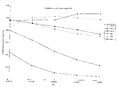
Figure 3 relates to quantitation of nucleic acids by amplifying a target
nucleic
acid sequence in the presence of an internal quantity control standard, as
described in
Example 3.
To aid in understanding the invention, several terms are defined below.
The terms "nucleic acid" and "oligonucleotide" refer to probes and oligomer
fragments to be detected, and shall be generic to polydeoxyribonucleotides
(containing
2-deoxy-D-ribose), to polyribonucleotides (containing D-ribose), and to any
other type of
polynucleotide which is an N glycoside of a purine or pyrimidine base, or
modified purine
or pyrimidine base. There is no intended distinction in length between the
terms "nucleic
2163388
-6-
acid" and "oligonucleotide", and these terms will be used interchangeably.
These terms refer
only to the primary structure of the molecule. Thus, these terms include
double- and single-
stranded DNA, was well as double- and single-stranded RNA.
The tenns "target region", "target sequence", and "target nucleic acid
sequence" refer
to a region of a nucleic acid which is to be detected.
The term "probe" refers to an oligonucleotide, typically labeled, that forms a
duplex
structure with a sequence of a target nucleic acid due to complementary base
pairing. The
probe will comprise a "hybridizing region", preferably consisting of 10 to 50
nucleotides,
more preferably 20 to 30 nucleotides, corresponding to a region of the target
sequence.
"Corresponding" means identical to or complementary to the designated nucleic
acid. In the
present invention, probe oligonucleotides are labeled with, i.e., bound to, a
fluorescent label
to enable detection.
The term "hybridization" refers the formation of a duplex structure by two
single-
stranded nucleic acids due to complementary base pairing. Hybridization can
occur between
fully complementary nucleic acid strands or between nucleic acid strands that
contain minor
regions of mismatch. Conditions under which only fully complementary nucleic
acid strands
will hybridize are referred to as "stringent hybridization conditions". Two
single-stranded
nucleic acids that are complementary except for minor regions of mismatch are
referred to as
"substantially complementary". Stable duplexes of substanrially complementary
sequences
can be achieved under less stringent hybridization conditions. Those skilled
in the art of
nucleic acid technology can deterniine duplex stability empirically
considering a number of
variables including, for example, the length and base pair concentration of
the oligo-
nucleotides, ionic strength, and incidence of mismatched base pairs.
The terms "sequence-specific oligonucleotide" and "SSO" refer to
oligonucleotide
probes wherein the hybridizing region is exactly complementary to the sequence
to be
detected. The use of stringent hybridization conditions under which the probe
will hybridize
only to that exactly complementary target sequence allows the detection of the
specific target
sequence. Stringent hybridization conditions are well known in the art, see,
e.g., Sambrook
et al 1985, Molecular Cloning - A Laboratory Manual, Cold Spring Harbor
Laboratory,
Cold Spring Harbor, New York. Stringent conditions are sequence dependent and
will be
different in different circumstances. Generally, stringent conditions are
selected to be about
5 C lower than the thermal melting point (Tm) for the specific sequence at a
defined ionic
strength and pH. The Tm is the temperature (under defined ionic strength and
pH) at which
50% of the base pairs have dissociated. Relaxing the stringency of the
hybridizing
2163388
-~-
conditions will allow sequence mismatches to be tolerated; the degree of
mismatch tolerated
can be controlled by suitable adjustment of the hybridization conditions.
The term "subsequence" refers herein to a nucleotide sequence contained within
another sequence.
The term "label", as used herein, refers to any atom or molecule which can be
attached to a nucleic acid, and which can be used either to provide a
detectable signal or to
interact with a second label to modify the detectable signal provided by the
second label.
Preferred labels are light-emitting compounds which generate a detectable
signal by
fluorescence, chemiluminescence, or bioluminescence.
The term "fluorophore" refers to a compound which is capable of fluorescing,
i.e.
absorbing light at one frequency and emitting light at another, generally
lower, frequency.
The term "bioluminescence" refers to a form of chemiluminescence in which the
light-emitting compound is one that is found in living organisms. Examples of
bioluminescent compounds include bacterial luciferase and firefly luciferase.
The term "quenching" refers to a decrease in fluorescence of a first compound
caused
by a second compound, regardless of the mechanism. Quenching typically
requires that the
compounds be in close proximity. As used herein, either the compound or the
fluorescence
of the compound is said to be quenched, and it is understood that both usages
refer to the
same phenomenon.
The term "reaction mixture" refers to a solution containing reagents necessary
to
carry out a given reaction. An "amplification reaction mixture", which refers
to a solution
containing reagents necessary to carry out an amplification reaction,
typically contains
oligonucleotide primers and a DNA polymerase in a suitable buffer. Reaction
mixtures for
specific reactions are well-known in the literature.
The methods of the invention are applicable to the detection of either
synthesis or
cleavage of oligonucleotides. Detection of the cleaved oligonucleotide is
carried out in a
solution containing a DNA binding compound that can interact with the label to
decrease the
light emission of the label. The change in the length of the labeled
oligonucleotide from
synthesis or cleavage results in a detectable change in the light emission of
the attached label.
Suitable light-en-iitting labels and DNA binding compounds that can interact
to modify the
light emission of the label are described below.
2163388
-8-
In the exemplified methods of the present invention, the emission of a
fluorescent
label bound to the single-stranded oligonucleotide is detected. A DNA binding
compound
quenches the label fluorescence to a degree that depends on the length of the
attached
oligonucleotide. Both the occurrence of in-solution quenching by a DNA binding
compound
of a fluorescent label bound to a single-stranded oligonucleotide and the
dependence of the
quenching on the length of the oligonucleotide are unexpected in view of the
prior art.
In a preferred embodiment, the DNA binding agent is polyethylenimine (PEI),
which
refers to a class of branched or unbranched polymers of ethylenimine of
various molecular
weights. Derivatives of PEI, such as hydroxyethylated PEI, may be suitable in
the present
methods. Other compounds which have been demonstrated to work in the methods
of the
present invention include spermine and spermadine.
Branched PEI is commercially available from Polysciences, Inc. (Warrington,
PA) in
molecular weights, as estimated by viscosity, ranging from 600 up to at least
60,000-
80,000. PEI of molecular weights 600, 1200, 1800, 10,000, and 60,000-80,000
have been
tested and shown to function in the methods of the present invention to
differentiate
fluorescently-labeled oligonucleotides of length 2 from those of length 33.
One of skill in the
art will be able to empirically determine which size of PEi is most suitable
for a given
application.
Suitable concentrations of the DNA binding compound are determined
empirically.
Typically, the optimum concentration of DNA binding compound is affected by
the type of
light-enutting label used and the concentration of reaction reagents. In
particular, salt
concentration has been observed to affect the optimum concentration of PEI. In
some
reactions, the addition of a chelator (for example, about 1.5 mM EDTA) into
the reaction
mixture has been observed to broaden the range of PEI over which the window is
near the
maximum value. Routine optimization of the concentration of DNA binding
compound
which provides the maximum difference between the fluorescences of the long
and short
labeled oligonucleotides can be carried out essentially as described in
Examples 1 and 2,
below.
Many light emitting compounds described in the art are suitable for use as
oligo-
nucleotide labels in the methods of the present invention. Ideally, a
fluorophore should have
a high Stokes shift (i.e. a large diff.erence between the wavelength for
maximum absorption
and the wavelength for maximum emission) to minimize interference by scattered
excitation
light. Suitable compounds which are well known in the art include, but are not
limited to,
fluorescein and derivatives such as fluorescein (FAM), hexachlorofluorescein
(HEX),
tetrachlorofluorescein (TET), and dichlorodimethylfluorescein (JOE); rhodamine
and
2163388'
-9-
derivatives such as Texas Red, rhodamine (ROX), and tetramethylrhodamine
(TAMRA);
Lucifer Yellow, and coumarin derivatives such as 7-Me2N-coumarin-4-acetate, 7-
OH-4-
CH3-coumarin-3-acetate, and 7-NH2-4-CH3-coumarin-3-acetate (AMCA). FAM, HEX,
TET, JOE, ROX , and TAMRA are marketed by-Perkin Elmer, Applied Biosystems
Division (Foster City, CA). Texas Red and many other suitable compounds are
marketed by
Molecular Probes (Eugene, OR). Examples of chemiluminescent and bioluminescent
compounds that may be suitable for use as the energy donor include luminol
(aminophthalhydrazide) and derivatives, and Luciferases.
An oligonucleotide can be prepared by any suitable method, including, for
example,
cloning and isolation of appropriate sequences using restriction enzymes and
direct chemical
synthesis by a method such as the phosphotriester method of Narang et al.,
1979, Meth.
Enzymol. 68:90-99; the phosphodiester method of Brown et al., 1979, Meth.
Enzymol.
68:109-151; the diethylphosphoramidite method of Beaucage et al., 1981,
Tetrahedron Lett.
22:1859-1862; and the solid support method of U.S. Patent No. 4,458,066.
Methods for
synthesizing labeled oligonucleotides are described in Agrawal and Zamecnik,
1990, Nucl.
Acids. Res. 18(18):5419-5423; MacMillan and Verdine, 1990, J. Org. Chem.
55:5931-
5933; Pieles et al., 1989, Nucl. Acids. Res. 17(22):8967-8978; Roget et al.,
1989, Nucl.
Acids. Res. 17(19):7643-7651; and Tesler et al., 1989, J. Am. Chem. Soc.
111:6966-6976.
A review of synthesis methods is provided in Goodchild, 1990, Bioconjugate
Chemistry
1(3):165-187.
The methods of the present invention are particularly suitable for the
detection of
amplified nucleic acids, either DNA or RNA. Suitable amplification methods in
addition to
the PCR (U.S. Patent Nos. 4,683,195; 4,683,202; and 4,965,188) include, but
are not
limited to, the following: Ligase Chain Reaction (LCR, Wu and Wallace, 1989,
Genomics
4:560-569 and Barany, 1991, Proc. Natl. Acad. Sci. USA 88:189-193); Polymerase
Ligase
Chain Reaction (Barany, 1991, PCR Methods and Applic. 1:5-16); Gap-LCR (Patent
Publication WO 90/01069); Repair Chain Reaction (European Patent Publication
No. 439,182), 3SR (Kwoh et al . 1989, Proc. Natl. Acad. Sci. USA 86:1173-1177;
Guatelli et al . 1990, Proc. Natl. Acad. Sci. USA 87:1874-1878; Patent
Publication
WO 92/08800), and NASBA (U.S. Patent No. 5,130,238). This invention is not
limited to
any particular amplification system. As other systems are developed, those
systems may
benefit by practice of this invention. A recent survey of amplification
systems was published
in Abramson and Myers, 1993, Current Opinion in Biotechnology 4:41-47.
A preferred embodiment of the invention provides improvements to the process
described in U.S. Patent No. 5,210,015, supra, and Holland et al., 1991, Proc.
Natl. Acad.
Sci. USA 88:7276-7280. The process uses the 5' to 3' exonuclease activity of a
CA 02163388 2002-08-02
-10-
thermostable DNA polymerase to cleave annealed iabeled oligonucleotide probes
from
hybridization duplexes and release labeledfragr=nents for detecaon. Cleavage
of the labeled
probes of the present invention by the 5' to 3' exonuclease-activiry of the
DNA polymerase
frees the labels into the reaction mixture. The in-solution signal quenching
by the DNA
binding compound is significantly greater when the fluorophore is bound to the
full-length
uncleaved oligonucleotide probe than when bound to the shortened cleaved
fragment. The
resulting increase in observed fluorescence indicates probe cleavage, which
necessarily
indicates both the presence of target sequences and the occurrence of
probe/target
hybridization.
In general, the nucleic acid in the sample will be a sequence of DNA, most
usually
genomic DNA. However, the present invention can also be practiced with other
nucleic
acids, such as messenger RNA, ribosomal RNA, viral RNA, or cloned DNA.
Suitable
nucleic acid samples include single or double-stranded DNA or RNA for use in
the present
invention. Those of skill in the art will recognize that, depending on which
reaction is used
to cleave the labeled oligonucleotide probes, whatever the nature of the
nucleic acid, the
nucleic acid can be detected merely by making appropriate and well recognized
modifications
to the method being used.
Sample preparation will vary depending on the source of the sample, the target
to be
detected, and the oligonucleodde-degrading reaction used in the assay. Each
assay requires a
target sample in a buffer that is compatible with the assay reagents. If the
target is amplified
either before or simultaneously with detecdon of probe cleavage, the target
nucleic acid must
be in a buffer compatible with the enzymes used to amplify the target. The
target nucleic acid
can be isolated from a variety of biological materials including tissues, body
fluids, feces,
sputum, saliva, plant cells, bacterial cultures, and the like.
Sample preparation methods suitable for each assay are described in the art,
see, for
example, Sambrook et al., supra. Simple and rapid methods of preparing samples
for the
PCR amplification of target sequences are described in Higuchi, 1989, in PCR
Technology
(Erlich ed., Stockton Press, New York), and in PCR Protocols, Chapters 18-20
(Innis et
al., ed.. Academic Press, 1990). One of skill in the an will be able to select
and empirically
optimize a suitable protocol.
The light emission of a label in a solution is measured in a
spectrofluorometer, such
as a Hitachi/Perkin Elmer Model 650-40*(Perkin Elmer, Norwalk, CT) or a PTI LS-
100 ~
Luminescence Spectrophotometer (Photon Technolo,v International, London,
Ontario,
Canada). A spectrofluorometer, depending on the features of the particular
maz:hine udlized,
offers the opportunity to set the excitation and enussion wavelenQth, as well
as"bandwidth.
* Trade-mark
- - --- ---------
2163388
-11-
One of ordinary skill in the art will know how to determine the wavelength and
bandwidth
settings appropriate for detecting the light emission from a particular label.
General guidance
is found in, for example, The Merck Index, (eds. Budavari et al., 1989, Merck
Co. Inc.
Rahway, NJ) and the Molecular Probes, Inc. (Eugene, Oregon) Catalog, 1990, by
Haugland. Although each label has a discrete light emission spectrum, a broad
range of
detection wavelengths are suitable for practicing the invention.
The change in light emission resulting from probe cleavage preferably is
measured
by comparing the light emission measured before and after probe cleavage.
Alternatively, the
change in light emission can be measured concurrent with probe-cleavage by
incorporating
the DNA binding agent into the reaction mixture and monitoring the probe
fluorescence
continuously or intermittently while the reaction is carried out. In this
case, the use of
reaction vessels which are also suitable for use in measuring light emission,
which allows
direct measurements of light-emission without the need to open the reaction
vessel, is
preferred. However, as particular DNA binding agents may inhibit particular
probe-cleaving
reactions, it may be necessary to omit the DNA binding agent from the reaction
mixture. In
this case, pre-reaction measurements can be carried out using a duplicate
reaction mixture.
Typically, the reaction mixture is divided prior to carrying out the reaction,
and a portion of
the reaction mixture which is not subject to the reaction conditions is used
for the pre-
reaction emission measurements. Typically, it is most convenient to measure
both the pre-
reaction light emission and the post-reaction light emission after completion
of the reaction.
In preferred methods in which the nucleic acid detection method is combined
with
PCR amplification, as described above, the amplification reaction is carried
out as an
automated process. Thermal cyclers are currently available from Perkin Elmer
(Norwalk,
CT) that uses a heat block capable of holding up to 48 or 96 reaction tubes.
Consequently,
up to 96 amplification reactions can be carried out simultaneously.
Suitable optical systems for measuring the light emission from all tubes in a
PCR
amplification are described in Higuchi et al., 1992, supra, Higuchi et al.,
1993, supra, and
European Patent Publication No. 512,334. In one such optical system, multiple
fiber optic
leads are used to transmit the excitation light from the source to the
reaction tube and
measures the emission light from each tube. Only a single spectrofluorometer
is needed to
read fluorescence from the reaction tubes, as each fiber optic can be read
rapidly one at a
time. An alternative optical system uses a video camera to measure the
fluorescence of
multiple reaction vessels simultaneously. It will be obvious to one of skill
in the art that
alternative detection apparatuses also are adaptable to the present methods.
2163388
- 12-
An alternative suitable detection scheme uses a 96-well microtiter format.
This type
of fonnat is frequently desirable in clinical laboratories for large scale
sample screening, for
example, for genetic analysis such as screening for sickle-cell anemia or the
AIDS virus in
blood bank screening procedures. The present invention is suitable for this
type of analysis
and eliminates the need for the numerous washing and extraction procedures
that are
required with known "in-well" assay procedures such as ELISA type formats or
other
optical density-based methods, see e.g. Kolber et al., 1988, J. Immun. Meth.
108:255-264,
Huschtscha et al 1989, In Vitro Cell and Dev. Biol. 25(1):105-108, and Voller
et al., 1979,
The Enzyme Linked Immunosorbent Assay, Dynatech Labs, Alexandria, VA. The
present
detection methods also allow direct light emission measurement using an
apparatus similar to
ELISA plate reader, but designed to excite and measure fluorescence. For
example, the
CytoFluorTM 2300 machine manufactured by Millipore (Bedford, MA) is suitable
in such a
method.
It will be obvious to one skilled in the art that the methods of the present
invention
are not limited to a particular detection method, thermal cycler or signal
measuring machines,
or number of reaction vessels.
The methods of the present invention can be used to simultaneously detect
multiple
target sequences. Probes specific to each target are present in the reaction
mixture. For each
target nucleic acid present in the sample, the corresponding probe will
hybridize and be
cleaved. In order to detect the cleaved probes separately, each species of
probe is labeled
with a label that emits light at a detectably distinct wavelength. Each
species of probe is then
detected separately by suitable selection of the measured wavelength.
Thus, the methods of the present invention are useful for detecting the
amplification
products in PCR co-amplification methods for detecting several targets in one
sample. The
invention is particularly useful for quantitative comparisons of two different
nucleic acid
targets in the same sample. Methods for quantitating nucleic acids are
described in U.S.
Patent No. 5,219,727. The quantitation methods described are PCR-based methods
using an
internal standard to detem-iine either the relative amount of a target or
accurately quantitate the
amount of target present prior to amplification, respectively.
The differential quenching of short and long labeled oligonucleotides may also
be
used to measure the incorporation of nucleotides into a synthesized
oligonucleotide. For
example, DNA polymerase activity assays measure the rate of incorporation of
nucleotides.
To measure the incorporation of nucleotides, a reaction mixture is provided
containing
labeled dNTPs in addition to the other reagents necessary for oligonucleotide
synthesis, such
as a DNA polymerase in a suitable buffer. The fluorescence of the reaction
mixture prior to
2163388
-13-
synthesis is measured in the presence of a DNA binding compound. Prior to
synthesis, all
labels are bound to single nucleotides and quenching is minimal. Following
synthesis, the
fluorescence of the reaction mixture is measured in the presence of the DNA
binding
compound. The synthesis of oligonucleotides results in a decrease in amount of
labeled
nucleotides. Because the longer, newly synthesized oligonucleotides are more
effectively
quenched by the DNA binding compound, the incorporation of dNTP into
oligonucleotides
results in a reduction of fluorescence.
The examples of the present invention presented below are provided only for
illustrative purposes and not to limit the scope of the invention. Numerous
embodiments of
the invention within the scope of the claims that follow the examples will be
apparent to
those of ordinary sldll in the art from reading the foregoing text and
following examples.
Example 1
Lenzth-dependent Quenchin by PET
This example describes the length-dependent quenching of labeled
oligonucleotides
in solution. Oligonucleotides of length 33 and length 2 were labeled with one
of a variety of
fluorescent labels and the fluorescence of each oligonucleotide was measure in
a solution
containing PEI. The measurements were repeated using solutions containing
various
concentrations of PEI in order to determine the optimal concentration of PEI
for which the
difference in signal between the oligonucleotides of length 33 and 2 is
maximized.
Probes were synthesized on an ABI 394 DNA synthesizer (Perkin Elmer ABD,
Foster City, CA) at a 1 micromole scale. Amidites of the fluorescent label
were used during
oligonucleotide synthesis to provide a 5'-labeled oligonucleotide.directly.
Probes of length
33 were synthesized with a P04 group at the 3' end, for use in the methods
described in
Example 2. The nucleic acid sequence of each of the labeled probes of length
33 was SK535
(SEQ ID NO: 1) 5'-AGAAGGTGAGATGACCAGAGGACTGAGTCCAAT. The nucleic
acid sequence of the probes of length 2 consisted of 5'-AG, which corresponds
to a
degradation product of the probes of length 33.
The fluorescent labels used are listed below, along with the excitation and
emission
wavelengths for each of the labels are shown below. The slit width of the
filter used for the
detection of each label, as described below, also is indicated.
CA 02163388 2002-08-02
-I4-
Fil ters [Jsgd For F] uorescence Measurements
(wavelength and width shown in nanometers)
Irabel Excitation Maximum Emission Maximum
fluorescein (FAM) 485 (20) 530 (25)
hexachlorofluorescein (HEX) 530 (25) 590 (35)
dichlorodimethylfluorescein (JOE) 490 (40) 580 (50)
rhodarrmine (ROX) 590 (40) 645 (40)
tetramethylrhodamine (TAMRA) 560 (20) 620 (40)
For each of the probes listed below, replicate assay solutions containing 1 M
of
probe in 50 1 of buffer (50 mM Bicine* 100 mM KOAc (pH 8.3), 3.6 mM Mn(OAc)2)
were added to the wells of microtiter plates. A series of solutions containing
PEI (molecular
weight 1200, from Polysciences Inc., Warrington, PA) at concentrations of
0.016%,
0.008%, 0.004%, 0.002%, and 0.001%, were prepared. A 50 1 volume of water was
added to one well to be used as a maximum signal control. A 50 1 of PEI
solution were
added to each of the remaining wells. Fluorescence measurements were carried
out in a
Millipore Cytofluor 2300 Microtiter plate reader using the excitation and
emission
wavelengths provided in the table, above.
The methods of the present invention rely on the difference in the quenching
of short
and lonz labeled oligonucleoddes. A measure of this difference in quenching,
referred to a
"window", was calculated from the individual fluorescence measurements as
follows.
Firstiy, the background level of quenching resulting from the buffer alone was
measured and
subtracted from each probe fluorescence measurement. Secondly, residual
fluorescence was
calculated as the ratio of the fluorescence of a probe in the presence of PEI
to the
fluorescence of the same probe without PEI, and expressed as a per cent of the
unquenched
fluorescence. Finally, the window was calculated as the difference between the
residual
fluorescence of the labeled oligonucleotide of length 2 and the residual
fluorescence of the
oligonucleotide of length 33. The window is a measure of the change in signal
which would
result from the degradation of labeled probes of length 33 into fragments of
length 2. A PEI
concentration which provides the maximum window provides the greatest
sensidviry in the
detection methods of the present invention.
The results are presented in Figure 1, wherein the windows obtained using the
various probe labels are plotted relative to the concentration of PEI. The
line designations
refer to olizonucleoddes labeled as follows:
hexx2: HEX-labeled oligonucleocides, wherein the label is attached to the 5'
end of
the olizonucleotides through a spacer.
" Trade-mark .
2163389
-15-
fam2: FAM-labeled oligonucleotides.
hex2: HEX-labeled probes, wherein the label is attached directly to the 5' end
of
the oligonucleotides.
joe: JOE-labeled oligonucleotides.
rox: ROX-labeled oligonucleotides.
tam: TAiVIRA-labeled oligonucleotides.
As can be seen from Figure 1, a significant window was observed with each
label at
some concentration of PEI. The size of the window obtained depended both on
the label and
the concentration of PEI. Attachment of a HEX label to the oligonucleotide
through a spacer
was observed to decrease the size of the window obtained within the tested
range of PEI
concentrations.
Example 2
The Effect of PEI Molecular Weiaht
In this example, the effect of the molecular weight and concentration of the
PEI on
the quenching of fluorescently-labeled probes are described. Experiments
essentially as
described in Example 1 were carried out using the FAM-labeled oligonucleotides
but using
PEI of various sizes and over a larger concentration range than in Example 1.
PEI with molecular weights of 600, 1200, 1800, and 10,000 kilodalton were
obtained from Polysciences Inc., Warrington, PA. Emission measurements were
carried out
in assay solutions containing buffer consisting of 50 mM Bicine, 115 mM KOAc,
potassium
acetate (pH 8.3), and 8% glycerol. For each size PEI, the PEI was incorporated
into the
assay solutions in concentrations of from 0.000013% to 0.02% (w/v). For each
PEI size
and concentration, the window was calculated as in Example 1.
The results are presented in Figure 2. Each size of PEI was seen to function
in the
methods of the present invention, although minor differences in window size
were obtained
using the different sizes of PEI. For each size of PEI, the optimal PEI
concentration can be
determined from Figure 2 as the PEI concentration which provides the largest
window. The
above experiment exemplifies the routine optimization of PEI size and
concentration which
one of skill in the art would carry out for each detection assay.
~~63388
- 16-
Example 3
Quantitation by PCR Amplification
This example describes the quantitation of a nucleic acid using the methods of
the
present invention. In this example, an target consisting of hepatitis C virus
(HCV) RNA was
amplified by an RT-PCR in the presence of fluorescently labeled probes. The
exonuclease
activity of the DNA polymerase cleaved labeled probes hybridized to the target
sequence
downstream from an amplification primer, thereby releasing small labeled
oligonucleotide
fragments of the probe into the reaction mixture. Following amplification, PEI
was added to
the reaction mixture and the fluorescence of the labeled probes measured. The
generation of
short labeled oligonucleotide degradation products results in an increase in
the measured
fluorescence because the degradation products are not quenched by the PEI as
much as full-
length, undegraded probe. Because probe degradation occurs concomitant with
target
amplification, the increase in fluorescence indicates amplification of the
target sequence.
A quantitative estimate of the initial target sequence copy number was
obtained by
comparing the increase in fluorescence resulting from the amplification of the
target sequence
to the increase in fluorescence obtained from the amplification of a second
nucleic acid
sequence. The second nucleic acid sequence is added to a reaction in a known
copy number,
thereby providing a internal quantitation standard (IQS) to which the
amplification of the
unknown sample is compared. Detection of the second nucleic acid sequence is
achieved
using a second fluorescent label which emi.ts light at a distinct wavelength.
Amplification
Two target RNA sequences were amplified in each reaction. Both target nucleic
acid
sequences were simultaneously amplified using a single primer pair, KY78 and
KY80. In
this example, the target RNA sequences were generated from transcription
plasmids. The
construction and use of a transcription plasniid containing an HCV sequence,
the
amplification primers, and the RT-PCR amplification are described in the
European Patent
Publication No. 529 493 and in Young et al., 1993, J. Clin. Microbiol.
31(4):882-886.
The first target sequence consisted of a region from the HCV 5' non-coding
region. The
second target nucleic acid sequence, which is used as an IQS, comprises the
same two
primer binding sites flanking a nucleic acid sequence which contains an
alternate probe
binding site. Consequently, both target sequences are amplified using the same
primer pair,
but the two target sequences can be detected independently using different
sequence-specific
probes. The construction and use of an IQS is described in Mulder et al.,
1994, J. Clin.
Microbiology 34(2):292-300.
CA 02163388 2002-08-02
-17-
Amplifications were carried out in-the presence of both=a FAM-labeled probe
specific
for the HCV taraet sequence and a HEX4abeled probe specific for the IQS. The
emission
maxima of the FAM and HEX =are at different wavelengths, which permits
independent
detection of the probes by suitable selection of the measured frequency. The
nucleic acid
sequences of the probes are shown in the 5' to 3' orientation. The probes were
synthesized
with the labels at the 5' end, as described above. Each probe was synthesized
to have a 3'-
P04 instead of a 3'-OH to block any extension by Taq polymerase during the
amplification
reaction. The sequence of the FA~,v1-labeled probe for the detection of the
HCV target
sequence was described in Young er al., 1993, supra, as KY88. The sequence of
the HEX-
labeled probe for the detection of the IQS is provided in Example 1, above
(SEQ ID NO: 1).
Amplificauons were carried out in 100 ul reactions containing the following
reagents:
HCV taraet sequence (copy number as described below)
1000 copies of the IQS
50 rrutil Bicine
100 mivl KOAc, pH 8.3
3.6 mM 2Mn(OAc)2
0.4 uti1 each primer
1 u:vf each probe
0.2 m-M each dUTP, dATP, dGTP, dCTP
20 units rTth DNA polymerase*
2 units Amperase Uracil-N-Glycosolase*
8% Glycerol
* Developed and manufactured by Hoffmann-La Roche and marketed by Perkin
Elmer,
Norwalk, CT.
Amplifications were carried out usina from 0 to 107 copies of HCV target. In
addition to the reaction mixtures subjected to PCR thermal cyclin; conditions,
two additional
reaction mixtures were made and stored (no temperature cycling) for use as
ineasurement
controls. Reaction mixtures were subjected to the followina amplification
scheme in a
GeneAmp 9600 Thermal Cycler (Perkin Elmer, Norwalk, CT):
50'C for 2 minutes
60'C for 30 minutes
95'C for 1 minute
2 cycles:
95'C for 15 seconds
60'C for 20 seconds
' Trade-mark
2163388-18-
46 cycles:
90 C for 15 seconds
60 C for 20 seconds
72 C for at least 5 to 10 minutes, up to 1 hour
Following amplification, reactions are held at 4 C until analyzed.
Analysis
Fifty l of 0.004% PEI (molecular weight 1200) solution containing 0.8 mM EDTA
were added to 50 l of each reaction mixture following amplification and also
to one of the
two control reaction mixtures not subject to amplification conditions. The
fluorescence was
measured in a CytoFluorTM 2300 (Millipore, Locarion) microtiter plate reader.
The
fluorescence of FAM-labeled probes was measured at room temperature using a
485 nm
excitation filter (20 nm band pass width) and 530 nm emission filter (25 nm
band pass
width). The fluorescence of HEX-labeled probes was measured at room
temperature using a
530 nm excitation filter (25 nm band pass width) and 590 nm emission filter
(35 nm band
pass width).
The measured fluorescence of each probe label was corrected by subtracting the
fluorescence of an uncycled reaction mixture without probe, both measured at
the
wavelength appropriate for the label. The results are presented in Figure 3,
plotted as the
logarithm (log) of the ratio of the fluorescences (HCV/IQS) versus the log of
the HCV target
copy number. A "standard curve" was generated by a least-squares linear fit of
the data.
The standard curve generated from the above experiment allows the quantitation
of
unknown HCV samples. For quantitation of an unknown HCV sample, the HCV
nucleic
acid is amplified with a known amount of IQS as described above. The log of
the ratio of the
measured changes in fluorescence is calculated, and the corresponding HCV copy
number is
determined from the equation of the standard curve.
2163388 _ 19-
SEQUENCE LISTING
(1) GENERAL INFORMATION:
(i) APPLICANT: F. Hoffmann-La Roche AG
(ii) TITLE OF INVENTION: Method for Detecting a Change in Length
of a Oligonucleotide Label labeled with a
light-emitting Label
(iii) NUMBER OF SEQUENCES: 1
(iv) CORRESPONDENCE ADDRESS:
(A) ADDRESSEE: F. Hoffmann-La Roche AG
(B) STREET: Grenzacherstrasse 124
(C) CITY: Basle
(D) STATE: BS
(E) COUNTRY: Switzerland
(F) ZIP: CH-4002
(v) TELECOMMUNICATION INFORMATION:
(A) TELEPHONE: 061 688 7493
(B) TELEFAX: 061 688 13 95
(2) INFORMATION FOR SEQ ID NO:l:
(i) SEQUENCE CHARACTERISTICS:
(A) LENGTH: 33 base pairs
(B) TYPE: nucleic acid
(C) STRANDEDNESS: single
(D) TOPOLOGY: linear
(ii) MOLECULE TYPE: DNA (genomic)
(xi) SEQUENCE DESCRIPTION: SEQ ID NO:1:
AGAAGGTGAG ATGACCAGAG GACTGAGTCC AAT 33