Note: Descriptions are shown in the official language in which they were submitted.
w ~510oG~6 PCTiCa9a~003.ii
..-. ,
2155921 _1_
APPARATDS AND METHOD FOR ALIGNING KNEE PROSTHESES
FIELD OF THE INVENTION
The present invention refers to a method and
apparatus for establishing the correct alignment and
orientation required for a knee prosthesis during total
knee arth=oplasty surgery. In particular, the invention
pertains to determining the correct position and
orientation of cutting guides with respect to a
patient's femur o: tibia so that the femur or tibia can
be cut to fit the knee prosthesis such that the
prosthesis will be implanted in an anatomically correct
orientation.
BACKGROUND OF TH~ INVENTION
During knee resurfacing arthroplasty, commonly
called knee replacement surgery, the distal surfaces of
the femur are cut away and replaced with a metal cap to
simulate the bearing surfaces of the femur. The
proximal surface of the tibia is modified in a similar
way, to provide a metal backed plastic bearing surface.
The metal femoral component of the new "artificial
joint" transfers the patient's weight to the tibial
component such that the joint can support the patient's
weight and provide a near-normal motion of the knee
joint. Several studies have indicated that the long
term survival of such an artif ical knae joint is
dependant on how accurately the components of the knee
joint are implanted with respect to the weight bearing
axis of the patient's leg. In a correctly functioning
knee, the weight bearing axis passes through the centre
of the head of the femur, the centre of the knee and the
centre of the ankle joint. This weight bearing axis is
typically located by analyzing an X-ray image of the
patient's leg, taken while the patient is standing. The
X-ray image is used to locate the centre of the head of
the femur and to calculate its position relative to
selected landmarks on the femur. The selected landmarks
are then found on the patient's femur during surgery and
the calculations used to estimate the actual position of
Wl ~I00076 PCTlCA94lOJ341
°" 2165921
-2-
the femoral head. These two pieces of information are
used to determine the correct alignment of the weight
bearing axis for the femur. To completely define the
correct position for the femoral component of the knee
prosthesis, the correct distance between the centre of.
the femoral head and the knee joint and the rotation of
the knee joint about the weight bearing axis must be
established. These two pieces of information are
determined from landmarks on the distal portion of the
femur. The correct alignment for the tibial component
of the knee prosthesis is determined by finding the
centre of the ankle joint and relating its position to
landmarks on the tibia. This point and the centre of
the proximal tibial plateau are used to define the
weight bearing axis of the tibia. The correct distance
between the ankle joint and the knee joint and the
rotation of the knee joint about the weight bearing axis
are determined by reference to the distal portion of the
femur and landmarks on the tibial plateau.
Various mechanical alignment instruments are
used to assist the surgeon in making cuts on the distal
femur and proximal tibia which will allow the femoral
and tibial components of the new knee joint to be
attached to the femur and tibia. These mechanical
alignment instruments permit the surgeon to f ix cutting
guides in place with respect to the selected landmarks
on the bones so that the cuts will be correctly oriented
with respect to the weight bearing axis determined from
the X-ray image.
There are two general types of alignment
instruments in common use. These are intramedullary and
extramedullary alignment systems. Intramedullary
alignment systems use the inside of the femur or tibia,
the medullary canal, as one of the selected landmarks
for establishing alignment. Extramedullary alignment
systems use only the external surfaces of the body to
establish alignment.
Vt 15100076 PCTlCa9.1~00341
'~ 2165921
-3-
A typical extramedullary alignment system
requires the surgeon to visually align a slender rod
with the centre of the knee and the centre of the
femoral head for alignment of the femoral component,
then align a similar rod with the centre of the ankle
and the centre of the tibial plateau for alignment of
the tibial component. The centers of the femoral head
and ankle are found by either palpitation or established
with an intraoperativs X-ray. If correctly placed, the
rods will lie parallel to, and offset from the weight
bearing axis. Once aligned, the rods are used as a
guide to fix the location of the cutting guides with
respect to the femur and tibia so that the cuts can be
performed.
A typical intramedullary alignment system
requires the surgeon to insert a rod into the medullary
canal of the femur and tibia. If properly placed, these
rods should lie on the axis of the bones. In the case
of the tibia, the weight bearing axis is very close to
the axis of the bone. In the case of the femur the axis
of the bone is quite different from the weight bearing
axis due to the offset nature of the hip joint, and this
difference must be measured from the pre-operative X-ray
and used to correct the alignment of the femoral cutting
jigs.
Both intramedullary and extramedullary
approaches to alignment have num. pus inherent drawbacks
and sources of error. Extrameduilary alignment depends
on accurate visual estimation of the alignment of the
extramedullary rods. Location of the femoral head by
palpitation is difficult and error prone, particularly
with obese patients. Use of intraoperative X-rays
improves the result somewhat, but this is time consuming
and exposes the patient and operating room personnel to
radiation. X-rays are also subject to distortion and
require visual interpretation and estimation to
correctly analyze, as they offer only one planar view in
two dimensions.
216592 1
-4-
Intramedullary alignment approaches provide only
slightly better results, in that the knee joint alignment
is still determined by estimating the difference between
the bone axis and the weight bearing axis from a
potentially distorted X-ray image. In addition,
intramedullary rods must be introduced very carefully, not
only to make sure they align correctly with the medullary
canal, but also to make sure that the insertion of the rods
does not create an embolism, which could kill or seriously
injure the patient.
An ideal alignment system finds the weight
bearing axis of the patient's leg directly, without the
need for preoperative or intraoperative X-rays, estimation,
calculation, location of hidden or obscured landmarks, or
surgical intervention outside of that required for access
to the knee joint surfaces. The ideal alignment system
depends only on the accepted definition that the weight
bearing axis passes through the centre of the head of the
femur, the centre of the knee joint and the centre of the
ankle, in order to locate the weight bearing axis.
SUMMARY OF THE INVENTION
The present invention provides apparatus and
method for locating the weight bearing axis of a patient's
limb by directly locating the centers of rotation of the
head of the femur and ankle. These centers of rotation are
combined with easily identified landmarks on the distal
femur and proximal tibia to simply and accurately define
the actual weight bearing axis of the patient's limb.
In one aspect, the invention provides apparatus
for aligning a surgical instrument to the mechanical axis
of the femur, said apparatus comprising (a) tension member
means for attachment to the femur for applying tension to
the femur in the vicinity of the knee joint at a point
chosen to be on the mechanical axis, said tension member
means allowing the femur to move freely into line with the
applied tension, and (b) means for engaging said surgical
instrument with said tension member means for using the
axis of the applied tension to indicate the mechanical axis
A
21 6592 1
-5-
of the femur in the medial-lateral plane. The invention
also provides method for locating the mechanical axis of a
femur in the medial-lateral plane for a supine patient,
. comprising the steps of (a) applying tension to the femur
in the vicinity of the knee joint at a point chosen to be
on the mechanical axis; (b) allowing the femur to move
freely into line with the applied tension; and (c) using
the axis of the applied tension to indicate the mechanical
axis of the femur in the medial-lateral plane.
BRIEF DESCRIPTION OF THE DRAWINGS
Figures lA and 1B are schematic representations
of the tension alignment system.
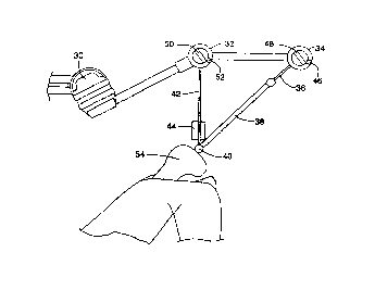
Figure 2 is a pictorial view of a tension
alignment system in accordance with the invention.
Figure 3 shows the alignment system of figure 2
with the carriage and bone clamp attached.
Figure 4 shows a cutting guide aligned with the
bone by the alignment system.
DESCRIPTION OF THE PREFERRED EMBODIMENT
Figure lA shows in schematic form how the tension
alignment system operates, as viewed in the coronal plane,
assuming that the patient is supine. Femur 10 is
constrained to rotate about the centre of femoral head 12.
Attachment point 14 is chosen to be at the centre of the
knee joint. Attachment point 14 will typically be located
approximately 1 cm anterior to the femoral attachment of
the posterior cruciate ligament. Tension member 18 is
connected between attachment point 14 and fixed point 16.
Tension member 18 may be of a flexible material capable of
applying only tension, or alternatively may be attached to
attachment points 14 and 16 such that the attachments are
free to rotate and no moments are supported by either
attachment point. To establish the alignment axis, tension
is applied to tension member 18. Femur 10 will rotate
about femoral
,.....
WC ,/00076 , PCTlCA941U0341
.~-.
2165921 -6-
head 12 until the axis of tension member 18 passes
through the centre of femoral head 12.
Figure ib shows in schematic form how the
tension alignment method operates as viewed from the
sagital plane, assuming that the patient is supine.
Attachment at or near attachment point 14 is weight
support line 20. Weight support line 20 is attached to
tension indicating device 24, which may be a spring type
scale. Tension indicating device 24 is connected to
fixed point 24, which is chosen to lie directly above
attachment point 14. To establish the alignment axis,
the tension required to counteract the moment about the
hip joint due to the weight of femur 10 is recorded from
tension indicating device 24. Tension is applied to
tension member 18. Weight support line 20 is then
tightened until tension indicating device 24 indicates
the same force as originally recorded. In this way, the
moment about the hip joint due to the weight of femur 10
is removed from tension member 18 so that tension member
18 will not be deflected by the weight of femur 10.
When the deflection is eliminated, the axis of tension
member 18 passes through the centre of femoral head 12.
Figure 2 shows one possible embodiment of an
apparatus which aligns a cutting guide to the mechanical
axis of the femur. Connection point 40 is a ball end
fitting attached to a threaded post. The threaded post
is screwed into the end of femur 54 at a point chosen to
be at the mechanical centre of the knee joint, usually
located approximately 1 cm anterior to the femoral
attachment of the posterior cruciate ligament. Attached
to connection point 40 is alignment rod 38, which is
attached so that it can freely rotate about connection
point 40. The distal end of alignment rod 38 is
attached to tension cable 36. Tension cable 36 is
connected to tension reel 34 such that tension cable 36
can be wound on to tension reel 34 with crank 48 to
increase the tension in tension cable 36. Tension reel
34 includes a ratchet mechanism to stop tension cable 36
WC~I000?6 ~ ~ PCTICA94100341
21 6592 1
from unreeling from the tension reel once tension is
applied. Ratchet release 46 allows a user to release
the ratchet and unreel tension cable 36 when desired.
Also connected to connection point 40 is
tension indicator 44, which in the preferred embodiment
is a spring scale with an indicating pointer calibrated
to measure tension in pounds and kilograms. Attached to
the distal end of tension indicator 44 is support cable
42. Support cable 42 is cone~cted to tension reel 32
such that support cable 36 can be wound on to tension
reel 32 with crank 50 to increase the tension in support
cable 42. Tension reel 32 includes a rachet mechanism
to stop support cable 42 from unreeling from the tension
reel once tension is applied. Rachet release 52 allows
a user to release the ratchet and unreel support cable
42 when desired.
The alignment system is connected to support
arm 30 so that it may be suspended generally above femur
54. Any fixed support located generally above femur 54
capable of supporting the weight of femur 54 and the
alignment system would be suitable. In the preferred
embodiment, the support arm is an Endex Endoscopy
Positioning System (Andronic Devices Ltd., Richmond,
B.C., Canada).
In use, connection point 40 is first screwed
into the distal femur at the point where the mechanical
axis of the femur passes through the knee joint.
Support arm 30 is moved into position above femur 54 so
that tension reel 32 is located directly over femur 54
and support cable 42 hangs vertically over connection
point 40. Tension indicator 44 is connected to
connection point 40. Excess length of support cable 42
is reeled onto tension reel 32 until the weight of femur
54 is fully supported. The tension indicated by tension
indicator 44 is then recorded. Tension rod 38 is then
connected to connection point 40. Excess length of
tension cable 36 is reeled onto tension reel 34 until a
tension of more than approximately 20 pounds is reached.
W 5100076 PCTICA941003s1
,.-
2165921 -8-
Tension reel 32 is then re-adjusted until the tension
indicated on tension indicator 44 is equal to the
previously recorded value. At this point, alignment rod
38 is aligned with the mechanical axis of femur 54 in
all planes.
Referring to Figure 3, carriage 56 is placed
over tension rod 38 and moved along tension rod 38 until
the proximal face of carriage 56 is in contact with the
distal face of femur 54 to establish a reference
distance between the femoral head and the knee joint.
Alignment skid 60 of carriage 56 is brought into contact
with the posterior face of the condyles of the femur to
establish rotational alignment of the distal femur about
the mechanical axis.
Attached to carriage 56 is bone clamp 58.
Bone clamp 58 is attached to femur 54 in the position
determined by carriage 56 using screws 62. In this way,
bone clamp 58 is attached to femur 54 aligned with
respect to the mechanical axis of femur 54, rotation
about the mechanical axis and the distance between the
femoral head and the knee joint.
Referring to Figure 4, the entire alignment
jig, consisting of tension reels 32 and 34, support
cable 42, tension indicator 44, carriage 56, tension rod
38 and tension cable 36 are removed from connection
point 40. Connection point 40 is then removed from
femur 54, leaving only the correctly aligned bone clamp
58 attached to femur 54. Advantageously, support art 30
may be connected to bone clamp 58 to support femur 54
during the course of the surgery.
To perform the cutting of femur 54, saw guide
64 is attached to bone clamp 58. Saw guide 64 provides
alignment and stability to~a saw blade during the
cutting operation.
Many alterations and adaptations may be made
to the embodiment described herein. Accordingly, the
invention is to be limited only by reference to the
appended claims. For example, although the embodiment
V~ l5l00076 PCT!CA94I00341
21 6 5 9 2 1 -9-
described is intended for use in alignment of the femur, 1
the same technique could be used to align the tibia
during total knee arthroplasty and may be applicable to
alignment of various bones for various surgical
procedures. Although cables and a tension indicator are
used to support the femur and eliminate the effect of
weight on the alignment system, other means for
eliminating the weight component, including constant
force springs, pneumatic cylinders or other means could
l0 achieve the same result. Cables used as pure tension
members in this embodiment could be replaced with rigid
members connected by universal joints without changing
the basic methods used for alignment. Although the
preferred embodiment describes a bone clamp which is
attached to the distal femur in exact alignment with the
mechanical ax..s, it may be advantageous to attach the
bone clamp in any convenient orientation and provide a
movable or otherwise adjustable attachment means on the
bone clamp Which may be aligned with the mechanical
axis.
