Note: Descriptions are shown in the official language in which they were submitted.
wO 95102686 2166102 PCT/US94/06675
- 1 -
REDIRECTION OF CELLULAR IMMUNITY BY RECEPTOR CHIMERAS
Field of the Invention
The invention concerns functional protein-tyrosine
kinase chimeras which are capable of redirecting immune
system function. More particularly, it concerns the
regulation of lymphocytes, macrophages, natural killer
cells or granulocytes by the expression in said cells of
chimeras which cause the cells to respond to targets
recognized by the chimeras. The invention also concerns
functional protein-tyrosine kinase chimeras which are
capable of directing therapeutic cells to specifically
recognize and destroy either cells infected with a
specific infective agent, the infective agent itself, a
tumor cell, or an autoimmune-generated cell. More
particularly, the invention relates to the production of
protein-tyrosine kinase chimeras capable of directing
cytotoxic T lymphocytes to specifically recognize and
lyse cells expressing HIV envelope proteins. The
invention therefore provides a therapy for diseases such
as AIDS (Acquired Immunodeficiency Syndrome) which are
caused by the HIV virus.
Backctround of the Invention
T cell recognition of antigen through the T cell
receptor is the basis of a range of immunological
phenomena. The T cells direct what is called cell-
mediated immunity. This involves the destruction by
cells of the immune system of foreign tissues or infected
cells. A variety of T cells exist, including "helper"
and "suppressor" cells, which modulate the immune
response, and cytotoxic (or "killer") cells, which can
kill abnormal cells directly.
A T cell that recognizes and binds a unique
antigen displayed on the surface of another cell becomes
WO 95102686 2166102 PCT/US94/06675
- 2 -
activated; it can then multiply, and if it is a cytotoxic
cell, it can kill the bound cell.
Autoimmune disease is characterized by production
of either antibodies that react with host tissue or
immune effector T cells that are autoreactive. In some =
instances, autoantibodies may arise by a normal T- and B-
cell response activated by foreign substances or
organisms that contain antigens that cross react with
similar compounds in body tissues. Examples of
clinically relevant autoantibodies are antibodies against
acetyicholine receptors in myasthenia gravis; and anti-
DNA, anti-erythrocyte, and anti-platelet antibodies in
systemic lupus erythematosus.
HIV and Immunopathogenesis
In 1984 HIV was shown to be the etiologic agent of
AIDS. Since that time the definition of AIDS has been
revised a number of times with regard to what criteria
should be included in the diagnosis. However, despite
the fluctuation in diagnostic parameters, the simple
common denominator of AIDS is the infection with HIV and
subsequent development of persistent constitutional
symptoms and AIDS defining diseases such as a secondary
infections, neoplasms, and neurologic disease.
Harrison's Principles of Internal Medicine, 12th ed.,
McGraw Hill (1991).
HIV is a human retrovirus of the lentivirus group.
The four recognized human retroviruses belong to two
distinct groups: the human T lymphotropic (or leukemia)
retroviruses, HTLV-1 and HTLV-2, and the human
imnnunodeficiency viruses, HIV-1 and HIV-2. The former
are transforming viruses whereas the latter are
cytopathic viruses.
is WO 95102686 2166102 PCT/US94106675
I- ,
- 3 -
HIV-1 has been identified as the most common cause
of AIDS throughout the world. Sequence homology between
= HIV-2 and HIV-1 is about 40% with HIV-2 being more
closely related to some members of a group of simian
immunodeficiency viruses (SIV). See Curran, J. et al.,
Science, 12,9:1357-1359 (1985); Weiss, R. et al., Nature,
I.aA:572-575 (1986).
HIV has the usual retroviral genes (env, gaa, and
gQ},) as well as six extra genes involved in the
replication and other biologic activities of the virus.
As stated previously, the common denominator of AIDS is a
profound immunosuppression, predominantly of cell-
mediated immunity. This immune suppression leads to a
variety of opportunistic diseases, particularly certain
infections and neoplasms.
The main cause of the immune defect in AIDS, has
been identified as a quantitative and qualitative
deficiency in the subset of thymus-derived (T)
lymphocytes, the T4 population. This subset of cells is
defined phenotypically by the presence of the CD4 surface
molecule, which has been demonstrated to be the cellular
receptor for HIV. Dalgleish et al., Nature, = :763
(1984). Although the T4 cell is the major cell type
infected with HIV, essentially any human cell that
expresses the CD4 molecule on its surface is capable of
binding to and being infected with HIV.
Traditionally, CD4+ T cells have been assigned the
role of helper/inducer, indicating their function in
providing an activating signal to B cells, or inducing T
lymphocytes bearing the reciprocal CD8 marker to become
cytotoxic/suppressor cells. Reinherz and Schlossman,
' gtll, J,9_:821-827 (1980); Goldstein et al., Immunol. Rev.,
1$:5-42, (1982).
' HIV binds specifically and with high affinity, via
a stretch of amino acids in the viral envelope (gp120),
WO 95/02686 PCT/US94/06675
2166102
- 4 -
to a portion of the V1 region of the CD4 molecule located
near its N-terminus. Following binding, the virus fuses
with the target cell membrane and is internalized. Once =
internalized it uses the enzyme reverse transcriptase to
transcribe its genomic RNA to DNA, which is integrated into the cellular DNA
where it exists for the life or the
cell as a "provirus."
The provirus may remain latent or be activated to
transcribe mRNA and genomic RNA, leading to protein
synthesis, assembly, new virion formation, and budding of
virus from the cell surface. Although the precise
mechanism by which the virus induces cell death has not
been established, it is felt that the major mechanism is
massive viral budding from the cell surface, leading to
disruption of the plasma membrane and resulting osmotic
disequilibrium.
During the course of the infection, the host
organism develops antibodies against viral proteins,
including the major envelope glycoproteins gp120 and
gp41. Despite this humoral immunity, the disease
progresses, resulting in a lethal immunosuppression
characterized by multiple opportunistic infections,
parasitemia, dementia and death. The failure of the host
anti-viral antibodies to arrest the progression of the
disease represents one of the most vexing and alarming
aspects of the infection, and augurs poorly for
vaccination efforts based upon conventional approaches.
Two factors may play a role in the efficacy of the
humoral-response to immunodeficiency viruses. First,
like other RNA viruses (and like retroviruses in
particular), the immunodeficiency viruses show a high
mutation rate in response to host immune surveillance.
Second, the envelope glycoproteins themselves are heavily
glycosylated molecules presenting few epitopes suitable
for high affinity antibody binding. The poorly antigenic
wO 95/02686 2166102 PCTIUS94/06675
~
- 5 -
target which the viral envelope presents, allows the host
little opportunity for restricting viral infection by
specific antibody production.
Cells infected by the HIV virus express the gp120
glycoprotein on their surface. Gp120 mediates fusion
events among CD4+ cells via a reaction similar to that by
which the virus enters the uninfected cells, leading to
the formation of short-lived multinucleated giant cells.
Syncytium formation is dependent on a direct interaction
of the gp120 envelope glycoprotein with the CD4 protein.
Dalgleish et al., supra; Klatzman, D. et al., Nature,
=:763 (1984); McDougal, J.S. et al., Science, 2,U:382
(1986); Sodroski, J. et al., Nature, M:470 (1986);
Lifson, J.D. et al., Nature, 323:725 (1986); Sodroski, J.
et al., Nature, =:412 (1986).
Evidence that the CD4-gp120 binding is responsible
for viral infection of cells bearing the CD4 antigen
includes the finding that a specific complex is formed
between gp120 and CD4. McDougal et al., sypra. Other
investigators have shown that the cell lines, which were
noninfective for HIV, were converted to infectable cell
lines following transfection and expression of the human
CD4 cDNA gene. Maddon et al., D,ell, 46:333-348 (1986).
Therapeutic programs based on soluble CD4 as a'
2~5 passive agent to interfere with viral adsorption and
syncytium-mediated cellular transmission have been
proposed and successfully demonstrated in vitro by a
number of groups (Deen et al., Nature, 3321:82-84 (1988);
Fisher et al., Nature, = :76-78 (1988); Hussey et al.,
Nature = :78-81 (1988); Smith et al., Science, = :1704-
1707 (1987); Traunecker et al., Nature, = :84-86
(1988)); and CD4 immunoglobulin fusion=proteins with
extended halflives and modest biological activity have
= subsequently been developed (Capon et al., Nature,
337:525-531 (1989); Traunecker et al. Nature, 3~, 68-70
WO 95/02686 PCT/US94/06675 =
2166102 - 6 -
(1989); Byrn et al., Nature, 2":667-670 (1990);
Zettlmeissl et al., DNA Cell Siol. 9_:347-353 (1990)).
Although CD4 immunotoxin conjugates or fusion proteins show potent
cytotoxicity for infected cells in vitro
(Chaudhary et al., Nature, 335:369-372 (1988); Till et =
al., Science, 2_4.Z:1166-1168 (1988)), the latency of the
immunodeficiency syndrome makes it unlikely that any
single-treatment therapy will be effective in eliminating
viral burden, and the antigenicity of foreign fusion
proteins is likely to limit their acceptability in
treatments requiring repetitive dosing. Trials with
monkeys affected with SIV have shown that soluble CD4, if
administered to animals without marked CD4 cytopenia, can
reduce SIV titer and improve in vitro measures of myeloid
potential (Watanabe et al., Nature, M:267-270 (1989)).
However a prompt viral reemergence was observed after
treatment was discontinued, suggesting that lifelong
administration might be necessary to prevent progressive
immune system debilitation.
Cell Surface Receptor-Associated Protein-Tyrosine Kinases
The initial impetus for engagement of cellular
effector programs in the immune system is often cell
recognition of clustered ligands. Among the receptors
known to transmit activating signals upon aggregation are
the B cell and T cell antigen receptors (DeFranco, 1992,
Eur. J. Biochem. 210:381-388; Weiss, 1991, Annu. Rev.
Genet. 25:487-510), members of the IgG and IgE Fc
receptor families (Fanger et al., 1989, Immtuiol. Today
10:92-99; Ravetch and Kinet, 1991, Annu. Rev. Immunol.
9:457-492) and a number of accessory receptors, including
CD2, CD4, CD8 and CD28 in T cells (Yokoyama and Shevach,
1989, Year Immunol. 4:110-146), CD19, CD20, CD21 and CD40
in B cells (Clark and Ledbetter, 1989, Adv. Cancer Res.
* WO 95/02686 2166tO2 PCT/US94/06675
- 7 -
52:81-149), and CD44, CD45 and CD58 in monocytes (Webb et
al., 1990, Science 249:1295-1297). In addition, a large
number of phospholipid linked proteins promote cellular
activation in an antigen receptor-dependent manner when
crosslinked on the surface of T cells (Balk and Terhorst,
1989, Immunol. Ser. 45:411-416; Kroczek et al., 1986,
Nature 322:181-184; Yeh et al., 1987, J. Immunol. 138:91-
97; Yokoyama and Shevach, 1989, Year Immunol. 4:110-146).
At present it is not clear how a simple physical
event, aggregation, results in a clearly distinguished
physiological signal. Engagement of cellular effector
programs mediated by the T cell and B cell antigen
receptors, and various forms of Fc receptor, can be
mimicked by crosslinking of chimeric proteins bearing the
intracellular domains of individual chains of the
receptor complexes (Irving and Weiss, 1991, Cell 64:891-
901; Kolanus et al., 1992, EMBO J. 11:4861-4868;
Letourneur and Klausner, 1991, Proc. Natl. Acad. Sci. USA
88:8905-8909; Letourneur and Klausner, 1992, Science
255:79-82; Romeo and Seed, 1991, Cell 64:1037-1046;
Wegener et al., 1992, Cell 68:83-95). The minimal
effective trigger element appears to require a
phylogenetically conserved (Reth, 1989, Nature 338:383-
384) peptide sequence containing two tyrosine residues
separated by 10 or 11 residues and embedded in a
hydrophilic, typically acidic context (Romeo et al.,
1992, Cell 68:889-897; Irving et al., 1993, J. Exp. Med.
177, 1093-1103). Clustering of receptors bearing this
element.initiates an activation cascade for which protein
tyrosine kinase (PTK) activity appears to be essential;
PTK inhibitors block both early events in B and T cell
activation such as calcium mobilization and the later
sequelae of cytokine release and cellular proliferation
(June et al., 1990, J. Immunol. 144:1591-1599; Lane et
al., 1991, J. Immunol. 146:715-722; Mustelin et al.,
WO 95/02686 PCTIUS94/06675 2166102
- 8 -
1990, Science 247:1584-1587; Stanley et al., 1990, J.
Immunol. 145:2189-2198). Although the more distal
consequences of receptor activation differ according to
cell type, the early events are strikingly similar among
cells from disparate hematopoietic lineages. For example
the rapid increases in PTK activity observed following
crosslinking of the B cell antigen receptor (Gold et al.,
1990, Nature 345:810-813; Campbell and Sefton, 1990, EMBO
J. 9:2125-2131), the T cell antigen receptor (June, C.H.,
et al. 1990, Proc. Natl. Acad. Sci. USA 87:7722-7726;
June, C.H., et al., 1990, J. Immunol. 144:1591-1599) and
the high affinity IgE receptor (Eiseman and Bolen, 1992,
Nature 355:78-80; Li et al., 1992, Mol. Cell. Biol.
12:3176-3182) all have among their early phosphorylation
targets the y isoform of phosphatidylinositol-specific
phospholipase C (Carter et al., 1991, Proc. Natl. Acad.
Sci. USA 88:2745-2749; Li et al., 1992, Mol. Cell Biol.
12:3176-3182; Park et al., 1991, J. Biol. Chem.
266:24237-24240; Park et al., 1991, Proc. Natl. Acad.
Sci. USA 88:5453-5456; Secrist et al., 1991, J. Biol.
Chem. 266:12135-12139; Weiss et al., 1991, Annu. Rev.
Genet. 25:487-510), which is directly activated by
tyrosine phosphorylation (Nishibe et al., 1990, Science
250:1253-1256).
The PTK activities known thus far to associate
with cell surface receptors fall in two classes: those
belonging to the family of Src proto-oncogene-related
kinases and those related to the recently characterized
Syk kinase. Among the former, the Fyn kinase has been
shown to associate with the T cell receptor (Gassmann et
al., 1992, Eur. J. Immunol. 22:283-286; Samelson et al.,
1990, Proc. Natl. Acad. Sci. USA 87:4358-4362), the Lyn,
Fyn, Blk and Lck kinases have been reported to associate
with the B cell IgM receptor, (Burkhardt et al., 1991,
Proc. Natl. Acad. Sci. USA 88:7410-7414; Campbell and
WO 95/02686 2~ ~ 6102 PCT/US94/06675
~
- 9 -
Sefton, 1992, Mol. Cell. Biol. 12:2315-2321; Yamanashi et
al., 1991, Science 251:192-194), and the Lyn and Yes
kinases have been shown to associate with the high
affinity IgE receptor (Eiseman and Bolen, 1992, Nature
355:78-80; Hutchcroft et al., 1992, Proc. Nati. Acad.
Sci. USA 89:9107-9111; Hutchcroft, J.E., et al., 1992, J.
Biol. Chem. 267:8613-8619). The mechanism of the
observed association has not been established in detail,
but preliminary data suggest that the intracellular
domains of receptor complex chains may physically
associate with Src family kinases (Clark et al., 1992,
Science 258:123-126; Timson Gauen et al., 1992, Mol.
Cell. Biol. 12:5438-5446). At present it is not clear
whether these associations are direct or indirect.
To date, the most compelling evidence for the
importance of Src family kinases in cell activation has
been developed from the study of the Fyn and Lck kinases
in T cells. Overexpression of Fyn in transgenic mice
leads to an antigen hyperresponsive phenotype in the
resulting T cells, while overexpression of a
catalytically inactive form blocks T cell receptor
mediated proliferation (Cooke et al., 1991, Cell 65:281-
291). Thymic T cells isolated from mutant mice lacking
Fyn kinase activity show a profound defect in the ability
to mount a proliferative response in response to
treatment with a combination of phorbol ester plus either
anti-CD3 antibody or Concanavalin A (Appleby et al.,
1992, Cell 70:751-763; Stein et al., 1992, Cell 70:741-
750). Splenic T cells isolated from such mice show a
less severe, but substantial, attenuation of the cell
activation response (Appleby et al., 1992, Cell 70:751-
763; Stein et al., 1992, Cell 70:741-750).
Sn T cells the Lck kinase associates indirectly
with the TCR through.the CD4 and CD8 coreceptors (Rudd et
al., 1988, Proc. Natl. Acad. Sci. USA 85:5190-5194; Shaw
WO 95/02686 PCT/US94/06675
2166102
- 10 -
et al., 1989, Cell 59:627-636; Turner et al., 1990, Cell
60:755-765; Veillette et al., 1988, Cell 55:301-308).
Overexpression of Lck in an antigen-responsive cell line
potentiates receptor sensitivity in similar fashion to
that seen with Fyn (Abraham and Veillette, 1990, Mol.
Cell. Biol. 10:5197-5206; Davidson et al., 1992, J. Exp.
Med. 175:1483-1492; Luo and Sefton, 1992, Mol. Cell.
Biol. 12:4724-4732). In a CD4-dependent murine T cell
hybridoma model, reconstitution of antigen-specific
helper function could be achieved only with CD4 molecules
which were capable of interacting with Lck (Glaichenhaus
et al., 1991, Cell 64:511-520).
However the strongest evidence for the direct
participation of the Lck kinase in antigen
receptor-mediated signalling comes from studies of mutant
cell lines which lack Lck. Two such lines have been
studied, one derived from the Jurkat human T cell
leukemia line (Goldsmith and Weiss, 1987, Proc. Natl.
Acad. Sci. USA 84:6879-6883; Straus and Weiss, 1992, Cell
70:585-593) and the other from the murine cytotoxic T
cell clone CTLL-2 (Karnitz et al., 1992, Mol. Cell. Biol.
12:4521-4530). Both Lck-negative mutant lines are
defective in TCR mediated signalling, and complementation
of either mutant line by transfection with an Lck
expression plasmid restores responsiveness to TCR
crosslinking stimuli (Karnitz et al., 1992, Mol. Cell.
Biol. 12:4521-4530; Straus and Weiss, 1992, Cell 70:585-
593).
.Recently members of a new family of tyrosine
kinases, initially represented by the closely related or
identical kinases Syk (Taniguchi et al., 1991, J. Biol.
Chem. 266:15790-15796) and PTK 72 (Zioncheck et al.,
1986, J. Biol. Chem. 261:15637-15643; Zioncheck et al.,
1988, J. Biol. Chem. 263:19195-19202), have been to shown
to associate with cell surface receptors. Although PTK
WO 95/02686 PCTIUS94/06675
= 21~6 10.2
- 11 -
72 and Syk have not been definitively proven to be
identical, they share a common tissue distribution
(thymus and spleen), molecular mass, and lability to
proteolysis. PTK 72 has been shown to associate with the
B cell IgM receptor (Hutchcroft et al., 1992, Proc. Natl.
Acad. Sci. USA 89:9107-9111; Hutchcroft, J.E., et al.,
1992, J. Biol. Chem. 267:8613-8619) and to be
phosphorylated upon crosslinking of the receptor with
anti-IgM (Hutchcroft et al., 1991, J. Biol. Chem.
266:14846-14849). A concomitant activation of the
enzyme, as measured by both autophosphorylation and
phosphorylation of an exogenous protein fragment, was
demonstrated following surface IgM crosslinking
(Hutchcroft et al., 1992, Proc. Natl. Acad. Sci. USA
89:9107-9111; Hutchcroft, J.E., et al., 1992, J. Biol.
Chem. 267:8613-8619). PTK 72 is also found associated
with the high affinity IgE receptor in a rat basophilic
leukemia cell line (Hutchcroft et al., 1992, Proc. Natl.
Acad. Sci. USA 89:9107-9111; Hutchcroft, J.E., et al.,
1992, J. Biol. Chem. 267:8613-8619).
A second member of the Syk family, ZAP-70, has
been shown to be a PTK associating with the zeta chain of
the T cell receptor following receptor crosslinkinq (Chan
et al., 1991, Proc. Natl. Acad. Sci. USA 88:9166-9170).
Although expression in coS cells of ZAP-70, Fyn or Lck
leads to modest increases in total cell tyrosine
phosphate, coexpression of ZAP-70 and either Lck or Fyn
leads to a dramatic increase in net tyrosine
phosphorylation (Chan et al., 1992, Cell 71:649-662). If
a CD8-zeta chain chimera is also present, the chimera
becomes phosphorylated and ZAP-70 is found associated
with it (Chan et al., 1992, Cell 71:649-662). At present
it is not clear whether ZAP-70 activates the Src family
kinases and/or vice versa, nor why coexpression of
kinases in COS cells should lead to an apparent
CA. 02166102 2007-06-12
- 12 -
constitutive activation. Nonetheless the active association
of ZAP-70 with crosslinked TCR suggests a role for this PTK
in the propagation of the receptor response.
Unlike the Src family kinases, Syk and ZAP-70 bear two
SH2 domains and no N-terminal myristoylation site (Taniguchi
et al., 1991, J. Biol. Chem. 266:15790-15796; Chan et al.,
1992, Cell 71:649-662). A natural expectation for the
mechanism of kinase-receptor association is that the two SH2
domains bind the two tyrosines of the antigen receptor
trigger motifs once they are phosphorylated_ However, at
present this remains merely a hypothesis.
Summary of the invention
Various embodiments of this invention provide an immune
cell which expresses a membrane-bound, proteinaceous chimeric
receptor, comprising (a) an intracellular portion of a Syk
family protein-tyrosine kinase which signals said immune cell
to destroy a receptor-bound target cell or a receptor-bound
target infective agent and (b) an extracellular portion which
specifically recognizes and binds said target cell or said
target infective agent, whereby said immune cell will
specifically recognize and destroy said target cell or target
infective agent.
Various embodiments of this invention provide an immune
cell which expresses a first membrane-bound, proteinaceous
chimeric receptor, comprising (a) an intracellular portion of
a ZAP-70 protein-tyrosine kinase which signals said immune
cell to destroy a receptor-bound target cell or a receptor-
bound target infective agent and (b) an extracellular portion
which specifically recognizes and binds said target cell or
said target infective agent and a second membrane-bound,
proteinaceous chimeric receptor, said second chimeric
receptor comprising (a) an intracellular portion of a Src
CA 02166102 2007-06-12
-12a-
kinase family protein-tyrosine kinase which signals said immune
cell to destroy a receptor-bound target cell or a receptor-bound
target infective agent and (b) an extracellular portion which
specifically recognizes and binds said target cell or said target
infective agent, whereby said immune cell will specifically
recognize and destroy a target cell or target infective agent.
in various embodiments of this invention, the ZAP-70 may
include human ZAP-70 Tyr 369.
Other embodiments of this invention provide DNA encoding a
chimeric receptor expressed by a cell of this invention, as well
as vectors comprising such chimeric receptor DNA.
Other embodiments of this invention provide use of a
plurality of immune cells of this invention for directing a
cellular immune response in a mammal.
Other embodiments of this invention provide use of a
plurality of immune cells of this invention for preparation of a
medicament for directing a cellular immune response in a mammal.
The present invention demonstrates the feasibility of
creating chimeras between the intracellular domain of a protein-
tyrosine kinase molecule and an extracellular domain which is
capable of fulfilling the task of target recognition. In
particular, clustering of chimeras bearing Syk or ZAP-70 kinase
sequences triggers calcium mobilization. Aggregation of Syk
chimera alone, or coaggregation of chimeras bearing Fyn or Lck
and zAP-70 kinases, suffices to initiate cytolytic effector
function. Such effector function facilitates the specific
recognition and destruction of undesirable target cells, for
example, pathogens, pathogen-infected cells, tumor cells, or
autoimmune cells.
Any number of useful chimeric molecules according to the
invention may be constructed. For example, the formation of
chimeras consisting of the intracellular portion of a protein-
tyrosine kinase joined to the extracellular portion of a suitably
engineered antibody molecule allows the target recognition
potential of an immune system cell to be specifically redirected
to the
WO 95/02686 2166102 PCT/US94/06675
- 13 -
antigen recognized by the extracellular antibody portion.
Thus with an antibody portion capable of recognizing some
= determinant on the surface of a pathogen, immune system
cells armed with the chimera would respond to the
presence of the pathogen with the effector program
appropriate to their lineage, e.g., helper T lymphooytes
would respond by cytotoxic activity against the target,
and B lymphocytes would be activated to synthesize
antibody. Macrophages and granulocytes would carry out
their effector programs, including cytokine release,
phagocytosis, and reactive oxygen generation. Similarly,
with an antibody portion capable of recognizing tumor
cells, the immune system response to the tumor would be
beneficially elevated. With an antibody capable of
recognizing immune cells having an inappropriate
reactivity with self determinants, the autoreactive cells
could be selectively targeted for destruction.
Although these examples draw on the use of
antibody chimeras as a convenient expository tool, the
invention is not limited in scope to antibody chimeras,
and indeed, the use of specific nonantibody extracellular
domains may have important advantages. For example with
an extracellular portion that is the receptor for a
virus, bacterium, or parasite, cells armed with the
chimeras would specifically target cells expressing the
viral, bacterial or parasitic determinants. The
advantage of this approach over the use of antibodies is
that the native receptor for pathogen may have uniquely
high selectivity or affinity for the pathogen, allowing a
greater degree of precision in the resulting immune
response. Similarly, to delete immune system cells which
inappropriately reabt with a self antigen, it may suffice
to join the antigen (either as an intact protein, in the
case of B cell depletion therapies, or as MHC complex, in
the case of T cell depletion therapies) to intracellular
WO 95/02686 2166102 PCT/US94/06675 ~
- 14 -
protein-tyrosine kinase chains, and thereby affect the
specific targeting of the cells inappropriately
responding to self determinants.
Another use of the chimeras is the control of cell
populations in vivo subsequent to other forms of genetic
engineering. For example, the use of tumor infiltrating
lymphocytes or natural killer cells to carry cytotoxic
principles to the site of tumors has been proposed. The
present invention provides a convenient means to regulate
the numbers and activity of such lymphocytes and cells
without removing them from the body of the patient for
amplification in vitro. Thus, because the intracellular
domains of the chimeric receptors mediate the
proliferative responses of the cells, the coordination of
the extracellular domains by a variety of aggregating
stimuli specific for the extracellular domains (e.g., an
antibody specific for the extracellular domain) will
result in proliferation of the cells bearing the
chimeras.
Although the specific embodiments of the present
invention comprise chimeras between the Syk or Syk and
Src families of protein-tyrosine kinases, any tyrosine
kinase having a similar function to these molecules could
be used for the purposes disclosed here. The
distinguishing features of desirable immune cell trigger
molecules comprise the ability to be expressed
autonomously, the ability to be fused to an extracellular
domain (directly or indirectly through a transmembrane
domain).such that the resultant chimera is present on the
surface of a therapeutic cell, and the ability to
initiate cellular effector programs upon aggregation
secondary to encounter with a target ligand.
At present the most convenient method for delivery
of the chimeras to immune system cells is through some
form of genetic therapy. However reconstituting immune
~~~f~~~~
WO 95102686 PCT/US94/06675
- 15 -
system cells with chimeric receptors by mixture of the
cells with suitably solubilized purified chimeric protein
would also result in the formation of an engineered cell
population capable of responding to the targets
recognized by the extracellular domain of the chimeras.
similar approaches have been used, for example, to
introduce the intact HIV receptor, CD4, into erythrocytes
for therapeutic purposes. In this case the engineered
cell population would not be capable of self renewal.
The present invention relates to functional
simplified protein-tyrosine kinase chimeras which are
capable of redirecting immune system function. More
particularly, it relates to the regulation of
lymphocytes, macrophages, natural killer cells or
granulocytes by the expression in said cells of chimeras
which cause the cells to respond to targets recognized by
the chimeras. The invention also relates to a method of
directing cellular response to an infective agent, a
tumor or cancerous cell, or an autoimmune generated cell.
The method for directing the cellular response in a
mammal comprises administering an effective amount of
therapeutic cells to said mammal, said cells being
capable of recognizing and destroying said infective
agent, tumor, cancer cell, or autoimmune generated call.
In another embodiment, the method of directing
cellular response to an infective agent comprises
administering therapeutic cells capable of recognizing
and destroying said agent, wherein the agent is a
specifi,c virus, bacteria, protozoa, or fungi. Even more
specifically, the method is directed against agents such
as HIV and Pneumocystis carinii.
To treat an HIV infection, an effective amount of
chimeric-receptor expression cytotoxic T lymphocytes are
administered to a patient; the lymphocytes are capable of
CA 02166102 2006-09-01
- 16 -
specifically recoqnizinq and lysing cells infected vith
HIV as well as circulatinq virus.
Thus, in one embodiment, there is provided
accordinq to the invention a method for directing
cellular response to HIV infected cells, comprising
administering to a patient an effective amount of
cytotoxic T lymphocytes vhich are capable of specifically
recognizing and lysinq calls infected vith HIV.
In yet another embodiment is provided the chimeric
receptor proteins which direct the cytotoxic T
lymphocytes to recognize and lyse the HIV infected cell.
Yet another embodiment of the invention comprises host
cells transformed with a vector cosprisinq the chimeric
receptors.
These and other non-limiting embodiments of the
present invention will be apparent to those of skill from
the following detailed description of the invention.
In the follovinq detailed description, reference
will be made to various methodologies known to those of
skill in the art of molecular bioloqy and immunoloqy.
Standard reference works setting forth the general
principles of recombinant DNA technology include Watson,
J.D. et al., Molecular Bioloav of the Gene, Volumes I and
II, the Beniamin/Cumminqs Publishing Company, Inc.,
publisher, Menlo Park, CA (1987); Darnell, J.F. et al.,
Molecuiar cell Bioloav, Scientific American Books, Inc.,
Publisher, Nev York, N.Y. (1986); Lewin, B.K., Genes iI,
John Wiley & Sons, publishers, New York, N.Y. (1985);
Old, R.W., et al., Princi2les of Gene Manioulation: An
Introduction to Genetic Enaineerina, 2d edition,
University of California Press, publisher, Berkeley, CA
~WO 95/02686 PCT/US94/06675
- 17 -
(1981); Maniatis, T., et al., Molecular Clonina: A
Laboratory Manual, 2nd Ed. Cold Spring Harbor Laboratory,
publisher, Cold Spring Harbor, NY (1989); and Current
Protocols in Molecular Bioloav, Ausubel et al., Wiley
Press, New York, NY (1989).
DEFINI I',ONS
By "cloning" is meant the use of in vitro
recombination techniques to insert a particular gene or
other DNA sequence into a vector molecule. In order to
successfully clone a desired gene, it is necessary to
employ methods for generating DNA fragments for joining
the fragments to vector molecules, for introducing the
composite DNA molecule into a host cell in which it can
replicate, and for selecting the clone having the target
gene from amongst the recipient host cells.
By "cDNA11 is meant complementary or copy DNA
produced from an RNA template by the action of RNA-
dependent DNA polymerase (reverse transcriptase). Thus a
"cDNA clonell means a duplex DNA sequence complementary to
an RNA molecule of interest, carried in a cloning vector.
By "cDNA library" is meant a collection of
recombinant DNA molecules containing cDNA inserts which
comprise DNA copies of mRNA being expressed by the cell
at the time the cDNA library was made. Such a cDNA
library may be prepared by methods known to those of
skill, and described, for example, in Maniatis et al.,
Molecular Cloning: A Labo atory Manual, sunra.
Generally, RNA is first isolated from the cells of an
orqanism from whose qenome it is desired to clone a
particular gene. Preferred for the purpose of the
present invention are mammalian, and particularly human,
lymphocytic cell lines. A presently preferred vector for
this purpose is the vaccinia virus WR strain.
WO 95/02686 PCT/US94/06675
2166102
- 18 -
By "vector" is meant a DNA molecule, derived,
e.g., from a plasmid, bacteriophage, or mammalian or
insect virus, into which fragments of DNA may be inserted =
or cloned. A vector will contain one or more unique
restriction sites and may be capable of autonomous =
replication in a defined host or vehicle organism such
that the cloned sequence is reproducible. Thus, by "DNA
expression vector" is meant any autonomous element
capable of directing the symthesis of a recombinant
peptide. Such DNA expression vectors include bacterial
plasmids and phages and mammalian and insect plasmids and
viruses.
By "substantially pure" is meant a compound, e.g.,
a protein, a polypeptide, or an antibody, that is
substantially free of the components that naturally
accompany it. Generally, a compound is substantially
pure when at least 60%, more preferably at least 75%, and
most preferably at least 90% of the total material in a
sample is the compound of interest. Purity can be
measured by any appropriate method, e.g., column
chromatography, polyacrylamide gel electrophoresis, or
HPLC analysis. in the context of a nucleic acid,
"substantially pure" means a nucleic acid sequence,
segment, or fragment that is not immediately contiguous
with (i.e., covalently linked to) both of the coding
sequences with which it is immediately contiguous (i.e.,
one at the 5' end and one at the 3' end).in the naturally
occurring genome of the organism from which the DNA of
the invention is derived.
By "functional derivative" is meant the
"fragments," "variants," "analogues," or "chemical
derivatives" of a molecule. A "fragment" of a molecule,
such as any of the cDNA sequences of the present
invention, is meant to refer to any nucleotide subset of 35 the molecule. A
"variant" of such molecule is meant to
~WO 95/02686 2166102 PCTIUS94/06675
- 19 -
refer to a naturally occurring molecule substantially
similar to either the entire molecule, or a fragment
thereof. An "analog" of a molecule is meant to refer to
a non-natural molecule substantially similar to either
the entire molecule or a fragment thereof. A molecule is
said to be "substantially similar" to another molecule if
the sequence of amino acids in both molecules is
substantially the same. Substantially similar amino acid
molecules will possess a similar biological activity.
Thus, provided that two molecules possess a similar
activity, they are considered variants as that term is
used herein even if one of the molecules contains
additional or fewer amino acid residues not found in the
other, or if the sequence of amino acid residues is not
identical. As used herein, a molecule is said to be a
"chemical derivative" of another molecule when it
contains additional chemical moieties not normally a part
of the molecule. Such moieties may improve the
molecule's solubility, absorption, biological half life,
etc. The moieties may alternatively decrease the
toxicity of the molecule, eliminate or attenuate any
undesirable side effect of the molecule, etc. Moieties
capable of mediating such effects are disclosed, for
example, in Reminaton's Pharmaceutical Sciences, 16th
et., Mack Publishing Co., Easton, Penn. (1980).
Similarly, a "functional derivative" of a receptor
chimera gene of the present invention is-meant to include
"fragments," "variants," or "analogues" of the gene,
which mhy be "substantially similar" in nucleotide
sequence, and which encode a molecule possessing similar
activity to a protein-tyrosine kinase chimera.
Thus, as used herein, a protein-tyrosine kinase
chimera protein is also meant to include any functional
derivative, fragments, variants, analogues, or chemical
derivatives which may be substantially similar to the
WO 95/02686 PCTIUS94/06675
2166102 - 20 -
"wild-type" chimera and which possess similar activity
(i.e., most preferably, 90%, more preferably, 70%,
preferably 40%, or at least 10t of the wild-type receptor
chimera's activity). The activity of a functional
chimeric receptor derivative includes specific binding
(with its extracellular portion) to a targeted agent or
cell and resultant destruction (directed by its
intracellular portion) of that agent or cell; such
activity may be tested, e.g., using any of the assays
described herein.
A DNA sequence encoding the chimera of the present
invention, or its functional derivatives, may be
recombined with vector DNA in accordance with
conventional techniques, including blunt-ended or
staggered-ended termini for ligation, restriction enzyme
digestion to provide appropriate termini, filling in of
cohesive ends as appropriate, alkaline phosphatase
treatment to avoid undesirable joining, and ligation with
appropriate ligases. Techniques for such manipulations
are disclosed by Maniatis, T., et al., suAra, and are
well known in the art.
A nucleic acid molecule, such as DNA, is said to
be "capable of expressing" a polypeptide if it contains
nucleotide sequences which contain transcriptional and
translational regulatory information and such sequences
are "operably linked" to nucleotide sequences which
encode the polypeptide. An operable linkage is a linkage
in which the regulatory DNA sequences and the DNA
sequence sought to be expressed are connected in such a
way as to permit gene expression. The precise nature of
the regulatory regions needed for gene expression may
vary from organism to organism, but shall in general
include a promoter region which, in prokaryotes, contains
both the promoter (which directs the initiation of RNA
transcription) as well as the DNA sequences which, when
is WO 95102686 2166102 PCrlUS94106675
- 21 -
transcribed into RNA, will signal the initiation of
protein synthesis. Such regions will normally include
those 5'-non-coding sequences involved with initiation of
transcription and translation, such as the TATA box,
capping sequence, CAAT sequence, and the like.
If desired, the non-coding region 3' to the gene
sequence coding for the protein may be obtained by the
above-described methods. This region may be retained for
its transcriptional termination regulatory sequences,
such as termination and polyadenylation. Thus, by
retaining the 3'-region naturally contiguous to the DNA
sequence coding for the protein, the transcriptional
termination siqnals may be provided. Where the
transcriptional termination signals are not
satisfactorily functional in the expression host cell,
then a 3' region functional in the host cell may be
substituted.
Two DNA sequences (such as a promoter region
sequence and a protein-tyrosine kinase chimera-encoding
sequence) are said to be operably linked if the nature of
the linkage between the two DNA sequences does not (1)
result in the introduction of a frame-shift mutation, (2)
interfere with the ability of the promoter region
sequence to direct the transcription of the receptor
chimera gene sequence, or (3) interfere with the ability
of the receptor chimera gene sequence to be transcribed
by the promoter region sequence. A promoter region would
be operably linked to a DNA sequence if the promoter were
capable.of effecting transcription of that DNA sequence.
Thus, to express the protein, transcriptional and
translational signals recognized by an appropriate host
are necessary.
The present invention encompasses the expression
of a protein-tyrosine kinase chimera protein (or a
functional derivative thereof) in either prokaryotic or
WO 95/02686 PCT/US94/06675
2166102
,:..
- 22 -
eukaryotic cells, although eukaryotic (and, particularly,
human lymphocyte) expression is preferred.
Antibodies according to the present invention may
be prepared by any of a variety of methods. For example,
cells expressing the receptor chimera protein, or a
functional derivative thereof, can be administered to an
animal in order to induce the production of sera
containing polyclonal antibodies that are capable of
binding the chimera.
In a preferred method, antibodies according to the
present invention are monoclonal antibodies. Such
monoclonal antibodies can be prepared using hybridoma
technology (Kohier et al., Nature 2.65 :495 (1975); Kohler
et al., Eur. J. Immunol. 6:511 (1976); Kohler et al.,
Eur. J. Imznunol. 6:292 (1976); Hammerling et al., In:
Monoclonal Antibodies and T-Cell Hybridomas, Elsevier,
N.Y., pp. 563-684 (1981)). In general, such procedures
involve immunizing an animal with the chimera antigen.
The splenocytes of such animals are extracted and fused
with a suitable myeloma cell line. Any suitable myeloma
cell line may be employed in accordance with the present
invention. After fusion, the resulting hybridoma cells
are selectively maintained in HAT medium, and then cloned
by limiting dilution as described by Wands, J.R., et al.
(6iastroenteroloav 1Q:225-232 (1981). The hybridoma cells
obtained through such a selection are then assayed to
identify clones which secrete antibodies capable of
binding the chimera.
=Antibodies according to the present invention also
may be polyclonal, or, preferably, region specific
polyclonal antibodies.
Antibodies against the chimera according to the
present invention may be used to monitor the amount of
chimeric receptor (or chimeric receptor-bearing cells) in
a patient. Such antibodies are well suited for use in
0 WO 95/02686 PCT/US94/06675
- 23 -
standard immunodiagnostic assay known in the art,
including such immunometric or "sandwich" assays as the
forward sandwich, reverse sandwich, and simultaneous
sandwich assays. The antibodies may be used in any
number of combinations as may be determined by those of
skill without undue experimentation to effect
immunoassays of acceptable specificity, sensitivity, and
accuracy.
Standard reference works setting forth general
principles of immunology include Roitt, I., Essential
Immunology, Sixth Ed., Blackwell Scientific Publications,
Publisher, Oxford (1988); Kimball, J. W., Introduction to
Immunoloav, Second Ed., Macmillan Publishing Co.,
Publisher, New York (1986); Roitt, I., et al.,
Immunoloav, Gower Medical Publishing Ltd., Publisher,
London, (1985); Campbell, A. , "Monoclonal Antibody
Technology," in, Burdon, R., et al., eds., Laboratorv
Techniaues in Biochemistrv and Molecular Bioloqy, Volume
13, Elsevier, Publisher, Amsterdam (1984); Klein, J.,
Immunolcqy: The Science of Self-Nonself Discrimination,
John Wiley & Sons, Publisher, New York (1982); and
Kennett, R., et al., eds., Monoclonal Antibodies.
jivbridoma: A New Dimension In Biological Analvses,
Plenum Press, Publisher, New York (1980).
By "detecting" it is intended to include
determining the presence or absence of a substance or
quantifying the amount of a substance. The term thus
refers to the use of the materials, compositions, and
methods.of the present invention for qualitative and
quantitative determinations.
The isolation of other hybridomas secreting
monoclonal antibodies of the same specificity as those
described herein can be accomplished by the technique of
anti-idiotypic screening (Potocmjak, et al., Science
~:1637 (1982)). Briefly, an anti-idiotypic antibody is
WO 95/02686 PCT/US94/06675
2166102
- 24 -
an antibody which recognizes unique determinants present
on the antibody produced by the clone of interest. The
anti-idiotypic antibody is prepared by immunizing an
animal of the same strain used as the source of the
monoclonal antibody with the monoclonal antibody of
interest. The immunized animal will recognize and
respond to the idiotypic determinants of the immunizing
antibody by producing antibody to these idiotypic
determinants (anti-idiotypic antibody).
For replication, the hybrid cells may be
cultivated both in vitro and in vivo. High in vivo
production makes this the presently preferred method of
culture. Briefly, cells from the individual hybrid
strains are injected intraperitoneally into pristane-
primed BALB/c mice to produce ascites fluid containing
high concentrations of the desired monoclonal antibodies.
Monoclonal antibodies of isotype IgM or IgG may be
purified from cultured supernatants using column
chromatography methods well known to those of skill in
the art.
Antibodies according to the present invention are
particularly suited for use in immunoassays wherein they
may be utilized in liquid phase or bound to a solid phase
carrier. In addition, the antibodies in these
immunoassays can be detectably labeled in various ways.
There are many different labels and methods of
labeling known in the art. Examples of the types of
labels which can be used in the present invention
include=, but are not limited to, enzymes, radioisotopes,
fluorescent compounds, chemiluminescent compounds,
bioluminescent compounds and metal chelates. Those of
ordinary skill in the art will know of other suitable
labels for binding to antibodies, or will be able to
ascertain the same by the use of routine experimentation.
Furthermore, the binding of these labels to antibodies
WO 95/02686 1'CT/US94106675
~ fr
- 25 -
can be accomplished using standard techniques commonly
known to those of ordinary skill in the art.
One of the ways in which antibodies according to
the present invention can be detectably labeled is by
linkinq the antibody to an enzyme. This enzyme, in turn,
when later exposed to its substrate, will react with the
substrate in such a manner as to produce a chemical
moiety which can be detected as, for example, by
spectrophotometric or fluorometric means. Examples of
enzymes which can be used to detectably label antibodies
include malate dehydrogenase, staphylococcal nuclease,
delta-v-steroid isomerase, yeast alcohol dehydrogenase,
aipha-qlycerophosphate dehydrogenase, triose phosphate
isomerase, biotinavidin peroxidase, horseradish
peroxidase, alkaline phosphatase, asparaginase, glucose
oxidase, p-galactosidase, ribonuclease, urease, catalase,
glucose-VI-phosphate dehydrogenase, glucoamylase and
acetylcholine esterase.
The presence of detectably labeled antibodies also
can be detected by labeling the antibodies with a
radioactive isotope which then can be determined by such
means as the use of a ganma counter or a scintillation
counter. Isotopes which are particularly useful for the
purpose of the present invention are 3H, 1251, 32p, 355,
14C, 51Cr, 36C1,, 57Co, 58CO, 59Fe and 75Se.
it is also possible to detect the binding of
detectably labeled antibodies by labeling.the antibodies
with a fluorescent compound. When a fluorescently
labeled.antibody is exposed to light of the proper
wavelenqth, its presence then can be detected due to the
fluorescence of the dye. Among the most commonly used
fluorescent labeling compounds are fluorescein,
isothiocyanate, rhodamine, phycoerythrin, phycocyanin,
allophycocyanin, o-phthaldehyde and fluorescamine.
WO 95/02686 Z1.66102 PCT/US94/06675
- 26 -
The antibodies of the invention also can be
detectably labeled using fluorescent emitting metals such
as 152Eu, or others of the lanthanide series. These
metals can be attached to the antibody molecule using
such metal chelating groups as diethyl-
enteriaminepentaacetic acid (DTPA) or
ethylenediaminetetraacetic acid (EDTA).
Antibodies also can be detectably labeled by
coupling them to a chemiluminescent compound. The
presence of the chemiluminescent-tagged antibody is then
determined by detecting the presence of luminescence that
arises during the course of the chemical reaction.
Examples of particularly useful chemiluminescent labeling
compounds are luminal, isoluminol, theromatic acridinium
ester, imidazole, acridinium salts, oxalate ester, and
dioxetane.
Likewise, a bioluminescent compound may be used to
label the antibodies according to the present invention.
Bioluminescence is a type of chemiluminescence found in
biological systems in which a catalytic protein increases
the efficiency of the chemiluminescent reaction. The
presence of a bioluminescent antibody is determined by
detecting the presence of luminescence. Important
bioluminescent compounds for purposes of labeling include
luciferin, luciferase aequorin.
The antibodies and substantially purified antigen
of the present invention are ideally suited for the
preparation of a kit. Such a kit may comprise a carrier
means being compartmentalized to receive in close
confinement therewith one or more container means such as
vials, tubes and the like, each of said container means
comprising the separate elements of the assay to be used.
The types of assays which can be incorporated in
kit form are many, and include, for example, competitive
and non-competitive assays. Typical examples of assays
~WO 93/02686 PCT/US94/06675
- 27 -
which can utilize the antibodies of the invention are
radioimmunoassays (RIA), enzyme immunoassays (EIA),
enzyme-linked immunosorbent assays (ELISA), and
immunometric, or sandwich, immunoassays.
By the term "immunometric assay" or "sandwich
immunoassay," it is meant to include simultaneous
sandwich, forward sandwich and reverse sandwich
immunoassays. These terms are well understood by those
skilled in the art. Those of skill will also appreciate
that antibodies according to the present invention will
be useful in other variations and forms of assays which
are presently known or which may be developed in the
future. These are intended to be included within the
scope of the present invention.
In=the preferred mode for performing the assays it
is important that certain "blockers" be present in the
incubation medium (usually added with the labeled soluble
antibody). The "blockers" are added to assure that non-
specific proteins, protease, or human antibodies to mouse
immunoglobulins present in the experimental sample do not
cross-link or destroy the antibodies on the solid phase
support, or the radiolabeled indicator antibody, to yield
false positive or false negative results. The selection
of "blockers" therefore adds substantially to the
specificity of the assays described in the present
invention.
It has been found that a number of nonrelevant
(i.e., nonspecific) antibodies of the same class or
subclass (isotype) as those used in the assays (e.g.,
IqGl, IgG2a, IqM, etc.) can be used as "blockers." The
concentration of the "blockers" (normally 1-100 g/fcl) is
, important, in order to maintain the proper sensitivity
yet inhibit any unwanted interference by mutually
occurring cross reactive proteins in human serum. In
addition, the buffer system containing the "blockers"
WO 95/02686 PCT/US94106675
2166102
-28-
needs to be optimized. Preferred buffers are those based
on weak organic acids, such as imidazole, HEPPS, MOPS,
TES, ADA, ACES, HEPES, PIPES, TRIS, and the like, at
physiological pH ranges. Somewhat less preferred buffers
are inorganic buffers such as phosphate, borate or
carbonate. Finally, known protease inhibitors should be
added (normally at 0.01-10 g/ml) to the buffer which
contains the "blockers."
There are many solid phase immunoadsorbents which
have been employed and which can be used in the present
invention. Well known immunoadsorbents include glass,
polystyrene, polypropylene, dextran, nylon and other
materials, in the form of tubes, beads, and microtiter
plates formed from or coated with such materials, and the
like. The immobilized antibodies can be either
covalently or physically bound to the solid phase
immunoadsorbent, by techniques such as covalent bonding
via an amide or ester linkage, or by absorption. Those
skilled in the art will know many other suitable solid
phase immunoadsorbents and methods for immobilizing
antibodies thereon, or will be able to ascertain such,
using no more than routine experimentation.
For in vivo, in vitro, or in situ diagnosis,
labels such as radionuclides may be bound to antibodies
according to the present invention either directly or by
using an intermediary functional group. An,intermediary
group which is often used to bind radioisotopes which
exist as metallic cations to antibodies is
diethylenetriaminepentaacetic acid (DTPA). Typical
examples of metallic cations which are bound in this
manner are: 99MI'C, 123I, 111IN, 1311, 97Ru, 67Cu, 67Ga and
68Ga. The antibodies of the invention.can also be labeled
with non-radioactive isotopes for purposes of diagnosis.
Elements which are particularly useful in this manner are
157Gd, 55Mn, 162Dy, 52Cr and 56Fe.
~WO 95102686 PCT/US94/06675
- 29 -
The antigen of the invention may be isolated in
substantially pure form employing antibodies according to
the present invention. Thus, an embodiment of the
present invention provides for substantially pure
protein-tyrosine kinase chimera, said antigen
characterized in that it is recognized by and binds to
antibodies according to the present invention. In
another embodiment, the present invention provides a
method of isolating or purifying the chimeric receptor
antigen, by forming a complex of said antigen with one or
more antibodies directed against the receptor chimera.
The substantially pure chimera antigens of the
present invention may in turn be used to detect or
measure antibody to the chimera in a sample, such as
serum or urine. Thus, one embodiment of the present
invention comprises a method of detecting the presence or
amount of antibody to protein-tyrosine kinase antigen in
a sample, comprising contacting a sample containing an
antibody to the chimeric antigen with detectably labeled
receptor chimera, and detecting said label. It will be
appreciated that immunoreactive fractions and
immunoreactive analogues of the chimera also may be used.
By the term "immunoreactive fraction" is intended any
portion of the chimeric antigen which demonstrates an
equivalent immune response to an antibody directed
against the receptor chimera. By the term
"immunoreactive analogue" is intended a protein which
differs from the receptor chimera protein by one or more
amino acids, but which demonstrates an equivalent
immunoresponse to an antibody of the invention.
By "specifically recognizes and binds" is meant
that the antibody recognizes and binds the chimeric
receptor polypeptide but does not substantially recognize
and bind other unrelated molecules in a sample, e.g., a
biological sample.
WO 95/02686 PCTIUS94/06675
2166102
- 30 -
By "autoimmune-generated cell" is meant cells
producing antibodies that react with host tissue or
immune effector T cells that are autoreactive; such cells
include antibodies against acetylcholine receptors
(leading, e.g., to myasthenia gravis) or anti-DNA, anti-
erythrocyte, and anti-placelet autoantibodies (leading,
e.g., to lupus erythematosus).
By "therapeutic cell" is meant a cell which has
been transformed by a chimera of the invention so that it
is capable of recognizing and destroying a specific
infective agent, a cell infected by a specific agent, a
tumor or cancerous cell, or an autoimmune-generated cell;
preferably such therapeutic cells are cells of the
hematopoietic system.
By a "target infective agent" is meant any
infective agent (e.g., a virus, bacterium, protozoan, or
fungus) which can be recognized by a chimeric receptor-
bearing therapeutic cell. By a "target cell" is meant
any host cell which can be recognized by a chimeric
receptor-bearing therapeutic cell; target cells include,
without limitation, host cells which are infected with a
virus, bacterium, protozoan, or fungus as well as tumor
or cancerous cells and autoimmune-generated cells.
By "extracellular" is meant having at least a
portion of the molecule exposed at the cell surface. By
"intracellular" is meant having at least a portion of the
molecule exposed to the therapeutic cell's cytoplasm. By
"transmembrane" is meant having at least a portion of the
molecule spanning the plasma membrane. An "extracellular
portion", an "intracellular portion" and a "transmembrane
portion", as used herein, may include flanking amino acid
sequences which extend into adjoining cellular
compartments.
By "oligomerize" is meant to complex with other
proteins to form dimers, trimers, tetramers, or other
~WO 95/02686 PCTlUS94106675
- 31 -
higher order oligomers. Such oligomers may be homo-
oliqomers or hetero-oligomers. An "oligomerizing
portion" is that region of a molecule which directs
complex (i.e., oligomer) formation.
By "cytolytic" is meant to be capable of
destroying a cell (e.g., a cell infected with a pathogen,
a tumor or cancerous cell, or an autoimmune-generated)
cell or to be capable of destroying an infective agent
(e.g., a virus).
By "immunodeficiency virus" is meant a retrovirus
that, in wild-type form, is capable of infecting T4 cells
of a primate host and possesses a viral morphogenesis and
morphology characteristic of the lentivirus subfamily.
The term includes, without limitation, all variants of
HIV and SIV, including HIV-1, HIV-2, SlVmac, SIVagm,
SIVmnd, SIVsmm, SlVman, SlVmand, and SlVcpz.
By "MHC-independent" is meant that the cellular
cytolytic response does not require the presence of an
MiC class IS antigen on the surface of the targeted cell.
By a "functional cytolytic signal-transducing
derivative" is meant a functional derivative (as defined
above) which is capable of directing at least 10%,
preferably 40%, more preferably 70%, or most preferably
at least 90% of the biological activity of the wild type
molecule. As used herein, a "functional cytolytic
signal-transducing derivative" may act by directly
signaling the therapeutic cell to destroy.a receptar-
bound agent or cell (e.g., in the case of an
intracellular chimeric receptor portion) or may act
indirectly by promoting oligomerization with cytolytic
signal transducing proteins of the therapeutic cell
(e.g., in the case of a transmembrane domain). Such
derivatives may be tested for efficacy, e.g., using the
JM y t o assays described herein.
WO 95/02686 PCT/US94/06675 ~
- 32 -
By a "functional HIV envelope-binding derivative"
is meant a functional derivative (as defined above) which
is capable of binding any HIV envelope protein. Functional derivatives may be
identified using, e.g., the
5IM vitro assays described herein.
THERAPEUTIC ADMINISTRATION
The transformed cells of the present invention may
be used for the therapy of a number of diseases. Current
methods of administering such transformed cells involve
adoptive immunotherapy or cell-transfer therapy. These
methods allow the return of the transformed immune-system
cells to the bloodstream. Rosenberg, S.A., Scientific
Aa~erican, 62 (May 1990); Rosenberg et al., The New
Enalanc7 Journal of Medicine, 323 (9) :570 (1990).
The pharmaceutical compositions of the invention
may be administered to any animal which may experience
the beneficial effects of the compounds of the invention.
Foremost among such animals are humans, although the
invention is not intended to be so limited.
Detailed Description
The drawings will first be described.
Description of the Drawings
FICi. 1A is a schematic diagram showing the
organization of receptor-kinase fusion proteins of the
invention. FIG. i8 shows flow cytometry results for
CD16/7/zeta, CD16/7/Lck, CD16/7/Fyn(T),CD16/7/Syk and
CD16/7/ZAP-70 expressed by vaccinia recombinants in
Jurkat cells. FIa. 1C shows in vitro kinase activity
assay of immunoprecipitated CD16/7/zeta (negative
control), CD16/7/Lck, CD16/7/Fyn(T), CD16/7/Syk and
CD16/7/ZAP-70; the lower molecular mass species appearing
in the Fyn chimera immunoprecipitate has yet to be
identified.
: O 95/02686 ~ 166102 PCT/US94/06675
- 33 -
FIQ. 2 shows the cytosolic calcium response
triggered by crosslinking of kinase chimeras in
TCR-negative Jurkat cells. The relative intracellular
calcium concentration of the positive population
(measured by the ratio of indo-i violet to blue
fluorescence) is shown. Jurkat cells infected with
vaccinia recombinants expressing the different fusion
proteins were exposed to anti-CD16 mAb 3G8 followed by
phycoerythrin conjugated goat F(ab')z antibodies to mouse
IgG at time 0. Chimeras based on TCR zeta chain and
FcRIIB2 serve as positive and negative controls
respectively.
FIG. 3 shows the premature engagement of calcium
response in TCR positive cells expressing Syk kinase
chimera. Infection and analysis were performed as
described above for Fig. 2. A substantial proportion of
cells expressing Syk chimera showed a high ratio of
violet to blue fluorescence prior to addition of primary
antibody.
FIG. 4A and 4B show an anti-hybridoma killing
assay.
FIG. 4A shows the percent 51Cr-chromate released from
hybridoma target cells shown as a function of the ratio
of effector cells (CTL expressing kinase chimera) to
target cells; cells expressing receptor chimeras bearing
the intracellular domains of TCR zeta,chain and FcRIIB2
serve as positive and negative controls respectively.
71G. 4B shows specificity of the killing (absence of
bystander killing). BW5147 cells (lacking surface
anti-CD16 antibody) were loaded with 51Cr-chromate and
exposed to CTL expressing kinase chimeras under the same
conditions as for a-parallel sample of chromate-loaded
3G8 cells (expressing anti-CD16 antibody). No detectable
release of chromate was observed from the BW5147 cells.
WO 95/02686 2PCT/US94/06675 =
~.~~io2 =
- 34 -
FIG. 5A, 5B, and 5C show that coexpression of
ZAP-70 and Fyn or Lck allows induction of cytolysis and
reduces latency for the calcium response. CTL were
coinfected with vaccinia recombinants expressing the
indicated chimeras and analyzed for cytolytic potential
or calcium mobilization. The efficacy of the chimeras is
underestimated by this analysis since the fraction of
cells expressing both chimeras was not independently
measured (the fraction of cells expressing at least one
chimera was used to normalize activity). FIG. 5A shows a
cytolysis assay using CTL expressing pairs of
CD16/7/kinase chimeras. FIG. 58 shows calcium response
of TCR negative cells expressing pairs of CD16/7/kinase
chimeras. FIG. SC shows a cytolysis assay of CTL
coexpressing a CD4/CD7/Fyn chimera and a CD16/CD7/ZAP-70
chimera. CD16/7/zeta chimera serves as the positive
control, while CD16/7/FcRIIB2 chimera serves as the
negative control.
FIG. 6A and 6H show that chimeras bearing kinase
deletions or point mutations are ineffective in calcium
mobilization and redirected cytolysis. Kinase negative
fusion protein variants were constructed by deletion (in
the case of Syk) or point mutation (in the case of
ZAP-70) and tested for calcium response and cytolysis.
PIG 6A shows calcium response in TCR negative cells.
FIG. 62 shows a redirected cytolysis assay.
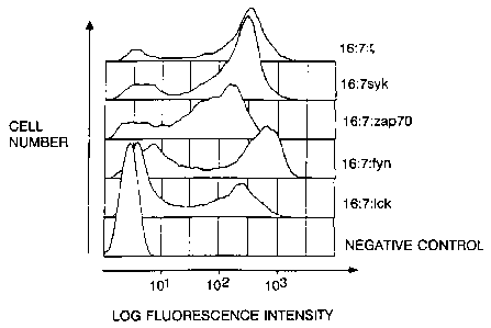
FIG. 7A, 7B, and 7C show that chimeras based on
human Syk are essentially equipotent with chimeras based
on porcine Syk. FIG. 7A is the sequence of human Syk and
comparison with porcine Syk; the first 11 and last 7
residues are determined by the primer sequences. !=a. 72
shows calcium mobilization analysis of TCR negative cells expressing human Syk
chimera. PIG. 7C shows a redirected
cytolysis assay of CTL expressing human Syk chimera.
WO 95/026M PCTNS94/06675
- 35 -
FIG. 8 shows changes in tyrosine phosphorylation
pattern following crosslinking of chimeric kinases. T
cell antigen receptor-negative Jurkat cells expressing
the indicated chimeras or pairs of chimeras were treated
with anti-CD16 and goat anti-mouse IgG second antibody
and then lysed, fractionated on a polyacrylamide gel,
transferred to nitrocellulose and probed with
anti-phosphotyrosine antibody. Lanes marked I+f
represent extracts from cells subjected to crosslinking,
while those marked 1-1 were lysed directly without prior
exposure to secondary antibody. Control lanes were
created by similar treatment of TCR-negative cells
expressing a CD16/7 fusion protein which did not contain
an intracellular domain. For comparison, the effects of
anti-CD3 treatment of TCR-positive Jurkat cells (with or
without wild type vaccinia virus infection) are shown at
right. The prominent bands in the vicinity of 100kD on
the left part of this panel correspond to the expected
molecular masses of the kinase chimeras.
FIG. 9 shows tyrosine phosphorylation of
phospholipase C-yl following aggregation of chimeras.
PLC-yl was immunoprecipitated from cells subjected to
antibody crosslinking and the immunoprecipitates were
fractionated on gels, transferred to nitrocellulose, and
probed with anti-phosphotyrosine antibody. A substantial
increase in phosphorylated PLC-yl was seen following
aggregation of Syk chimeras, whereas a more limited but
easily detectable increase is seen following
coaggregation of Fyn and ZAP-70 chimeras.
FIG. 1OA and 1O8 show in vitro kinase assays.
Cells expressing chimeric kinases were subjected to
antibody-mediated chimera crosslinking, after which the
kinases were immunoprecipitated and the
immunoprecipitates evaluated for phosphorylation of
endogenous substrate. FIG. l0A shows a comparison of the
PCT/US94106675
WO 95/0Z686 2166102
- 36 -
activity of immunoprecipitated kinase chimeras over an
incubation period of ten minutes, using
immunoprecipitates isolated from crosslinked (+) or
uncrosslinked (-) cells. FIG. 10B shows a time course of
assimilation of phosphate label into endogenous
substrates by Syk kinase chimera, with (+) or without (-)
crosslinking.
T Ceil Activation by Clustered Tyrosine Kinases
There now follows a description of particular
embodiments of the invention. In this description, it is
demonstrated that nonreceptor kinases are activated by
simple clustering events. Artificial receptor kinases
were created whose intracellular domains consisted of the
complete Src or Syk family kinase sequences and examined
for the consequences of aggregation by external
crosslinking stimuli. A clear distinction emerged
between the Syk and Src family kinase activities:
crosslinking the latter did not lead to significant
cellular activation, while crosslinking the former led to
the appearance of free intracellular calcium ion and, in
the case of Syk, of cytolytic potential. The failure of
ZAP-70 chimeras to induce distal receptor mediated
programs could be overcome by coclustering ZAP-70 chimera
with either Fyn or Lck kinase chimeras. The examples now
described are provided for the purpose of.illustrating,
not limiting, the invention.
Construction of Protein-Tyrosine Rinase Chimeras and
Demonstration of Efficacy
Gene fusions encoding proteins resembling cell
surface receptor kinases were constructed by appending a
DNA fragment encoding the extracellular domain of the
~WO 95102686 2166102 PCT/US94/06675
- 37 -
CD16 molecule to a short spacer segment encoding the
juxtamembranous and transmembrane domains of CD7 joined
in turn to the complete coding sequences of the human Lck
(Koga at al., 1986, Eur. J. Immunol. 16:1643-1646),
murine Fyn (T) (Cooke and Perlmutter, 1989, New. Biol.
1:66-74), porcine Syk (Taniquchi et al., 1991, J. Biol.
Chem. 266:15790-15796) and human ZAP-70 (Chan et al.,
1992, Cell 71:649-662) tyrosine kinases (Fiq. 1A). The
resultinq tripartite gene fusions were introduced into
recombinant vaccinia viruses by homologous recombination
and selection for coexpression of the E. coli qpt gene
product. Infection of cells with the recombinants
resulted in the efficient cell surface expression of all
four kinase chimeras (Fig. 1B). Immunoprecipitation of
the resulting protein chimeras with anti-CD16 antibodies
revealed the presence of molecular species of the
expected masses which were active in an in vitro
phosphorylation assay (Fig. 1C).
We next examined whether crosslinking of the
fusion proteins would allow the accumulation of free
intracellular calcium in a fashion similar to that found
with fusion proteins based on T cell antigen receptor
intracellular domains. To do this we infected various
cells with vaccinia recombinants and measured the
relative cytoplasmic calcium concentration following
crosslinking of the extracellular domains with
antibodies. Both spectrofluorimetric (bulk population)
and flow cytometric (sinqle cell) measurements were
performed, with cells loaded with the dye Indo-1
(Grynkiewicz et al., 1985, J. Biol. Chem. 260:3440-3450;
Rabinovitch et al., 1986, J. Immunol. 137:952-961). Flow
cytometric analyses were performed on data obtained from
cells whose cell surface expression of CD16, as
determined by phycoerythrin fluorescence intensity, fell
within a relatively narrow predefined ranqe. Although
WO 95102686 PCT/US94106675
2166102
- 38 -
minor variations in mean fluorescence intensity were
still observed within this range (due to differences in
the underlying distribution of chimeras expressed by the
cells), this approach allowed us to contrast the
responses of cells bearing approximately the same number
of receptors. Figure 2 shows an analysis of data
collected from cells of a mutational variant of the
Jurkat human T cell leukemia line lacking T cell antigen
receptor (Weiss and Stobo, 1984, J. Exp. Med. 160:1284-
1299). In these cells neither Lck nor Fyn chimeras had
the capacity to mobilize calcium following crosslinking.
In several experiments clustering of the Lck fusion
protein resulted in a slight decrease in resting calcium
concentration, relative to the negative control, a fusion
protein based on the low affinity IgG receptor FcRII82
intracellular domain (Kolanus et al., 1992, EMBO J.
11:4861-4868). Aggregation of fusion proteins based on
both ZAP-70 and Syk was highly effective in promoting the
appearance of free cytoplasmic calcium ion, roughly as
effective as aggregation of a similar chimera bearing the
intracellular domain of the T cell receptor zeta chain.
A slight delay in onset of the calcium response was seen
with both ZAP-70 and Syk kinase chimeras, relative to the
time of onset of calcium mobilization by zeta chimera.
In T cell receptor positive cells (Fig. 3), flux
evaluation of Syk chimeras was partially confounded by a
high resting concentration of free calcium ion,
suggestive of a constitutive engagement of the calcium
regulatcry apparatus.
Introduction of the chimeras into a cytolytic T
cell line then allowed us to assess the fusion proteins'
potential to engage*effector function. In this assay,
anti-CD16 hybridoma cells which express cell surface IqG
antibody against CD16 (Fleit et al., 1982, Proc. Natl.
Acad. Sci. USA 79:3275-3279; Shen et al., 1989, Mol.
~WO 95/02686 2166102 PCTIUS94/06675
- 39 -
Immunol. 26:959-969) are used as target cells. The
hybridoma cells are labeled by incorporation of
5iCr-chromate and cosedimented with effector cells,
prepared by infection of a human allospecific cytotoxic T
lymphocyte (CTL) line with vaccinia recombinants
expressing the CD16/CD7/kinase fusion proteins.
Expression of the chimeric receptor kinases allows the
infected CTL cells to bind to the target cells, and if
competent, to lyse them, a process which is measured by
release of the incorporated 51Cr-chromate. The relative
potency of the armed CTL is determined by comparison of
the ratio of effector to target cells needed to achieve a
given proportion of 51Cr release. Fig. 4A shows that CTL
expressing chimeric receptors comprising the Src family
kinases Lck or Fyn (T), or the Syk family kinase ZAP-70,
are incapable of mediating cytolysis against anti-CD16
hybridoma targets. However CTL expressing a kinase
chimera based on the Syk protein product were essentially
as effective as CTL expressing a chimera composed of CD16
fused in the same manner to the intracellular domain of
the T cell receptor zeta chain. The cytolytic activity
directed by the CD16/CD7/kinase chimeras could not be
ascribed to a nonspecific release of cyototoxic granules
because cosedimentation of an irrelevant chromium-laden
25' target with the kinase-armed CTL did not result in
detectable release of labeled chromium (Fig. 4B).
The disparity between syk and ZAP-70 activities in
the cytolysis assay was unexpected in light of the
similar activities of the two chimeras in the calcium
response assay. In light of the demonstration that
coexpression of nonchimeric ZAP-70 and Src-family kinases
led to activation in COS cells (Chan et al., 1992, Cell
71:649-662), we undertook an evaluation of the relative
potential of pairs of kinase chimeras to effect
cytolysis. CTL effectors were coinfected with
WO 95/02686 PCT/US94/06675
- 40 -
recombinant vaccinia viruses encoding ZAP-70 and Lck
chimeras, ZAP-70 and Fyn (T) chimeras, or Lck and Fyn (T)
chimeras. Fig. 5A shows that the coexpression of ZAP-70
and Fyn (T) chimeras, or ZAP-70 and Lck chimeras, endowed
CTL with an activity essentially equipotent with that of
CTL expressing CD16/CD7/Syk kinase chimeras alone, an
activity in turn as potent as that displayed by
CD16/CD7/zeta chimeras. Coexpression of Lck and Fyn(T)
chimeras did not allow significant cytolytic potential to
be redirected against anti-CD16 target cells (Fig. 5A).
Evaluation of the calcium mobilization potential of cells
coinfected with pairs of kinase fusion proteins showed
that coexpression of ZAP-70 and Src family kinase
chimeras increased the rapidity with which calcium was
mobilized in response to receptor crosslinking and that
coexpression of Lck and Fyn(T) chimeras did not result in
a significant accumulation of free intracellular calcium
(Fig. 5B). To further explore the role of the Fyn
chimera in the activation response induced by
coaggregation, we prepared a Fyn chimera consisting of
the extracellular and transmembrane domains of CD4 fused
to Fyn in a manner similar to that of the CD16 chimeras.
Fig. 5C shows that the effectiveness of this chimera in
there directed cytolysis assay is ten to twenty fold
lower than that of the comparable CD16 chimera,
suggesting that physical association of the chimeric
kinases is important for activation. However the
cytolytic activity of cells expressing the two chimeras
is significantly greater than would be observed for cells
expressing ZAP-70 chimera alone. Because of the
relatively high level of kinase expression in this
system, it cannot be excluded that the residual activity
reflects spontaneous random association of the CD4/Fyn
chimera with CD16/ZAP-70.
WO 93102686 PCT/U394106675
- 41 -
To establish that the activation seen in both
calcium response and cytolysis assays was directly
attributable to the relevant kinase activity and not to
passive association of the kinases with existing signal
transduction elements whose indirect aggregation then
initiated the activation response, we created kinase
negative variants of both the porcine Syk and human
ZAP-70 receptor chimeras. Fig. 6 shows that receptor
chimeras lacking either substantially all of the kinase
domain (in the case of Syk) or bearing a point mutation
abrogating phosphotransferase activity (in the case of
ZAP-70) lacked in vitro kinase activity and were
incapable of mediating calcium mobilization following
crosslinking, or of mediating receptor redirected
cytolysis.
Because the interaction of porcine Syk with the
human cellular apparatus might not be identical to the
interaction of human Syk, we also constructed similar
protein chimeras based on human Syk, after isolating
human Syk sequences by PCR with primers corresponding to
the amino and carboxyl termini of the porcine protein
sequence. Fig. 7A shows that pig and human Syk are
strikingly similar proteins, as suggested by analysis of
PCR products corresponding to portions of the kinase and
second SH2 domains (Chan et al., 1992, Cell 71:649-662).
Consonant with this, human syk chimeric receptor proteins
behaved essentially identically to the porcine constructs
in calcium release and cytolysis assays (Fig. 7B and 7C).
.To establish whether aggregation of the chimeric
tyrosine kinases results in a significant change in the
abundance of phosphotyrosine proteins, T cell receptor
negative cells were infected with vaccinia recombinants
encoding the chimeric kinases. The extracellular domains
of the chimeras were crosslinked with antibodies, and
total cellular lysates of the activated cells were
WO 95/02686 PCT1US94/06675
2166102 - 42 -
fractionated by electrophoresis, transferred to membranes
and analyzed with an antibody recognizing
phosphotyrosine. Lysates were prepared by disruption of
cells in nonionic detergents in the presence of vanadate
with or without EDTA, or by lysis in the presence of
sodium dodecyl sulfate, followed by sonication to shear
the liberated DNA. The pattern of phosphotyrosine
proteins was different in each case and lysates prepared
with vanadate but without EDTA showed additional species
not present in lysates prepared in SDS alone, suggesting
that EDTA may inhibit the postlysis action of tyrosine
kinases as well as phosphatases. The use of direct lysis
in SDS was found to lead to more reproducible patterns of
protein tyrosine phosphorylation than lysis with nonionic
detergents"in the presence of EDTA and vanadate. Fig. 8
shows that aggregation of chimeras bearing Syk, ZAP-70,
or Fyn plus ZAP-70 results in the appearance or increased
phosphorylation of several protein species comigrating
with proteins which show increased phosphorylation
following antigen receptor crosslinking. In particular,
the pattern of bands induced by Syk chimera clustering is
very similar to the pattern induced by anti-CD3 antibody
in T cell receptor-positive cells (Fig. 8). Among these
is an approximately 150 kD protein induced by aggregation
of Syk chimera, but also induced by coaggregation of Fyn
and ZAP-70 chimeras. in preliminary experiments this
phosphoprotein was observed to comigrate with
phospholipase C-y (data not shown).
=To establish the effects of kinase chimera
clustering on PLC-y directly, we crosslinked chimeras,
precipitated PLC-y with a mixture of monoclonal
antibodies, and analyzed the resulting immunoprecipitates
for the presence of phosphotyrosyl proteins. Fig. 9
shows that clustering of Syk results in a substantial
increase in the tyrosine phosphate content of PLC-y,
~WO 95/02686 21.66102 PCT/US94/06675
- 43 -
while cocrosslinking of Fyn plus ZAP-70 chimeras results
in a less dramatic but easily detectable increase in
tyrosine phosphate. Cells expressing Fyn chimera alone
showed a modest basal phosphorylation of PLC-y which did
not increase following receptor aggregation (Fig. 9).
Whether the same tyrosine residues are phosphorylated by
Syk, Fyn, or ZAP-70 plus Fyn, is presently unknown.
Because cells expressing Fyn chimera showed neither
resting nor induced calcium mobilization, the
phosphotyrosine signal seen in these cells may represent
utilization of other sites on PLC-y than those which
mediate phospholipase activation.
In a preliminary attempt to account for the
changes in phosphotyrosine pattern we evaluated the
activity of the various kinases following clustering in
an in vitro autophosphorylation assay. Chimeras were
aggregated, immunoprecipitated, and evaluated for their
ability to incorporate phosphate label into the protein
species present in the immunoprecipitate. Fig. 10A shows
that under these conditions no increase in kinase
activity was detected following crosslinking when the
kinase assay was carried out for ten minutes. Although
incorporation of labeled phosphate continues to increase
for up to 30 minutes in this assay, it is unclear what
factors limit activity; in particular the observed
kinetics may be dominated by the rate of dissociation of
the immune complexes, allowing kinase diffusion to
unutilized substrate. In an attempt to control for this
effect,'as well as to maximize the sensitivity of the
assay, we also evaluated Syk kinase activity with or
without prior crosslinking over a very brief time course
from 5 to 30 seconds; however here too there was no
significant increase in kinase activity (Fig. lOB).
Although at present we cannot exclude the possibility
that an increase in activity would be demonstrable with
w0 95/02696
2166102 PCT/US94/06675 -44-
an appropriate substrate, an aggregation-induced increase'
in Syk chimera activity was also not observed when an
exogenous peptide substrate (a Y 19 K substitution of cdo
2 residues 6-20) was used to measure kinase activity.
Bossib].e Mechaaisms
The mechanism by which a simple physical stimulus,
receptor aggregation, results in the transmission of a
distinctive chemical signal to immune system cells
remains unknown. Previous studies have established that
Src-family kinases can be found associated with many of
the important aggregation-activated receptors of the
immune system, and that crosslinking of such receptors
frequently leads to increased kinase activity. More
recently, the related Syk and ZAP-70 kinases have been
found to associate either stably (in the case of Syk) or
transiently (in the case of ZAP-70) with the B and T cell
antigen receptors, respectively, and at least in the case
of syk, receptor crosslinking has been reported to result
in increased kinase activity.
In this work we have shown that aggregation of Syk
family kinases, but not the Src family kinases Lck or
Fyn, leads to a calcium response in cells lacking T cell
receptor. The response appears not to be due to an
indirect association with other T call receptor or signal
transduction components because kinase negative mutants
can not induce the calcium response. Aggregation of
chimeras containing the Syk kinase is sufficient to allow
induction of specific cytolysis, while induction of
similar cytolysis by ZAP-70 chimera requires the
additional participation of a Src family kinase. At
present it is unclear which of the Syk family kinases is
likely to play a more important role in T cell
activation: both ZAP-70 and Syk are expressed in T cells,
WO 95/0686 PCT/US94/06675
~- 2166102
- 45 -
including the cell lines used in this study, and at least
one responsive human T cell line contains Syk but not
ZAP-70 as judged by specific PCR amplification products.
The pattern of increased protein tyrosine phosphorylation
seen following Syk chimera clustering is very similar to
the pattern seen after crosslinking of the T cell antigen
receptor.
one simple model for activation of immune system
cells by nonreceptor tyrosine kinases invokes a
receptor-associated kinase whose aggregation leads to
activation either by physical association (e.g., by
forming active kinase dimers) or by mutual enzymatic
action (e.g., crossphosphorylation); the activated enzyme
then acts on key intracellular substrates required for
calcium mobilization and inositolphosphate synthesis.
Support for this sequence of events can be found in
studies reporting increases in receptor-associated kinase
activity following receptor crosslinking (e.g., Bolen et
al., 1991, Adv. Cancer Res. 57:103-149; Burkhardt et al.,
1991, Proc. Natl. Acad. Sci. USA 88:7410-7414; Eiseman
and Bolen, 1992, Nature 355:78-80; Hutchcroft et al.,
1992, Proc. Natl. Acad. Sci. USA 89:9107-91111/J. Biol.
Chem. 267:8613-8619; Tsygankov et al., 1992, J. Biol.
Chem. 267:18259-18262; Wong et al., 1992, Oncogene
7:2407-2415). However the reported changes in kinase
activity are in most cases modest, and contrast with the
dramatic changes in phosphotyrosyl protein pattern seen
in vivo. Because it is difficult to unambiguously rule
out activation by weak allosteric interactions using in
vitro kinase assays as a tool, we cannot at this point
make a definitive statement about the relative importance
of kinase activation in the initiation of signal
transduction. But the data presented here suggest that
aggregation-induced repartitioning of enzyme and
substrate may be an important factor in directing an
WO 95/02686 2166102 PCTIUS94/06675
- 46 -
existing activity toward the appropriate physiological
target.
Aggregation of chimeras based on Syk family
kinases led to a calcium response which was slightly
delayed but similar in amplitude to the response seen
following aggregation of zeta receptor chimeras in cells
lacking endogenous T cell receptor. A more profound
delay in the appearance of free calcium ion was observed
following ZAP-70 chimera crosslinking, and this delay
could be substantially abolished by cocrosslinking ZAP-70
and Fyn chimeras. At present the explanation for the
observed latency is unclear. Because cocrosslinking
accelerated calcium mobilization, it is tempting to
ascribe the delay to the relative inefficacy of ZAP-70
for aggregation-mediated autoactivation. But other
factors may be equally important, and the tethering of
ZAP and Syk kinases at the cell surface may actually be
an impediment to activation when compared to the normal
process; for example if in the normal course of events
Syk family kinases are transiently recruited to clustered
receptors, activated and then released to diffuse to
their substrates, the permanent linkage of the kinase
domain to the plasma membrane could be restrictive by
hindering access to substrate and/or limiting signal
amplification due to the inability of the chimeric
receptors to function as a kind of catalytic center for
kinase activation.
A second peculiarity of the calcium response was
the finding that T cell receptor positive cells
expressing chimeras based on human or porcine Syk showed
a high baseline concentration of free calcium ion,
suggesting that calcium release had been spontaneously
triggered. A similar finding was not observed in
receptor negative cells. This result runs counter to a
general trend we have observed, in which T cell receptor
~WO 95/02686 PCT/US94/06675
- 47 -
negative mutants of human T cell tumor lines typically
are hyperresponsive to exogenously introduced trigger
molecules. To account for the apparent requirement for T
cell receptor in the spontaneous activation process, we
propose two possible and related explanations. One is
that chimeric Syk kinase may act constitutively on T cell
receptor intracellular domains to create phosphotyrosine
targets for the SH2 domains of the receptor chimera,
leading to intracellular aggregation through a
multivalent T cell receptor bridge, in turn promoting
kinase activation. Another possibility is that the T
cell receptor negative cell line may have lower levels of
a membrane associated kinase required for activation of
Syk, either because a global regulatory circuit results
in decreased de novo synthesis of the hypothetical
kinase, or because the absence of antigen receptor
results in a disregulated intracellular trafficking.
In B cells, the Syk kinase has been reported to be
constitutively associated with the intracellular elements
of the IgM antigen receptor (Hutchcroft et al., 1992,
Proc. 1Nat1. Acad. Sci. USA 89:9107-91111; Hutchcroft et
al., 1992, J. Biol. Chem. 267:8613-8619). The exact
mechanism of this association is unclear, but one
possibility is that phosphotyrosine is not required for
interaction of Syk SH2 domains with the tyrosine trigger
motif present in the cytoplasmic domain of the mb-1 and B
29 receptor chains. A partial precedent for this
suggestion is the report that the Philadelphia chromosome
breakpoint cluster region gene product BCR binds to a
variety of SH2 domains in a phosphotyrosine independent
manner (Pendergast et al., 1991, Cell 66:161-171; Muller
et al., 1992, Mol. Cell. Biol. 12:5087-5093). In this
case, though, it appears likely that phosphoserine andJor
phosphothreonine residues play a critical role in the
interaction. Alternatively, Syk may associate with rgM
WO 95/02686 PCT/US94/06675 ~
2166102
- 48 -
receptor intracellular motifs through the unique region
located between the Syk SH2 elements and the catalytic
domain. A third possibility is that only a very small ,
amount of tyrosine phosphorylated peptide is necessary to
recruit functionally important levels of Syk to the inner
face of the plasma membrane, and this low level of
tyrosine phosphate has thus far escaped detection.
Although in B cells the requirement for a Src
family kinase in activation has not been definitively
established, in T cells two kinases, Lck and Fyn (T) have
been shown by somatic or organismic genetics to play
important roles (Appleby et al., 1992, Cell 70:751-763;
Karnitz et al., 1992, Mol. Cell. Biol. 12:4521-4530;
Stein et al., 1992, Cell 70:741-750; Straus and Weiss,
1992, Cell 70:585-593). At present we cannot
conclusively establish whether the action of these
kinases normally precedes or follows the action of the
Syk family kinases. One hypothesis accounting for the
action of ZAP-70 in T cell activation invokes a
receptor-associated Src family kinase whose aggregation
permits a transitory phosphorylation of receptor chains
in turn leading to association of ZAP-70 and subsequent
cellular activation. The initial phosphorylation of
receptor chains proposed in this model must be
distinguished from the stable phosphorylation of zeta
seen at longer times following receptor crosslinking.
In murine T cells a small proportion of T cell
receptor complexes contain a zeta-related molecule called
eta (BaTiiyash et al., 1988, J. Biol. Chem. 263:9874-9878)
which represents an alternatively spliced form (Clayton
et al., 1991, Proc. Natl. Acad. Sci. USA 88:5202-5206).
Eta differs from zeta at the carboxyl terminus, and lacks
the most distal of the six tyrosines found in murine zeta
(Jin et al., 1990, Proc. Nat1. Acad. Sci. USA 87:3319-
3323). Although the phosphorylation of the zeta chain of
~WO 95102686 PCl'/US94/06675
- 49 -
murine TCR zeta-zeta isoforms can readily be detected
following antibody-mediated receptor crosslinking, under
. similar circumstances the TCR eta chain is not detectably
phosphorylated (Bauer et al., 1991, Proc. Natl. Acad.
Sci. USA 88:3842-3846). Stable phosphorylation of the
zeta chain appears to require two closely apposed zeta
chains since TCR isoforms bearing zeta-eta heterodimers
are not phosphorylated following receptor crosslinking
(Bauer et al., 1991, Proc. Natl. Acad. Sci. USA 88:3842-
3846). Despite the differences in phosphorylation, cell
lines comprising TCR isoforms consisting solely of eta
homodimers are functionally indistinguishable from cell
lines bearing only zeta homodimers (Bauer et al., 1991,
Proc. Nati. Acad. Sci. USA 88:3842-3846). Thus
phosphorylation of zeta, as observed 30 minutes after
antibody mediated receptor aggregation, does not
correlate with activation. in a separate study,
examination of the time course of accumulation of
phosphotyrosine phosphoproteins following TCR aggregation
has shown that the earliest observable species are two
proteins of mass 135 and 100 kD, whose phosphorylation is
first detectable at five and fifteen seconds after
crosslinking, respectively, and whose half maximal
phosphorylation, in both cases at approximately 30
seconds, precedes the times of half maximal calcium
mobilization and inositol phosphate formation (June et
al., 1990, Proc. Natl. Acad. Sci. USA 87:7722-7726; June
et al., 1990, J. Immunol. 144:1591-1599). By contrast,
the rate of phosphorylation of zeta is substantially
slower, leading to half maximal substitution at
approximately three to five minutes post-stimulation,
well after calcium accumulation and liberation of
inositol phosphates are observed (June et al., 1990,
Proc. Natl. Acad. Sci. USA 87:7722-7726; June et al.,
1990, J. Immunol. 144:1591-1599).
WO 95102686 ' PCTI[JS94/06675
2166102 0
- 50 -
Thus if the two step model is correct, the
tyrosine phosphorylation necessary to recruit ZAP-70 to
the inner face of the plasma membrane must be a more
rapid, presumably transitory, event than observed in the
studies above. Recently it has been suggested that
tyrosine phosphorylation with an appropriately fast (ca.
fifteen second) onset can be detected on both zeta and
CD3 epsilon chains following receptor crosslinking (Wange
et al., 1992, J. Biol. Chem. 267:11685-11688), and that a
70 kD protein bearing tyrosine phosphate can be found
associated with both zeta and epsilon chains. It is not
presently clear whether a stable association with
phosphorylated receptor chains is a prerequisite for
successful T cell activation.
As a general assertion, though, the results
reported here suggest that Syk family kinases act more
directly on the effector apparatus of T cells than Src
family kinases. An increasing body of evidence suggests
that Src family kinases can associate with a number of
cell surface molecules which are not members of the
antigen/Fc receptor family, including CD2 (Bell et al.,
1992, Mol. Cell. Biol. 12:5548-5554), CD23 (Sugie et al.,
1991, Proc. Nati. Acad. Sci. USA 88:9132-9135), CD36
(Huang et al., 1992, J. Biol. Chem. 267:5467-5473), IL-2
receptor beta chain (Hatakeyama et al., 1991, Science
252:1523-1528) and various phosphatidylinositol anchored
proteins (Stefanova at al., 1991, Science 254:1016-1019;
Thomas and Samelson, 1992, J. Biol. Chem. 267:12317-
12322),.some of which are known to require the additional
presence of the antigen receptor to promote activation in
T cells. A simple explanation for the latter requirement
maybe that the trigger motifs on the antigen receptor act =
as substrates for Src family kinases, allowing the
subsequent docking of Syk family kinases, followed
perhaps by some modifying event promoting their
WO 95/02686 2166102 PCT/US94106675
- 51 -
activation. Given the difficulty of establishing a
causal chain of phosphorylation and activation it may
also be that the trigger motifs have only a transitory
role, to act as a kind of catalytic center for the
recruitment, activation, and release of effector kinases.
Src family kinases are broadly distributed
throughout nonhematopoietic lineages, and recent studies
using Fyn negative mice have shown a role for Fyn in the
sustenance of long term potentiation, the phenomenon of
facilitated synaptic transmission thought to underlie the
initial consolidation of associative memory (Grant et
al., 1992, Science 258:1903-1910). If similar activation
pathways are mediated by Src family kinases in other cell
types, Syk family kinases may also prove to be more
extensively distributed throughout the extrahematopoietic
compartments.
8xperimental Methods
Construction of chimeras. The entire coding regions of
the human Lok (Koga et al., 1986, Eur. J. immunol.
16:1643-1646), murine Fyn (T) (Cooke and Perlmutter,
1989, New. Biol. 1:66-74), porcine Syk (Taniguchi at al.,
1991, J. Biol. Chem. 266:15790-15796) and human ZAP-70
(Chan et al., 1992, Cell 71:649-662) kinases were
attached to the intracellular domain of a chimeric
transmembrane protein consisting of the CD16
extracellular domain joined to a short 'stalk' segment
and transmembrane domain of CD7. The CD7 intracellular
domain was truncated at the stop transfer sequence by
addition of an Mlu site. The various kinases were
adapted with an Mlu site in the appropriate reading frame
to allow a tripartite fusion protein to be expressed.
The pig Syk sequence was obtained by reverse
transcription and PCR of total pig lymphocyte RNA using
WO 95/02686 2166162 PCT/US94/06675
- 52 -
primers bearing appropriate restriction sites. ZAP-70
sequences were similarly obtained by PCR from a human T
cell cDNA library. Several isolates were sequenced in
parallel and a mutation-free coding sequence was derived
for each kinase by restriction fragment interchange. The
resulting coding sequences were inserted into a vaccinia
virus expression vector downstream from the CD16/CD7
sequences. Human Syk was isolated from a natural killer
cell cDNA library and from a Daudi cell library using
primers corresponding to the ends of the porcine
sequence. The forward primer bore the sequence atg gca
gac agt gcc aac cac ttg ccc ttc ttc t and the reverse
primer bore the sequence cgc ggg gcg gcc gct tta att cac
cac gtc gta gta gta. After initial amplification
revealed the presence of bands of the expected size a
reamplification (10 cycles) was performed using an
extension primer at the 5' end having the sequence cgc
ggg acg cgt acc atg gca gac agt gcc aac, allowing the
fragment to be ligated to an Mlu I-cut vector.
Calcium mobilization analysis. Flow cytometric and bulk
spectrophotometric analyses were conducted on cells
expressing recombinant kinases using the calcium
sensitive fluorophore Indo-1 as previously described
(Romeo and Seed, 1991, Cell 64:1037-1046; Romeo et al.,
1992, Cell 68:889-897). Briefly, cells of the Jurkat
mutant subline JRT 3.T 3.5 (Weiss and Stobo, 1984, J.
Exp. Med. 160:1284-1299) were infected with recombinant
vaccinia viruses for one hour in serum free IMDM at an
moi of 10 and incubated for three to nine hours in IMDM,
10% FBS. Cells were collected by centrifugation and
resuspended at 3 x 10⁶/ml in complete medium containing
1mM Indo-1 acetomethoxyester (Grynkiewicz et al., 1985,
J. Biol. Chem. 260:3440-3450) (Molecular Probes, Eugene,
OR) and incubated at 37 C for 45 minutes. The Indo-1
loaded cells were pelleted and resuspended at 1 x 10⁶/ml
WO 95/02686 216610. 2 PCTlUS94/06675
~ . ,
- 53 -
in serum free INDM and stored at room temperature in the
dark. Cells were analyzed for free calcium ion by
simultaneous measurement of the violet and blue
fluorescence emission by flow cytometry (Rabinovitch at
al., 1986, J. Immunol. 137:952-961). To initiate calcium
flux, either unconjugated 3G8 (anti-CD16) monoclonal
antibody (at 1 g/ml) was added to the cell suspension
followed by 10 g/ml of phycoerythrin (PE)-conjugated Fab
02 goat anti-mouse IgG at time 0, or a PE-conjugated
anti-CD4 antibody (Leu-3a, Becton Dickinson) was added,
followed by unconjugated second antibody. Histograms of
the violet/blue emission ratio were collected from the PE
positive (infected) cell population, which typically
represented 40-80% of the cells. The violet/blue
emission ratio prior to the addition of antibody was used
to establish the normalized initial ratio, set equal to
unity.
Lvmphocyte cytolysis assay. A CD8+ CD4- HLA B44
restricted cytolytic line (WH3) was maintained in nIDM,
10% human serum with 100 U/ml of IL-2 and was
periodically stimulated with irradiated (3000 rad)
mononuclear cells having the HLA B44 haplotype. Cells
were grown for at least 10 days following stimulation
before use in cytotoxicity assays. The cells were
infected with recombinant vaccinia at a multiplicity of
infection of at least 10 for one hour in serum free
medium, followed by incubation in complete medium for
three hours. Cells were harvested by centrifugation and
resuspended at a density of 1 x 10⁷/ml. 100 l were
added to each well of a U-bottom microtiter plate
containing 100 l/well of complete medium. Cells were
diluted in two-fold,serial steps. Two wells for each
sample did not contain lymphocytes, to allow spontaneous
chromium release and total chromium uptake to be
measured. An aliquot of 106 3G8 10-2 target cells (Shen
CA 02166102 2006-09-01
- 54 -
et al., 1989, Mo1. Immunol. 26:959-969) or was
centrifuqed and resuspended in 50 l of sterile 51Cr
sodium chromate (1 a Ci/ml, DuPont) for one hour at 374C
with intermittent mixing, then washed three times with
PBS. loo l of labelled cells resuspended in medium at
10S/ml were added to each well. The microtiter plate was
spun at 750 x g for 1 minute and incubated for 4 hours at
370C. At the end of the incubation period the cells in
each well were resuspended by gentle pipetting, a sample
removed to determine the total counts incorporated, and
the microtiter plate was spun at 750 x g for 1 minute.
100 l aliquots of supernatant were removed and counted
in a qamma rays cintillation counter. The effector to
target ratio was corrected for the percent of effector
cells infected (usually >70%).
Creation of mutant kinase chimeras. A porcine Syk
kinase neqative fusion protein variant was created by
cleavaqe of the chimera with Stu I and Not I (the latter
lyinq just 31 to the carboxyl terminus), fillinq in the
Not I site, and liqating the ends together. The
resultinq sequence joined the first 298 residues of piq,
Syk to 4 extraneous residues (GPRL) before terminatinq.
A point mutation (K369G) in the ATP bindinq site of
ZAP-70 was created by insertion of a duplex
oligonucleotide fragment between the Ball and Earl sites
located between nucleotide residues 1319 and 1345 of the
top strand of the sequence reported by Chan et al. (1992,
Cell 71:649-662). The resulting sequence encoded glycine
at residue 369 in place of lysine.
Immunogrecipitation and kinase assay. Approximately 2 x
106 Hela S3 cells were infected for one hour in serum
free DME medium with recombinant vaccinia at a
multiplicity of infection of at least ten. 5 hrs after
infection the cells were harvested, washed twice with
phosphate buffered saline and lysed in 1t Triton X-100'''",
CA 02166102 2006-09-01
- 55 -
0.15 M NnCl, 0.02 H HEPES pH 7.3, 5 mM EDTA, 5 mM NaF,
0.2 m MNav03, 10 g/mi leu peptin, 10 g/ml a protinin
and 1mM PMSF. After a 10 min. incubation on ice, the
nuclei were removed by centrifugation and the CD16 fusion
proteins immunoprecipitated with antibody BMA209/2 and
protein-A sepharoseT". The fusion protein loaded resin was
washed 3 times with lysis buffer followed by a final wash
with 20 mM Hapas pH 7.3. To each sample was added 10 l
of kinase buffer (20 mM Hepes pH 7.3, 10 mM MqClZ1 10 mM
MnC1z) containing 10 Ci of [Y-32PjATP (>3000 Ci/mmole).
The reactions were allowed to incubate at room
temperature for 10 min. and terminated by the addition
of 20 l of 2X sample loading buffer (4% SDS, 100 mM Tris
pH6.8, 20% qlycerol, and 10% P-mercaptoethanol). After
the samples were boiled for 3 min., aliquots were run on
a 4-15% gradient gel. Kinase assays with a soluble
peptide substrate corresponding to positions 6-20 of cdc
2, in which tyr 20 was replaced with lys, were performed
according to the manufacturer's recommendations (UBI).
Immunoblot analysis of mrotein tvrosine 2hQgpbQrylAtiQp.
TCR negative 3.5 calls were infected with recombinant
virus stocks (moi of at least 10) for one hour. Calls
were subsequently incubated at 370C for 8-12h,
centrifuged, washed and resusupended in Iscove's medium
without serum at 107 calls per ml. Aliquots of cells
were incubated with anti-CD16 mAb (3G8, Medarex or
B1dA209/2, Behrinqwerke) at 1 g antibody per 2 - 3 x 10⁶
cells. Stimulated samples were further incubated with a
3-5 fold excess of an affinity purified anti-mouse IqG 1
antibody (Southern Biotechnology) for 5 min. Cells were
subsequently processed according to Secrist, J.P., Burns,
L.A., Karnitz, L., Koretzky, G.A., and Abraham, R.T. (J.
Biol. Chem. 268,5886-5893, 1993). with slight
modifications. Incubations were terminated by adding SDS
to a final concentration of 1t and samples were boiled
CA 02166102 2006-09-01
-56-
for three minutes. DNA was sheared by sonication for 1
min using a Heat Systems Ultrasonics, Inc., 2X sample
buffer was added and aliquots corresponding to 10s to 2.5
x 10S cells were separated on polyacrylamide gels and the
proteins transferred by semidry electroblotting (Hoefer)
onto nitrocellulose (Schleicher and Schuell BA45).
Filters were blocked for one hour in Tris Buffared Saline
with 0.05% Tween-20*m (TBST) containing 1.5% chicken
ovalbumin (Sigma), washed in TBST, and transferred into
solution containinq anti-phosphotyrosine antibody 4G10
(U8I) at a 1:10000 dilution and incubated at 229 for
1-2h. After TBST washes, filters wore incubated in TBST
and a 1:5000 dilution of anti-mouse horseradish
peroxidase conjugate for 1 h. Phosphorylated protein
bands were detected by chemiluminescence (ECL, Amersham).
Exposure times varied between 29 and 5 min.
Coastructioa of suffiaa IqaisProtaia-Tyrosiaa =iaase
chiseras
Chimeric molecules may be produced which include
the extracellular domain of an antibody molecule specific
for a target cell and the intracellular domain of a
protein-tyrosine kinase (e.q., those kinases described
herein). To produce such a molecule, human IqGi heavy
chain sequences are prepared by joininq sequences in the
Cs3 domain to a cDNA fraqment derived from the 3' and of
the transmembrane form of the antibody mRNA. The 31' end
fraqment is obtained by polymerase chain reaction using a
tonsil cDNA library as substrate, and oligonucleotides
having the sequences:
CGC GGG GTG ACC GTG CCC TCC AGC AGC TTG GGC and
CGC GGG GAT CCG TCG TCC AGA GCC CGT CCA GCT CCC
CGT CCT GGG CCT CA,
corresponding to the 5' and 3' ends of the desired DNA
fraqments respectively. The 5' oliqo is complementary to
WO 95/02686 PCT/US94/06675
-57-
a site in the CA1 domain of human IgGi, and the 3' oligo
is complementary to a site just 511 of the sequences
encoding the membrane spanning domain. The PCR product
is digested with BstXI and BamHI and ligated between
BstXI and BamH2 sites of a semisynthetic IgGi antibody
gene bearing variable and constant regions. Following
the insertion of the BstXI to BamHI fragment, the
amplified portions of the construct are replaced up to
the Smal site in Ca3 by restriction fragment interchange,
so that only the portion between the SmaI site and the 3'
oligo is derived from the PCR reaction.
To create a human IgGl chimeric receptor, the
heavy chain gene ending in a BamHI site is joined to the
kinase intracellular domain of interest by standard
techniques: Levels of chimeric receptor expression may
be determined by flow cytometry. Increases in expression
may be accomplished by coexpression of a plasmid encoding
an antibody light chain cDNA.
To create a single transcription unit which would
allow both heavy and light chains to be expressed from a
single promoter, a plasmid encoding a bicistronic mRNA is
created from heavy and light chain coding sequences, and
the 5' untranslated portion of the mRNA encoding the 78kD
glucose regulated protein, otherwise known as grp78, or
BiP. grp78 sequences are obtained by PCR of human
genomic DNA using primers having the sequences:
CGC GGG CGG CCG CGA CGC CGG CCA=AGA CAG CAC and
CGC GTT GAC GAG CAG CCA GTT GGG CAG CAG CAG
at the 5' and 3' ends respectively. Polymerase chain
reactions with these oligos are performed in the presence
of 10% dimethyl sulfoxide. The fragment obtained by PCR
is digested with Notl and HincIl and inserted between
Notl and HpaI sites downstream from human IgGl coding
sequences. Sequences encoding a human IgG kappa light
chain cDNA are then inserted downstream from the grp78
WO 95/02686 PCT/US94/06675
2166102 - 58 -
leader, using the HincIi site and another site in the
vector. The expression plasmid resulting from these
manipulations consists of the semisynthetic heavy chain
gene, followed by the grp78 leader sequences, followed by
the kappa light chain cDNA sequences, followed by
polyadenylation signals derived from an SV40 DNA
fragment. We have previously demonstrated that
transfection of COS cells with this expression plasmid
gave markedly improved expression of heavy chain
determinants, compared to transfection of plasmid
encoding heavy chain determinants alone.
To create a bicistronic gene comprising a heavy
chain/receptor chimera and a light chain, the upstream
heavy chain sequences can be replaced by any chimeric
heavy chain/ receptor gene described herein.
Once constructed, the IgG-tyrosine kinase chimeras
may be cloned into an expression vector, introduced into
a host cell, and tested by any of the assays described
herein (e.g., by calcium mobilization or cytolysis
assays).
Construction of CU-Tyrosine Rinase Chimeras
Chimeric molecules may be produced which include
the extracellular domain of the CD4 molecule and the
intracellular domain of a protein-tyrosine kinase (e.g.,
those kinases described herein). To produce such a
molecule, the tyrosine kinase-encoding sequence (for
example, cDNA) is isolated (for example, as described
above). This sequence is then joined to the
extracellular domain of an engineered form of CD4
possessing a BamHI site just upstream of the membrane
spanning domain (Aruffo et al., Proc. Natl. Acad. Sci.
I=,14,:8573-8577 (1987b); Zettlmeissl et al., DNA Cell
Biol., 2.347-353 (1990)) by standard techniques. To form
40 95/02686 2 t 66102 PCT/US94/06675
- 59 -
this fusion protein, a BamHI site may be engineered into
the sequence at the appropriate location (again, by
standard techniques). The gene fusions are introduced
into a vaccinia virus expression plasmid (as described
herein), and inserted into the genome of the vaccinia WR
strain by homologous recombination and selection for
growth in mycophenolic acid (Falkner et al., J. Virol.,
62:1849-1854 (1988); Boyle et al., Gene, ~U:123-128
(1988)'). Flow cytometric analysis is used to examine
expression by the vaccinia recombinants of the CD4-
tyrosine kinase fusion proteins at the cell surface.
Immunoprecipitation of cells infected with the vaccinia
recombinants is used to confirm the results (as described
above).
The efficacy of CD4 chimeras may be tested by any
of the calcium mobilization or cytolysis assays described
herein. In one particular example, a model
target:effector system based on CD4 recognition of the
HIV envelope gpl20/gp41 complex is created. HeLa cells
are infected with recombinant vaccinia viruses expressing
gp120/gp41 (Chakrabarti et al., Nature, 320:535-537
(1986); Earl et al., J. Virol., 64:2448-2451 (1990)) and
labeled with 51Cr. The labeled cells are incubated with
cells from a human allospecific (CDB+, CD4-) cytotoxic T
lymphocyte line which has been infected with vaccinia
recombinants expressing the CD4-tyrosine kinase chimera,
and examined to specific lysis.
To control for the possibility that vaccinia
infection might promote artefactual recognition by CTL,
similar cytolysis experiments are performed with target
cells infected with vaccinia recombinants expressing the
phosphatidylinositol linked form of CD16 (CD16pI) and
labeled with 51Cr, and with CTL infected with control
recombinants expressing CD16 chimeras.
WO 95/02686 2166102 PCT/US94/06675
=
- 60 -
In another example, neutrophilic granulocytes,
which have a very short lifespan (a 4h) in circulation
and are intensely cytolytic, are attractive cells for
expression of CD4-tyrosine kinase chimeras. Infection of
neutrophils with HIV is not likely to result in virus
release, and the abundance of these cells (the most
prevalent of the leukocytes) should facilitate host
defense. Another attractive possiblity for host cells
are mature T cells, a population presently accessible to
retroviral engineering (Rosenberg, S.A. Sci. Am., =:62-
69 (1990)). With the aid of recombinant IL-2, T cell
populations can be expanded in culture with relative
ease, and the expanded populations typically have a
limited lifespan when reinfused (Rosenberg et al., ~
Enal. J. Med., M:570-578 (1990)).
Under the appropriate conditions, HiV recognition
by cells expressing CD4 chimeras should also provide
mitogenic stimuli, allowing the possibility that the
armed cell population could respond dynamically to the
viral burden. Although we have focused here on the
behavior of the fusion proteins in cytolytic T
lymphocytes, expression of the chimeras in helper
lymphocytes might provide an HIV-mobilized source of
cytokines which could counteract the collapse of the
helper cell subset in AIDS. Recent description of
several schemes for engineering resistance to infection
at steps other than virus penetration (Friedman et al.,
Nature, =:452-454 (1988); Green et al., Cell, U:215-
223 (1989); Malim et al., Ce11, 8:205-214 (1989); Trono
et al., Cell, U:113-120 (1989); Buonocore et al.,
Nature, 2AI:625-628 (1990)) suggests that cells bearing
CD4 chimeras could be designed to thwart virus production
by expression of appropriate agents having an
intracellular site of action.
O 95102686 2166102 PCTIUS94/06675
~~ = '
- 61 -
The ability to transmit signals to T lymphocytes
through autonomous chimeras also provides the ability for
the regulation of retrovirally engineered lymphocytes ja
vivo. crosslinking stimuli, mediated for example by
specific IgM antibodies engineered to remove complement-
binding domains, may allow such lymphocytes to increase
in number in situ, while treatment with similar specific
IgG antibodies (for example recognizing an amino acid
variation engineered into the chimeric chain) could
selectively deplete the engineered population. we have
previously determined that anti-CD4 IgM antibodies do not
require additional crosslinking to mobilize calcium in
Jurkat cells expressing CD4:Z chimeras. The ability to
regulate cell populations without recourse to repeated
extracorporeal amplification may substantially extend the
range and efficacy of current uses proposed for
genetically engineered T cells.
Q,ther Embodiments
To create other chimeras consisting of protein
kinase intracellular sequences, cDNA or genomic sequences
encoding an extracellular domain of the receptor can be
endowed with a restriction site introduced at a location
just preceding the extracellular domain of choice. The
extracellular domain fragment terminating in the
restriction site can then be joined to the protein kinase
sequences. Typical extracellular domains may be derived
from receptors which recognize complement, carbohydrates,
viral proteins, bacteria, protozoan or metazoan
parasites, or proteins induced by them. Similarly,
ligands or receptors expressed by pathogens or tumor
cells can be attached to protein kinase sequences to
direct immune responses against cells bearing receptors
recognizing those ligands.
To identify the minimal protein kinase sequences
necessary for cytolysis, a series of deletion mutants may
WO 95/02686 PCT/1JS94/06674
2166102
- 62 -
be prepared by standard techniques in which successively
more of the kinase intracellular domain is removed. Such
deletion mutants are tested for efficacy in any of the
assays described herein. Useful intracellular domains
for the Syk protein kinase include, for example, amino
acids 336-628 of the porcine Syk sequence and amino acids
338-630 of the human Syk sequence.
while the invention has been described in
connection with specific embodiments thereof, it will be
understood that it is capable of further modifications
and this application is intended to cover variations,
uses, or adaptations of the invention and including such
departures from the present disclosure as come within the
art to which the invention pertains and as may be applied
to the essential features hereinbefore set forth as
follows in the scope of the appended claims.
WO 95/02686 PCT/US94/06675
- 63 -
BEOUENCE LISTING
(1) (iENERAL INFORIlATIONs
(i) APPLICANTs Brian Seed
Charles Romeo
Waldemar Kolanus
(ii) TITI+a OF INVENTIONs REDIRECTION OF CELLULAR IMMUNITY
BY RECEPTOR CHIMERAS
8
(iii) NMER OF SEQUENCES:
(iv) CORRESPONDENCE ADDRESS:
(A) ADDRESSEEs Fish & Richardson
(B) STREETs 225 Franklin Street
(C) CITY: Boston
(D) STATI:s Massachusetts
(E) COUNTRYs U.S.A.
(F) ZIPs 02110-2804
(v) COMPUTER READABLE FORN s
(A) NEDIU1t TYPLs 3.5" Diskette, 1.44 Mb
(B) COIPUTERs IBM PS/2 Model 50Z or 55SX
(C) OPERATING SYSTEM: MS-DOS (Vereion 5.0)
(D) SOFTWARE: WordPerfect (Version 5.1)
(vi) CURRENT APPLICATION DATAs
(A) APPLICATION NUMBER: 08/093,210
(B) FILING DATSs July 16, 1993
(C) CLASSIFICATION:
(viii) ATTORNEY/AGENT INFORNATIONs
(A) NA1Es Paul T. Clark
(B) REaISTRATION NUMBER: 30,162
(C) REFER=CB/DOCKET NUlD;ERs 00786/195001
(ix) TELECOIONNICATION INFORMATION:
(A) TELEPSONEs (617) 542-5070
(B) TBLEP]1Zs (617) 542-8906
(C) TEI.EZs 200154
WO 95/02686 2166102 PCT/US94/06675
- 64 -
(2) INFORIf7lTION FOR SEQUENCE IDENTIFICATION NUM&ER: 1:
(i) SEQIISNCB CHARACTERISTICSs
(A) IaNOT8: 33
(B) TYPEs nucleic acid
(C) STRANDEDNESS: single
(D) TOPOLOGYs linear
(xi) SEQUENCL DESCRIPTIONs SEQ ID NO: 1:
CGCGGGGTGA CCGTGCCCTC CAGCAGCTTG GGC 33
(2) IWFOR1a17.'IoM FOR SEQUmCE IDENTIFICATION NWfBERt 2s
(i) SEQVENCB CHARACTERISTICSs
(A) IWNG7.'S: 50
(B) T7tPEt nucleic acid
(C) STRANDEDNESS: sinqle
(D) TOPOLO3Ys linear
(xi) SEQUFatCS DESCRIPTION: SEQ ID NO: 2:
CGCGGGGATC CGTCGTCCAG AGCCCGTCCA GCTCCCCGTC CTGGGCCTCA 50
(2) INFORWITION FOR SBQIIwCB IDENTIFICATION NOl03BRt 3:
(i) SEQLTENCE CSARACTERISTICSs
(A) Z.ENGS: 33
(B) TYPEs nucleic acid
(C) STRIINDSDNSSSs single
(D) TOPOLOGY: linear
(xi) SEQUENCE DESCRIPTIONt SEQ ID NOs 3s
CGCGGGCGGC CGCGACGCCG GCCAAGACAG CAC 33
(2) INFORI0ITION FOR SLQOWCB IDEnTIFICATION NO! ERs 4 s
(i) SEQULNCa CHARACTERISTICSt
(A) I.ENOl'8: 33
(B) TYFBs nucleic acid
,(C) STRI:NDEDNESSs single
(D) TOPOI.OaYs linear
(xi) SSQVENCB DESCRIPTIONs SEQ ID NO: 4:
CGCGTTGACG AGCAGCCAGT TGGGCAGCAG CAG 33 (2) INFORMATION FOR SEQUENCE
IDENTIFICATION NUNBER: 5:
00 951N686 2166102 PCT/US94/06675
- 65 -
(i) SEQUlaTCE CHARACTERISTICSt
(A) LENaTHt 630
(8) T7[PEt atnino acid
(C) STRANDMDNESSt N/A
(D) TOPOLOGYt linear
(xi) SEQVENCE DESCRIPTION: SEQ ID NO: 5:
Met Ala Asp Ser Ala Aen His Leu Pro Phe Phe Phe Gly His Ile Thr
1 5 10 15
Arg Glu Glu Ala Olu Asp Tyr Leu Val Gln Gly Gly Met Ser Asp Gly
20 25 30
Leu Tyr Lou Leu Arq Gln Ser Arq Aen Tyr Lou Gly Gly Phe Ala Lou
35 40 45
Sar Val Ala His Gly Arg Lys Ala His Asn Tyr Thr Ile Glu Arg Glu
50 55 60
Lou Asn Gly Thr Tyr Ala Ile Ala Gly Gly Arg Thr His Ala Ser Pro
65 70 75 80
Ala Asp Leu Cys Asn Tyr Hie Ser Gln Glu Ser Asp Gly Leu Val Cys
85 90 95
Lou Lou Lys Lys Pro Phe Asn Arg Pro Gln Gly Val Gln Pro Lys Thr
100 105 110
Gly Pro Phe Glu Asp Leu Lys Glu Asn Leu Ile Arg Glu Tyr Val Lys
115 120 125
Gln Thr Met Asn Leu Gln Gly Gln Ala Leu Glu Gln Ala Ile Ile Ser
130 135 140
Gln Lys Pro Gln Leu Glu Lys Leu Ile Ala Thr Thr Ala His Glu Lys
145 150 155 160
Met Pro Trp Phe His Gly Lys Ile Ser Arg Glu Ile Ser Thr Gln Ile
165 170 175
Val.Leu Ile Gly Ser Lys Thr Asn Gly Lys Phe Leu Ile Arg Ala Arg
180 185 190
Asp Asn Asn Gly Ser Tyr Ala Leu Cys Lou Lou His Ile Gly Lys Val
195 200 205
Leu His Tyr Arg Ile Asp Lys Asp Lys Thr Gly Lys Lou Ser Ile Pro
210 215 220
Glu Gly Lys Lys Phe Asp Thr Leu Trp Gln Lou Val Glu His Tyr Ser
225 230 235 240
Tyr Lys Ala Asp Gly Leu Leu Arg Val Leu Thr Val Pro Cys Gin Lys
245 250 255
Ile Gly Thr Gln Gly Asn Val Asn Phe Gly Gly Arg Pro Gln Lou Phe
260 265 270
Gly Ser His Pro Ala Thr Hie Ser Ala Gly Gly Ile Ile Ser Arg Ile
275 280 285
Lys Ser Tyr Ser Phe Pro Lys Pro Gly His Arg Lye Ser Ser Pro Ala
290 295 300
Gln Gly Asn Arg Gln Glu Ser Thr Val Ser Phe Asn Phe,Tyr Glu Pro
305 310 315 320
Glu Lou Ala Pro His Ala Ala Asp Lye Gly Pro Gin Arg Ile Ala Leu
325 330 335
Pro Met Asp'Thr Glu Val Tyr Glu Ser Pro Tyr Ala Asp Pro Glu Glu
340 345 350
Ile Arg Pro Lys Glu Val Tyr Lou Asp Arg Lys Lou Lou Thr Lou Glu
355 360 365
Asp Lys Glu Lou Gly Ser Gly Asn Phe Gly Thr Val Lys Lys Gly Tyr
370 375 380
Tyr Gln Met Lys Lye Val Val Lys Thr Val Ala Val Lys Ile Lou Lys
385 390 395 400
Asn Glu Ala Asn Asp Pro Ala Leu Lys Asp Glu Leu Leu Ala Glu Ala
405 410 415
Asn Val Met Gln Gln Lou Asp Aen Pro Tyr Ile Val Arg Met Ile Gly
420 425 430
WO 95/02686 PCT/US94/06675
- 66 -
Ile Cys Glu Ala Glu Ser Trp Met Leu Val Met Glu Met Ala Glu Leu
435 440 445
Gly Pro Leu Asn Lys Tyr Leu Gln Gln Asn Arg His Val Lys Leu Lys
450 455 460
Asn Ile Ile Glu Leu Val His Gln Val Ser Met Gly Met Lys Tyr Leu
465 470 475 480
Glu Glu Ser Asn Phe Val His Arg Asp Leu Ala Ala Arg Asn Val Leu
485 490 495
Leu Val Thr Gln His Tyr Ala Lys Ile Ser Asp Phe Gly Leu Ser Lys
500 505 510
Ala Leu Arg Ala Asp Glu Asn Tyr Tyr Lys Ala Gin Thr His Gly Lys
515 520 525
Trp Pro Val Lys Trp Tyr Ala Pro Glu Cys Ile Asn Tyr Tyr Lys Phe
530 535 540
Ser Ser Lys Ser Asp Val His Ser Phe Gly Val Leu Met Asn Glu Ala
545 550 555 560
Phe Ser Tyr Gly Gln Lys Phe Tyr Arg Gly Met Lys Gly Ser Glu Val
565 570 575
Ile Ala Met Leu Glu Lys Gly Glu Arg Met Gly Cys Pro Ala Gln Cys
580 585 590
Pro Arg Glu Met Tyr Asp Leu Met Asn Leu Cys Trp Thr Tyr Asp Val
595 600 605
Glu Asn Arg Pro Gly Phe Ala Ala Val Glu Leu Arg Leu Arg Asn Tyr
610 615 620
Tyr Tyr Asp Val Val Asn
625 630
(2) INFORMATION FOR SEQUENCE IDENTIFICATION NUMBER: 6:
(i) SEQUENCE CHARACTERISTICS:
(A) LENGTH: 628
(B) TYPE: amino acid
(C) STRANDEDNESS: N/A
(D) TOPOLOGY: linear
(xi) SEQUENCE DESCRIPTION: SEQ ID NO: 6:
Met Ala Asp Ser Ala Asn His Leu Pro Phe Phe Phe Gly Gln Ile Thr
1 5 10 15
Arg Glu Glu Ala Glu Asp Tyr Leu Val Gln Gly Gly Met Ser Asp Gly
20 25 30
Leu Tyr Leu Leu Arg Gln Ser Arg Asn Tyr Leu Gly Gly Phe Ala Leu
35 40 45
Ser Val Ala Tyr Asp Arg Lys Ala His His Tyr Thr Ile Glu Arg Glu
50 55 60
Leu Asn Gly Thr Tyr Ala Ile Ser Gly Gly Arg Thr His Gly Ser Pro
65 70 75 80
Ala Giu Leu Cys His Tyr His Ser Gln Glu Leu Asp Gly Leu Val Cys
85 90 95
Leu Leu Lys Asn Pro Phe Asn Arg Pro Pro Gly Val Gln Pro Lys Thr
100 105 110
Gly Pro Phe Glu Asp Leu Lys Glu Asn Leu Ile Arg Glu Tyr Val Lys
115 120 125
Gin Thr His Asn Leu Gln Gly Gln Ala Leu Glu Gln Ala Ile Ile Ser
130 135 140
Gln Lys Pro Gln Leu Glu Lys Leu Ile Ala Thr Thr Ala His Glu Lys
145 150 155 160
Met Pro Trp Phe His Gly Lys Ile Ser Arg Asp Glu Ser Glu Gln Ile
165 170 175
Val Leu Ile Gly Ser Lys Ile Asn Gly Lys Phe Leu Ile Arg Ala Arg
180 185 190
O 95/02686 PCT/US94/06675
- 67 -
Asp Met Gly Ser Tyr Ala Leu Gly Leu Leu His Ile Gly Lys Val Leu
195 200 205
Met Tyr Arg Ile Asp Lys Asp Lys Thr Gly Lys Leu Ser Ile Pro Gly
210 215 220
Gly Lys Asn Phe Asp Thr Leu Trp Gln Leu Val Glu Lys Tyr Ser Tyr
225 230 235 240
Lys Ser Asp Gly Leu Leu Arg Val Leu Thr Val Pro Cys Gln Lys Ile
245 250 255
Gly Giy Gln Thr Gly Asn Asp Ser Phe Arg Pro Gln Leu Phe Ser Ala
260 265 270
His Ser Ala Thr Trp Ser Ala Gly Gly Ile Ile Ser Arg Ile Lys Ser
275 280 285
Tyr Ser Phe Pro Lys Pro Gly His Arg Lys Ala Ser Ser Pro Gln Gly
290 295 300
Asn Arg Pro Glu Ser Leu Val Ser Tyr Asn Phe Tyr Glu Ser Asp Arg
305 310 315 320
Gly Phe Trp Ala Asn Glu Arg Glu Ala Gln Arg Giu Ala Leu Pro Met
325 330 335
Asp Thr Glu Val Val Glu Ser Pro Tyr Ala Asp Pro Glu Glu Ile Arg
340 345 350
Pro Lys Glu Val Tyr Leu Asp Arg Lys Leu Leu Thr Leu Glu Asp Lys
355 360 365
Glu Leu Gly Ser Gly Asn Phe Gly Thr Val Lys Lys Gly Tyr Tyr Gln
370 375 380
Met Lys Lys Val Val Lys Thr Val Ala Val Lys Ile Leu Lys Asn Glu
385 390 395 400
Ala Asn Asp Pro Ala Leu Lys Asp Glu Leu Leu Ala Glu Ala Asn Val
405 410 415
Met Gin Gln Leu Asp Asn Pro Tyr Ile Val Arg Met Ile Gly Ile Cys
420 425 430
Glu Ala Ser Ser Trp Met Leu Val Met Glu Met Ala Glu Leu Giy Pro
435 440 445
Leu Asn Lys Tyr Leu Gln Gln Asn Arg His Val Lys Asp Lys Asn Ile
450 455 460
Ile Glu Leu Val His Gln Val Ser Met Gly Met Lys Tyr Leu Glu Glu
465 470 475 480
Cys Asn Pro Val His Arg Asp Leu Ala Ala Arg Asn Val Leu Leu Val
485 490 495
Thr Gin His Tyr Ala Lys Ile Ser Asp Phe Gly Leu Ser Lys Ala Leu
500 505 510
Arg Ala Asp Glu Asn Tyr Tyr Lys Ala Gin Thr His Gly Lys Trp Pro
515 520 525
Val Lys Trp Tyr Ala Pro Glu Cys Ile Asn Tyr Tyr Lys Phe Ser Ser
530 535 540
Lys Ser Asp Val Asn Ser Phe Gly Vai Leu Met Trp Glu Ala Phe Ser
545 550 555 560
Tyr Gly Gln Lys Phe Tyr Arg Gly Met Lys Gly Ser Glu Val Ser Ala
565 570 575
Met Leu Glu Lys Gly Glu Arg Met Gly Cys Phe Phe Gly Cys Phe Arg
580 585 590
Glu Met Tyr Glu Leu Asn Thr Leu Cys Asn Thr Tyr Asp Val Glu Asn
595- 600 605
Arg Pro Gly Phe Val Ala Val Glu Leu Arg Leu Arg Asn Tyr Tyr Tyr
610 615 620
Asp Val Val Asn
625
WO 95/02686 2166102 PCT/US94/06675 ~
- 68 -
(2) INFORMATION FOR SEQUENCE IDENTIFICATION NUMBER: 7:
(i) SEQUENCE CHARACTERISTICS:
(A) LENGTH: 34
(B) TYPE: nucleic acid
(C) STRANDEDNESS: single
(D) TOPOLOGY: linear
(xi) SEQUENCE DESCRIPTION: SEQ ID NO: 7:
ATGGCAGACA GTGCCAACCA CTTGCCCTTC TTCT 34
(2) INFORMATION FOR SEQUENCE IDENTIFICATION NUMBER: 8:
(i) SEQUENCE CHARACTERISTICS:
(A) LENGTH: 39
(B) TYPE: nucleic acid
(C) STRANDEDNESS: single
(D) TOPOLOGY: linear
(xi) SEQUENCE DESCRIPTION: SEQ ID NO: 8:
CGCGGGGCGG CCGCTTTAAT TCACCACGTC GTAGTAGTA 39