Note: Descriptions are shown in the official language in which they were submitted.
- 21~7 47~
TEST ELEMENT AND METHOD FOR
QUANTITATIVE NIR SPECTROSCOPIC ANALYSIS
Field of Invention
This invention relates to the assaying of
biological liquids for analytes using NIR spectroscopy
and a dried slide test element.
BA~K~KOUND OF THE INVENTION
For the last 11 years or more, the
conventional dried reagent approach to clinical
chemistry (which does not use liquid or reconstituted
liquid reagents in a cuvette) has been the dried slide
test element technology such as that used by Eastman
Kodak Company under the trademark "Ektachem" slides.
In such technology, a dried reagent is supplied along
with a binder in a reagent layer on a slide-like
element. To assist in spreading the liquid uniformly
into an area that then flows into the reagent layer, a
spreading layer is usually provided above the reagent
layer, for example, that taught in U.S. Patent No.
3,992,158. However, the reading of the elements is via
reflectance from the opposite side, and every effort is
made to keep the detection light from penetrating the
spreading layer, since the detectable color change
occurs in the reagent layer(s).
Although such test elements work admirably,
as evidenced by the billions of slide elements sold
since Eastman Kodak introduced the product, it would be
advantageous to provide a dried test element that does
not require the various reagents required in, e.g., the
aforesaid '158 patent and the related test elements.
Some efforts have been made to develop
spectroscopic analysis of liquids into a quantitative
science. Attempts were made to use impervious supports
on which a liquid sample is spread. However, for
quantitative analysis of most biological liquids, this
7 ~7 ~
--2--
is unsatisfactory since the constancy of the light-path
as required by Beer's law cannot be easily maintained
on an impervious support. A similar objection exists
for early pervious supports that were tried, e.g.,
paper and thin layer chromatography elements. That is,
although the depth of the liquid scanned is more
maintainable in such pervious supports, they lack
uniform three-dimensional porosity since the liquid
does not spread uniformly in all directions throughout
a known volume.
The most recent attempt at spectroscopic
analysis is the use of microporous polymers to create a
porous sheet for IR spectroscopy, for example, as
taught in WO 93/00580. Although this purports to
provide for wavelengths of detection that cover NIR as
well as IR (p. 1, lines 25-28~, where NIR as used
therein is from 750 nm to 3000 nm, in fact the rest of
the teaching is for IR only (above 3000 nm). The
reason is, that it is known in the literature that the
sheet materials listed in WO 93/00580 are unable to
function quantitatively in the NIR, particularly in the
reflective mode where the sheet must be at least 95%
reflective. All of those materials listed
(polyethylene, polypropylene, poly(tetrafluoroethylene)
(PTFE), ethylene/propylene copolymers, poly(vinyl
fluoride), polyester, chlorotrifluorethylene polymer,
and Nylon), with the exception of PTFE, are not at
least 95% reflective. PTFE does not however have the
porosity required - it is not "sufficiently porous" as
set forth in the Summary hereinafter.
~7 ~7 0
-
--3--
SUMMARY OF THE INVENTION
We have discovered a microporous material
that avoids the problems noted above.
More specifically, in accordance with one
aspect of the invention, there is provided a test
element for analyzing analytes in patient samples,
comprising
a support, and
a substantially constant light-path defining
layer comprising a diffusely-reflecting material that
is a) sufficiently porous in all directions as to allow
a liquid to spread uniformly in all directions, and b~
homogeneously reflects at least 95% of NIR radiation,
the element being substantially free of:
i) reagents capable of reacting with the analyte, and
ii) a hydrophilic polymer layer that a) is separate
from the light-path defining layer, and b) has a
thickness equal to or greater than 1 micron.
In accordance with another aspect of the
invention, there is provided a method of quantitatively
analyzing an analyte of a biological sample by near
infrared (NIR) reflective or scattering spectroscopic
quantitative analysis, comprising the steps of
a) placing an aliquot of sample onto a test
element comprising
a support, and
a substantially constant light-path defining
layer comprising a diffusely-reflecting material that
is a) sufficiently porous in all directions as to allow
a liquid to spread uniformly in all directions, and b)
homogeneously reflects at least 95% of NIR radiation,
~ ) illuminating the sample within the sheet
with light of NIR wavelengths,
c) detecting light of such wavelength that
is reflected or scattered by the sheet, and
2167470
--4--
d) spectroscopically analyzing the detected
light for signals that are characteristic of the
analyte being analyzed.
BRIEF DESCRIPTION OF THE DRAWINGS
Fig. 1 is a schematic section view of a prior
art test element with dried reagents, and the method
used to analyze for the analyte of choice; and
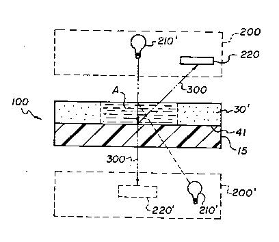
Fig. 2 is a schematic view similar to that of
Fig. 1, but of the invention;
Fig. 3 is a partially schematic plan view of
a useful spectroscope;
Figs. 4-7 are plots of actual analyte
concentration versus predicted concentration resulting
from the use of the invention; and
Fig. 8 is the absorption plot of gelatin at
NIR wavelengths, showing why gelatin is unacceptable in
significant thicknesses in the light-path defining
layers of the test element.
DESCRIPTION OF THE PREFERRED EMBODIMENT
This invention is based on the discovery that
the spreading layer described in the aforesaid '158
patent is ideally suited for quantitative NIR
spectroscopic analysis, when it comprises the light-
path defining layer, unlike the construction in the
'158 patent.
Referring to Fig. 1, the conventional slide
element 10 taught by the '158 patent comprises a
transparent plastic support 15, one or more reagent
layers 20, and a uniformly porous spreading layer 30.
Layer 30 provides for uniform spreading of a drop of
sample (shown in phantom) into a volume of dispersed
area A in layer 30 (shown with liquid), which then
migrates downward into reagent layer 20. (The reagents
of layer 20 are indicated by a heavier stipple.)
Importantly, the element is read from the opposite side
2167~7~
-
--5--
for reflectance, using a light source 40 and a detector
42 in a reflectometer 50. The spreading layer, in
turn, is provided with a screen material to make it
reflective, such as, by incorporating TiO2. As a
result, the light-path 60 is ideally defined by only
layers 15 and 20 and not by the spreading layer, so as
to detect a color change in layer 20. (Although some
examples of "Ektachem"~ brand slides made in
accordance with the teaching of the '158 patent may in
fact have some light transmitted into the spreading
layer, it is shown below that many "Ektachem" slides in
fact have less than 1% of the light-path operationally
defined by that layer, that is, within the normal
operating range of the slide. In any event, such
slides have always been taught for use with detection
reagents.)
Our invention is based on the discovery that
the spreading layer by itself makes an excellent porous
sheet for conducting quantitative NIR spectroscopic
analysis, when the light-path is in fact defined by the
spreading layers rather than any adjacent layers. The
reason is that the sufficiently porous nature of the
spreading layer makes it ideal for providing a constant
predictable path length of light during the period of
use, unlike the binders used in the reagent layers of
Fig. 1 that define the light-path within that slide
element.
As used herein, "NIR" spectroscopy or
radiation means, using the wavelengths of from 750 to
3000 nm. "Substantially constant light-path" means,
with deviations of no more than 1%. "Quantitative"
means, measurement of actual quantities based upon
accuracy and precision that is acceptable in a clinical
laboratory.
21~7 ~7 ~
-6-
"Sufficiently porous" means, in all
directions so that a liquid spreads within the layer
uniformly in all directions. As noted, this latter is
a property already known to exist in the spreading
layer of the element taught in the '158 patent. Hence,
the most preferred level of "sufficient porosity", as
used herein, is any layer of material that is as porous
in all directions as the spreading layers of the '158
patent. The TiO2 layer of Ex. 1 that follows is a
satisfactory example thereof.
To test for other materials having suitable
porosity, a useful procedure is that set forth in col.
6, line 22 to col. 7, line 7 of the aforesaid '158
patent which is expressly incorporated herein by
reference. However, volumetric and thickness
requirements set forth therein need not be followed,
nor must a gelatin sublayer be used. Alternatively,
the gelatin can be used since this is just a test for
porosity.
Thus it will be readily apparent that the
basic components of this invention are conventional:
the components and construction of the test element
light-path defining layer(s), and the NIR analysis for
detection of signal peaks representative of certain
analytes, e.g., total protein, glucose, total
cholesterol, albumin, globulin, triglycerides, urea,
creatinine, HDL, and LDL.
The invention resides in the discovery that
the test element of the '158 patent, when properly
constructed with substantially no reagents and without
the reagent layer, will allow adequate quantitative
detection of analytes in biological liquids using NIR
spectroscopy. For these reasons, except as stated
herein, details are not provided for the construction
of the test element or the conduction of NIR analysis,
as such are well known to those skilled in the
21~7~7~
-
--7--
respective art. (Representative useful peaks for ,
e.g., total protein, include 2050, 2160, 2170, and
2180 nm, as is well-known.)
Although the description herein is of
preferred embodiments featuring reflective NIR assays,
using the spreading layer of U.S. Patent No. 3,992,158,
the invention is not limited thereto. It is also
useful with scattering-type NIR analysis, and with a
light-path defining layer of some other material having
the same NIR capabilities and porosity as noted above
in the Summary.
The preferred slide test element is element
100, Fig. 2. This preferably comprises a layer 30'
that is substantially the same as any of the spreading
layers of the aforesaid '158 patent, provided that it
is free of detection reagents. Optionally it is
attached to NIR-transparent plastic support 15 by a
thin conventional subbing layer 41, or by corona-
treating or plasma-treating the support. Supports
described in the '158 patent are useful. "Thin" as
used herein for the subbing layer means, a thickness
less than 1 micron, since greater thicknesses tend to
interfere with the NIR reflection, especially if the
subbing layer comprises a hydrophilic polymer.
Alternatively, if the composition of layer
30' is self-adhering to layer 15, layer 41 can be
omitted.
Detection of analytes in a liquid deposited
in area A is by a conventional spectrophotometer 200,
using an appropriate NIR energy source 210 and an NIR
detector 220. (Although a preferred mode of analysis
is from the front-side as shown, it can also be from
the back side, shown in phantom.)
2l6747a
-
--8--
As is known from the '158 patent, preferred
materials for use in layer 30' of element 100 are
selected from the group consisting of a blush polymer,
microcrystalline particles, and adhered particles, all
with or without reflective pigments present.
When doing the NIR analysis from the top side
of test element 100, layer 41 optionally includes a
mirrored surface, which can be provided in the form of
a metallized layer, a thin particulate ceramic layer, a
fine metal oxide, or metal dispersed in a transparent
polymer. This enhances the reflection of path 300,
since without the mirrored surface, the NIR energy from
source 210 is more likely to be partially absorbed at
or near layers 41 and 15.
As noted, the NIR analysis is conventional.
In brief, it is done as follows:
A slide element is attached to a support and
placed in the position shown as "S" in Fig. 3. A
spectrum of the slide is collected using conventional
spectrometer 400 and wavelengths generated by light
source 402, chopper 404, monochromator grating 406,
focusing lenses 408, and detectors 410. The
wavelengths are selected from the full spectrum from
1200 to 2400 nm to achieve a blank reading. The
element is then removed from the spectrometer and
spotted with the liquid to be assayed, in a preferred
amount, such as 300 microliters. The liquid is allowed
to spread into the slide for 30 to 90 sec. and the
slide then returned to the spectrometer. A new
spectrum of the slide and liquid is generated and
collected using spectrometer 400. The results are then
analyzed using the 2nd derivative of the absorbence
followed by multiple linear regression (MLR) to
discriminate the analyte from background. Further
details are shown in the following examples.
2167~7~
g
Examples
The following examples are illustrative only,
and are not an exhaustive list of the embodiments of
the invention.
In the following examples, the slide test
element had the following construction:
(top layer)
TiO2 or BaSO4
Cellulose Acetate
(subbing layer)
Binder = poly(vinyl pyrrolidone)
(Coverage = less than or = 1 mg/cm2)
poly(ethylene terephthalate)
support
The protein examples which follow were
calibrated and read at 2057.5 nm and 2180.5 nm light.
Additionally, however, the regions 2050-2250 nm and/or
1500-1800 nm are also useful. The spectroscope used
was a "Quantum 1200 Plus" (TM) spectroscope available
from LT Industries, schematically shown in Fig. 3. The
2nd derivative of the absorbance followed by multiple
linear regression (MLR) was used to discriminate the
analyte from background. The second derivative is
preferred because it removes nonchemical differences
such as baseline shifts and instrument noise, and
enhances shoulders and inflections of the curves of the
raw absorption spectra. See, e.g., U.S. Patent No.
5,197,470 for an example of the process used herein.
2~67~7~
- -10-
Alternatively, principal component analysis and/or
regression and/or partial least squares can be
substituted for the multiple linear regression
analysis.
Example 1 - Total Serum Protein On A TiO~ Slide
The slide construct shown above using a 2-6
mil thick TiO2 matrix at a coverage of 1-6 g/m squared,
was spotted with 300 ~L of a calibrator fluid spiked
with amounts of total protein that are on the
horizontal axis of Fig. 4. The spectroscopic readings
were processed so that the MLR predictions are plotted,
Fig. 4, on the vertical axis, and the a priori actual
values plotted on the horizontal axis, yielding an r
value, where "r" is the regression correlation value,
of 0.997 and a standard deviation of 0.11. Clearly,
this result is well within laboratory standards of a
quantitative assay.
These results also constitute the calibration
curve used with subsequent examples.
Example 2 - Total Serum Protein On A TiO~ Slide
The experiment of Example 1 was repeated,
except that the calibration curve of Fig. 4, Example 1,
was used to assay other known concentrations of analyte
not used in preparing the calibration curve of Ex. 1.
The results are plotted in Fig. 5. The "r" value was
0.996 and the standard deviation, 0.12, indicating that
the results of Ex. 1 could be used as a calibration
curve with "unknowns".
Example 3 - Total Cholesterol on a saso4 Slide Test
Element
The experiment of Ex. 1 was repeated, except
that the light-path defining layer (other than the
support) comprised BaSO4 at 1 to 6 g/cm2 coverage,
instead of TiO2. The calibrators had spiked in the
- 2167~70
--11--
levels of cholesterol listed on the horizontal axis of
Fig. 6. The predicted values obtained
spectroscopically were those plotted on the vertical
axis, producing a result of r = 0.99 and a standard
deviation of 9 mg/dl, which is well within acceptable
limits for this concentration range of cholesterol.
Example 4 - Total Cholesterol on a BaSO4 Slide Test
Element
Example 2 was repeated, except that the assay
was for total cholesterol, using the calibration curve
generated in Ex. 3. The results appear in the plot of
Fig. 7, with r values and a standard deviation
identical to that of Fig. 6.
Comparative Example No. 1
Fig. 8 is included solely to demonstrate that
any substantial thickness [greater than 1 micron] in
the slide test element comprising gelatin, will not be
reflective of at least 95% of NIR radiation. The
reason of course is due to the substantial absorption
occurring particularly from 1500 nm to 2400 nm, a major
portion of the NIR range of the invention. (The type
of gelatin is of no consequence in this regard.)
Hence, gelatin is to be eschewed, at least in
thicknesses ~ 1 micron.
Comparative Example No. 2
To demonstrate that the conventional
"Ektachem" (TM) slide for, e.g., glucose, does not
permit passage of the reflectometer light into the
spreading layer for ranges of analyte for which the
slide is intended, such a glucose slide was tested in a
conventional Perkin Elmer "Lambda Nine" (TM)
spectrometer with a known concentration of glucose.
The peak absorbance of the dye was 1.22 absorbance
units which is approximately a 7% transmission, so that
` - 2167Q70
-12-
the light contributed by reflectance within the spread
layer (above that dye layer) is 0.07 x 0.07 = .0049, or
only 0.3% to 0.5% of the signal, instead of the
required at-least 95%. That is, the conventional
"Ektachem" slide is not designed to transmit light into
and through the spreading layer at the ranges of
analyte concentration intended for use.
