Note: Descriptions are shown in the official language in which they were submitted.
CA 02167692 2003-06-27
~ ~. .
METHOD FOR DIAGNOSING A DISORDER BY DETERMINING
EXPRESSION OF GAGE TUMOR REJECTION ANTIGEN
PRECURSORS
FIAL2 4r mm 3rtnMr=ort
This invention relates to a nucleic acid molecule which
codes for a tumor rejection antigen precursor. More
particularly, the invention concerns genes, whose tumor
rejection antigen precursor is processed, inter alia, into at
least one tumor rejection antigen that is presented by HLA-Cw6
molecules. The genes in question do not appear to be related
to other known tumor rejection antigen precursor coding
sequences.
13AC1UiROUND AND PRIf,1R ART
The process by which the mammalian immune system
recognizes and reacts to foreign or alien materials is a
complex one. An important facet of the system is the T
lymphocyte, or "T cell" response. This response requires that
T cells recognize and interact with complexes of cell surface
molecules, referred to as human leukocyte antigens ("HLA" ), or
major histocompatibil.ity complexes ("MHCs"), and peptides.
The peptides are derived from larger molecules which are
processed by the cells which also present the HLA/MHC
molecule. See in this regard Male et al.,Advanced Tmmunoloav
(J.P. Lipincott Company, 1987), especially chapters 6-10. The
interaction of T cells and HLA/peptide complexes is
restricted, requiring a1' cell specific for a particular
combination of an HLA molecule and a peptide. If a specific
T cell is not present, there is no T cell response even if its
partner complex is present.. 5ima.larly, there is no response
if the specific complex is absent, but the T cell is present.
T:his mechanism is involved in the immune system's response to
foreign materials, in autoiumune pathologies, and in responses
CA 02167692 2008-09-17
2
to cellular abnormalities. Much work has focused on the
mechanisms by which proteins are processed into the HLA
binding peptides. See, in this regard, Barinaga, Science 257:
880 (1992); Fremont et al., Science 257: 919 (1992); Matsumura
et al., Science 257: 927 (1992); Latron et al., Science 257:
964 (1992).
The mechanism by which T cells recognize cellular
abnormalities has also been implicated in cancer. For
example, in PCT application PCT/US92/04354, filed May 22,
1992, published on November 26, 1992,
a family of genes is disclosed, which are processed
into peptides which, in turn, are expressed on cell surfaces,
which can lead to lysis of the tumor cells by specific CTLs
cytolytic T lymphocytes, or "CTLs" hereafter. The genes are
said to code for "tumor rejection antigen precursorsn or
"TRAP" molecules, and the peptides derived therefrom are
referred to as "tumor rejection antigens" or "TRAs". see
Traversari et al., Immunogenetics 35: 145 (1992); van der
Bruggen et al., Science 254: 1643 (1991), for further
information on this family of Qenes. Also, see U.S. patent
No. 5,342,774.
In U.S. Patent No. 5,405,940, it is
explained that the MAGE-1 gene codes for a tumor rejection
antigen precursor which is processed to nonapeptides which
are presented by the HLA-A1 molecule. The reference teaches
that given the known specificity of particular peptides for
particular HLA molecules, one should expect a particular
peptide to bind to one HLA molecule, but not to others. This
is important, because different individuals possess different
HLA phenotypes. As a result, while identification of a
particular peptide as being a partner for a speoif3.c HLA
molecule has diagnostic and therapeutic ramifications, these
are only relevant for individuals with that particular HLA
phenotype. There is a need for further work in the area,
because cellular abnormalities are not restricted to one
CA 02167692 2008-09-17
3
particular HLA phenotype, and targeted therapy requires some
knowledqe of the phenotype of the-abnormal cells at issue.
In U.S. Patent No. 5,558,995, the fact that
the MAGE-1 expression product is processed to a second TRA is
disclosed. This second TRA is presented by HLA-C clone 10
molecules. The disclosure shows that a given TRAP can yield
a plurality of TRAs.
In U.S. Patent No. 6,284,476,
teaches that tyrosinase, a molecule which is produced by some
normal cells (e.g., melanocytes), is processed in tumor cells
to yield,peptides presented by HLA-A2 molecules.
In U.S. Patent No. 5,620,886,
a second TRA, not~ derived from tyrosinase is taught to be
presented by HLA-A2 molecules. The TRA is derived from a
TRAP, but is coded for by a non-MAGE gene. This disclosure
shows that a particular HI,A molecule may present TRAs derived
from different sources.
In U.S. Patent No. 5,571,711, an
unrelated tumor rejection antigen precursor, the so-called
"BAGE" precursor is described. The BAGE precursor is not
related to the MAGE family.
The work which is presented by the papers, patent, and
patent applications cited su ra deals, in large part, with the
MAGE family of genes, and the unrelated BAGE gene. It has now
been found, however, that additional tumor rejection antigen
precursors are expressed by cells. These tumor rejection
antigen precursors are referred to as "GAGE" tumor rejection
antigen precursors. They do not show homology to either the
MAGE family of genes or the BAGE gene. Thus the present
invention relates to genes encoding such TRAPs, the tumor
rejection antigen precursors themselves as well as
applications of both.
The invention is elaborated upon further in the
CA 02167692 2008-09-17
4
disclosure which follows.
BRiEI+' DEBCRIPTION OF THE FIGIIRES
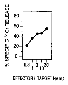
Figure 1 sets forth lysis studies using CTL clone 76/6. 5
Figure 2 shows tumor necrosis factor ("TNF") release assays
obtained with various transfectants and controls.
DETAILED DEBORIPTION OF PREFERRED EMBODIMENTS
Examole 1
A melanoma cell line, MZ2-MEL was established from
melanoma cells taken from patient MZ2, using standard
methodologies. This cell line is described, e.g., in PCT
Application PCT/US92/04354, filed May 22, 1992, published
November 26, 1992,
once the cell line was established, a sample
thereof was irradiated, so as to render it non-proliferative.
These irradiated cells were then used to isolate cytolytic T
cell clones ("CTLs") specific thereto.
A sample of peripheral blood mononuclear cells ("PBMCsll)
was taken from patient MZ2, and contacted to the irradiated
melanoma cells. The mixture was observed for lysis of the
melanoma cells, which indicated that CTLs specific for a
complex of peptide and HLA molecule presented by the melanoma
cells were present in the sample.
The lysis assay employed was a chromium release assay
following Herin efiaj., Int. J. Cancer 39:390-396 (1987).
The assay,
however, is described herein. The target melanoma c@lls were
grown in vitro, and then resuspended at 107 cells/mi in DMEM,
supplemented with 10 mM HEPES and 30% FCS, and incubated for
45 minutes at 37 C with 200 Ci/ml of Na(51Cr)04. Labelled
cells were washed three times with DMEM, supplemented with 10
mM Hepes. These were then resuspended in DMEM supplemented with 10 mM Hepes
and 10t FCS, after which 100 ul aliquots
containing 103 cells, were distributed into 96 well
.
CA 02167692 2003-06-27
microplates. Samples of PBLs were added in 100 ul of the same
medium, and assays were carried out in duplicate. Plates were
centrifuged for 4 minutes at 100g, and incubated for four
hours at 37 C in a 8t COZ atmosphere.
5 Plates were centrifuged again, and 100 ul aliquots of
supernatant were collected and counted. Percentage of 51Cr
release was calculated as follows:
$ S'Cr release - (ER-SR) x 100
(MR-SR)
where ER is observed, experimental SlCr release, SR is
spontaneous release measured by incubating 103 labeled cells
in 200 ul of medium alone, and MR is maximum release, obtained
by adding 100 ul 0.3% Triton X-100* to target calls.
Those mononuclear blood samples which showed high CTL
activity were expanded and cloned via limiting dilution, and
were screened again, using the same methodology. The CTL
clone MZ2-CTL 76/6 was thus isolated. The clone is referred
to as "76/6" hereafter.
The same method was used to test target R562 cells, as
weil as the melanoma cell line. Figure 1 shows that this CTL
clone recognizes and lyses the melanoma cell line, i.e. MZ2-
MEL but not K562. The clone was then tested against other
melanoma cell lines and autologous EBV-transformed B cells in
the same manner described sZpra. Figure 1 shows that
autologous B cells, transformed by Epstein Barr Virus ("EBV")
were not lysed, and that while MZ2-MEL 3.0 was lysed by CTL
clone 76/6, the cell line MZ2--MEL.4F` avari.ant which does
not express antigen F was not. Hence, the clone appears to be
specific for this antigenr
The results presented Euozaare inconclusive as to which
J;LA molecule presents the. TRA. The lysed cell line, i.e.,
MZ2-MEL, is known to express IiLA-A1, HLA-A29, HLA--B37, HLA-
B44, HLA-Cw6, and HLA-C clone 10. In experiments not reported
here but which follow the protocol of this example, a subline
of MZ2-MEL was tested, which had lost expression of HLA
molecules A29, 844, and C clone 10. The subline was lysed,
thus indicating that the px esenting molecule should be one of
Trademark
, , ~ ...,., ..- ..~..~... . ...._... _ _ .___.-....~._._._...- ~
CA 02167692 2008-09-17
6
Al, B37, or Cw6.
SUMle 2
Further studies were carried out to determine if 76/6
also produced tumor necrosis factor ("TNF") when contacted
with target cells. The method used was that described by
Traversari at al., Immunogenetics 35: 145-152 (1992).
Briefly, samples of the CTL line were combined with samples
of a target cell of interest in culture medium. After 24
hours, supernatant from the cultures was removed, and then
tested on TNF-sensitive WEHI cells. Cell line MZ2-MEL 43, a
subclone of the MZ2-MEL cell line discussed supra as well as
in the cited references, gave an extremely strong response,
and was used in the following experiments.
~amnle 3
The results from Example 2 indicated that M22.MEL.43
presented the target antigen of interest. As such, it was
used as a source of total m1tNA to prepare a cDNA library.
Total RNA was isolated from the cell line. The mRNA was
isolated using an oligo-dT binding kit, following well
recognized techniques. Once the mRNA was secured, it was
transcribed into cDNA, via reverse transcription, using an
oligo dT primer containing a Notl site, followed by second
strand synthesis. The cDNA was then ligated to a BstXI
adaptor, digested with Notl, size fractionated on a Sephacryl *
S-500 HR column, and then cloned, undirectionally into the
BstXI and Not I sites of pcDNA-I-Amp. The recombinant plasmid
was then electroporated into DHSa E. a" bacteria. A total
of 1500 pools of lo0 recombinant bacteria were seeded in
microwells. Each contained about 100 cDNAs, because nearly
all bacteria contained an insert.
Each pool was amplified to saturation and plasmid DNA was
extracted by alkaline lysis and potassium acetate
precipitation, without phenol extraction.
Examnle 4
* Trademark
CA 02167692 2008-09-17
7
Following preparation of the library described in Example
3, the cDNA was transfected into eukaryotic cells. The
transfections, described herein, were carried out in
' duplicate. Samples of COS-7 cells were seeded, at 15,000
cells/well into tissue culture flat bottom microwells, in
Dulbecco's modified Eagles Medium ("DMEM") supplemented with
10% fetal calf serum. The cells were incubated overnight at
37 C, medium was removed and then replaced by 50 l/well of
DMEM medium containing 10% Nu serum, 400 g/ml DEAE-dextran,
and 100 M chloroquine, plus 100 ng of the plasmids. ' As was
indicated eutira, the lysis studies did not establish which HLA
molecule presented the antigen. As a result, cDNA for each of
the HLA molecules which could present the antigen (Al, B37,
Cw6) was used, separately, to cotransfect the cells.
Specifically, one of 28 ng of cDNA for HLA-A1, cloned into
pCD-SRcz was used, as were 50 ng of cDNA for HLA-B37 in pcDNA-
I-Amp, or 75 ng of cDNA for HLA-Cw6 in pcDNA-I Amp, using the
same protocol as was used for transfection with the library.
Transfection was made in duplicate wells, but only 500
pools of the HLA-Cw6 transfectants could be tested in single
weils. Following four hours of incubation at 37 C, the
medium was removed, and replaced by 50 l of PBS containing
10%, DMSO. This medium was removed after two minutes and
replaced by 200 l of DMEM supplemented with 10% FCS.
Following this change in medium, COS cells were incubated
for 24-48 hours at 37 C. Medium was then discarded, and 1000-
3000 cells of CTL clone 76/6 were added, in 100 l of iscove
medium containing 10% pooled human serum supplemented with 20-.
U/mi of IL-2. Supernatant was removed after 24 hours, and
30 TNF content was determined in an assay on WEHI cells, as
described by Traversari et al., Immunogenetics 35: 145-152
(1992),
The 1500 pools transfected with HLA-A1, and the 1500
pools transfected with HLA-B37 stimulated TNF release to a
concentration of 15-20 pg/ml, or 2-6 pg/ml, respectively.
Most of the HLA-Cw6 transfectants yielded 3-20 pg/ml, except
* Trademark
2167692
WO 95/03422 PCT/US94/078780
8
for one pool, which yielded more than 60 pg/mi. This pool was
selected for further work.
Example 5
The bacteria of the selected pool were cloned, and 600
clones were tested. Plasmid DNA was extracted therefrom,
transfected into a new sample of COS cells in the same manner
as described supra, and the cells were again tested for
stimulation of CTL clone 76/6. Ninety-four positive clones
were found. One of these, referred to as cDNA clone 2D6 was
tested further. In a comparative test COS cells were
transfected with cDNA clone 2D6 and the HLA-Cw6, HLA-Cw6
alone, or 2D6 alone. Control cell lines MZ2-MEL F" and MZ2-
MEL F+ were also used. TNF release into CTL supernatant was
measured by testing it on WEHI cells, as referred to supra.
The optical density of the surviving WEHI cells was measured
using MTT. Figure 2 shows that the COS cells transfected with
HLA-Cw6 and cDNA-2D6, and the cell line MZ2-MEL F+ stimulated
TNF release from CTL clone 76/6, indicating that HLA-Cw6
presented the subject TRA.
ExampJle 6
The cDNA 2D6 was sequenced following art known
techniques. A sequence search revealed that the plasmid
insert showed no homology to known genes or proteins.
SEQUENCE ID NO: 1 presents cDNA nucleotide information for the
identified gene, referred to hereafter as "GAGE". A putative
open reading frame is located at bases 51-467 of the molecule.
Examnle 7
Following sequencing of the cDNA, as per Example 6,
experiments were carried out to determine if cells of normal
tissues expressed the gene. To determine this, Northern
blotting was carried out on tissues and tumor cell lines, as
indicated below. The blotting experiments used cDNA for the
complete sequence of SEQ ID NO: 1. PCR was then used to
confirm the results.
OVO 95/03422 2167692 PCTIUS94/07878
9
Table 1= Expression of aene GAGE
Normal tissues
PHA-activated T cells -
CTL clone 82/30 -
Liver -
Muscle -
Lung -
Brain -
Kidney -
Placenta -
Heart -
Skin -
Testis +
Tumor cell lines
Melanoma 7/16
Lung Carcinoma 1/6
Sarcoma 0/1
Thyroid medullary carcinoma 0/1
Tumor samples
Melanoma 1/1
Example 8
Detailed analysis of normal tissues and tumors was
carried out by applying polymerase chain reaction ("PCR") and
the GAGE gene information described supra.
First, total RNA was taken from the particular sample,
using art recognized techniques. This was used to prepare
cDNA. The protocol used to make the cDNA involved combining
4 ui of reverse transcriptase buffer 5x, 1 ul of each dNTP,
(10 mM), 2 ul of dithiothreitoi (100 mM), 2 ul of dT-15 primer
(20 um), 0.5 ul of RNasin (40 units/ul) , and 1 ul of M-MLV
reverse transcriptase (200 units/ul). Next, 6.5 ul of
template RNA (1 ug/3.25 ui water, or 2 ug total template RNA)
was added. The total volume of the mixture was 20 ul. This
was mixed and incubated at 42 C for 60 minutes, after which it
was chilled on ice. A total of 80 ul of water was then added,
to 100 ul total. This mixture was stored at -20 C until used
in PCR.
To carry out PCR, the primers
51'-AGA CGC TAC GTA GAG CCT-3J'
CA 02167692 2003-06-27
(sense)
and
51-CCA TCA GGA CCA TCT TCA-3"
(antisense)
5 SEQ ID NOS: 2 and 3, respectively, were used. The reagents
included 30.5 ul water, 5 ul of PCR buffer lOx, 1 ul of each
dNTP (10 UN), 2.5 ul of each primer (20 uM), and 0.5 ul of
polymerizing enzyme nDynazyme (2 units/ul). The total volume
as 45 ui. A total of 5 ul of., cDNA was added (this
10 corresponded to 100 ng total RNA). The mixture was combined,
and layered with one drop of mineral oil. The mixture was
transferred to a thermocycler block, preheated to 94 C, and
amplification was carried out for 30 cycles, each cycle
consisting of the following:
first denaturation: 94 C, 4 min.
denaturation: 94 C, 1 min.
annealing: 55 C, 2 min.
extension: 72 C, 3 min.
final extension: 72 C, 15 min.
Following the cycling, 10 ul aliquots were run on a 1.5%
agarose gel, stained with ethidium bromide.
cDNA amplified using the primers set forth supra yields
a 238 base pair fragment. There is no amplification of
contaminating genomic DNA, if present.
The results are presented in Table 2, which follows.
They confirm that the only normal tissue which expresses GAGE
is testis, whereas a number of tumors, including melanoma,
lung, breast, larynx, pharynx, sarcoma, testicular seminoma,
bladder and colon express the gene. Thus, any one of these
tumors can be assayed for by assaying for expression of the
GAGE gene.
* Trademark
. . _ . ...... _ .... . ..., . , ..., . õ ..., . . .. . .. ... .... .. . . _,
, .. . ... ... _. ... _....,_,.,,,.,.M,..,,,.._,... w_,.. ...~ .. _ _ . .
00 95/03422 11 216 7 6 9 2 pCTlUS94/07878
Table 2
RT-PCR analysis of the expression of gene GAGE
NORMAL TISSUES
Heart
B rai n -
Liver -
Lung -
lb Kidney
Spleen -
Lymphocytes -
Bone marrow -
Skin -
Naevus -
Melanocytes -
Fibroblasts -
Prostate -
Testis +
Ovary Breast -
Adrenals -
Muscle -
Placenta -
Umbilical Cord -
TUMORS
Cell lines Tumor samples
Melanoma 40/63 46/146 (32%)
Lung cancer
Epidermoid carcinoma 10/41 (24%)
Adenocarcinoma 4/18
Small Cell Lung Cancer 6/23 0/2
Breast cancer 15/146 (10%)
Head and Neck tumor
Larynx 6/15 (40%)
Pharyax 3/13
Sarcoma 1/4 6/18 (33%)
_;0 Testicular seminoma 6/6 (100%)
Bladder cancer 5/37 (14%)
Prostate cancer 2/20
Colon carcinoma 5/13 0/38
Renal cancer 0/6 0/45
5 Leukemia 3/6 0/19
2167692
WO 95/03422 PCT/i7S94/07878*
12
The foregoing examples show the isolation of a nucleic
acid molecule which codes for a tumor rejection antigen
precursor. This "TRAP" coding molecule, however, is not
homologous with any of the previously disclosed MAGE and BAGE
coding sequences described in the references set forth sunra.
Hence, one aspect of the invention is an isolated nucleic acid
molecule which comprises the nucleotide sequence set forth in
SEQ ID NO: 1. This sequence is neither a MAGE nor a BAGE
coding sequence, as will be seen by comparing it to the
sequence of any of these genes as described in the cited
references. Also a part of the invention are those nucleic
acid sequences which also code for a non-MAGE and non-BAGE
tumor rejection antigen precursor but which hybridize to a
nucleic acid molecule containing the described nucleotide
sequence, under stringent conditions. The term "stringent
conditions" as used herein refers to parameters with which the
art is familiar. More specifically, stringent conditions, as
used herein, refers to hybridization in 1M NaCl, 1% SDS, and
10% dextran sulfate. This is followed by two washes of the
filter at room temperature for 5 minutes, in 2xSSC, and one
wash for 30 minutes in 2xSSC, 0.1% SDS. There are other
conditions, reagents, and so forth which can be used, which
result in the same or higher degree of stringency. The
skilled artisan will be familiar with such conditions, and,
thus, they are not given here.
It will also be seen from the examples that the invention
embraces the use of the sequences in expression vectors, as
well as to transfect host cells and cell lines, be these
prokaryotic (e.g., E. coli), or eukaryotic (e.g., CHO or COS
cells). The expression vectors require that the pertinent
sequence, i.e., those described su , be operably linked to
a promoter. As it has been found that human leukocyte antigen
HLA-Cw6 presents a tumor rejection antigen derived from these
genes, the expression vector may also include a nucleic acid
sequence coding for HLA-Cw6. In a situation where the vector
contains both coding sequences, it can be used to transfect a
cell which does not normally express either one. The tumor
2167692
4v0 95/03422 PCT/US94/07878
13
rejection antigen precursor coding sequence may be used alone,
when, e.g., the host cell already expresses HLA-Cw6. Of
course, there is no limit on the particular host cell which
can be used. As the vectors which contain the two coding
sequences may be used in HLA-Cw6 presenting cells if desired,
and the gene for tumor rejection antigen precursor can be used
in host cells which do not express HLA-Cw6.
The invention also embraces so called expression kits,
which allow the artisan to prepare a desired expression vector
or vectors. Such expression kits include at least separate
portions of each of the previously discussed coding sequences.
Other components may be added, as desired, as long as the
previously mentioned sequences, which are required, are
included.
To distinguish the nucleic acid molecules and the TRAPs
of the invention from the previously described MAGE and BAGE
materials, the invention shall be referred to as the GAGE
family of genes and TRAPs. Hence, whenever "GAGE" is used
herein, it refers to the tumor rejection antigen precursors
coded for by the previously described sequences. "GAGE coding
molecule" and similar terms, are used to describe the nucleic
acid molecules themselves.
-The invention as described herein has a number of uses,
some of which are described herein. First, the invention
permits the artisan to diagnose a disorder characterized by
expression of the TRAP. These methods involve determining
expression of the TRAP gene, and/or TRAs derived therefrom,
such as a T12P, presented by HLA-Cw6. In the former situation,
such determinations can be carried out via any standard
nucleic acid determination assay, including the polymerase
chain reaction, or assaying with labelled hybridization
probes. In the latter situation, assaying with binding
partners for complexes of TRA and HLA, such as antibodies, is
especially preferred. An alternate method for determination
is a TNF release assay, of the type described sunra. To carry
out the assay, it is preferred to make sure that testis cells
are not present, as these normally express GAGE. This is not
WO 95/03422 216 7 6 9 2 pCT/US94/07878
14
essential, however, as one can routinely differentiate between
testis and other cell types. Also, it is practically
impossible to have testis cells present in non-testicular
sample.
The isolation of the TRAP gene also makes it possible to
isolate the TRAP molecule itself, especially TRAP molecules
containing the amino acid sequence coded for by SEQ ID NO: 1.
These isolated molecules when presented as the TRA, or as
complexes of TRA and HLA, such as HLA-Cw6, may be combined
with materials such as adjuvants to produce vaccines useful in
treating disorders characterized by expression of the TRAP
molecule. In addition, vaccines can be prepared from cells
which present the TRA/HLA complexes on their surface, such as
non-proliferative cancer cells, non-proliferative
transfectants, etcetera. In all cases where cells are used as
a vaccine, these can be cells transfected with coding
sequences for one or both of the components necessary to
provide a CTL response, or be cells which express both
molecules without transfection. Further, the TRAP molecule,
its associated TRAs, as well as complexes of TRA and HLA, may
be used to produce antibodies, using standard techniques well
known to the art.
When "disorder" is used herein, it refers to any
pathological condition where. the tumor rejection antigen
precursor is expressed. An example of such a disorder is
cancer, melanoma in particular. Melanoma is well known as a
cancer of pigment producing cells.
Therapeutic approaches based upon the disclosure are
premised on a response by a subject's immune system, leading
to lysis of TRA presenting cells, such as HLA-Cw6 cells. One
such approach is the administration of CTLs specific to the
complex to a subject with abnormal cells of the phenotype at
issue. It is within the skill of the artisan to develop such
CTLs 'tn vitro. Specifically, a sample of cells, such as blood
cells, are contacted to a cell presenting the complex and
capable of provoking a specific CTL to proliferate. The
target cell can be a transfectant, such as a COS cell of the
WO 95/03422 2167692 PCT/US94/07878
type described supra. These transfectants present the desired
complex on their surface and, when combined with a CTL of
interest, stimulate its proliferation. COS cells, such as
those used herein are widely available, as are other suitable
5 host cells.
To detail the therapeutic methodology, referred to as
adoptive transfer (Greenberg, J. Immunol. 136 (5) : 1917 (1986) ;
Riddel et al., Science 257: 238 (7-10-92); Lynch et al., Eur.
J. Immunol. 21: 1403-1410 (1991); Kast et al., Cell 59: 603-
10 614 (11-17-89)), cells presenting the desired complex are
combined with CTLs leading to proliferation of the CTLs
specific thereto. The proliferated CTLs are then administered
to a subject with a cellular abnormality which is
characterized by certain of the abnormal cells presenting the
15 particular complex, where the complex contains the pertinent
HLA molecule. The CTLs then lyse the abnormal cells, thereby
achieving the desired therapeutic goal.
The foregoing therapy assumes that at least some of the
subject s abnormal cells present the relevant HLA/TRA complex.
This can be determined very easily, as the art is very
familiar with methods for identifying cells which present a
particular HLA molecule, as well as how to identify cells
expressing DNA of the pertinent sequences, in this case a GAGE
sequence. Once cells presenting the relevant complex are
identified via the foregoing screening methodology, they can
be combined with a sample from a patient, where the sample
contains CTLs. If the complex presenting cells are lysed by
the mixed CTL sample, then it can be assumed that a GAGE
derived, tumor rejection antigen is being presented, and the
subject is an appropriate candidate for the therapeutic
approaches set forth supra.
Adoptive transfer is not the only form of therapy that is
available in accordance with the invention. CTLs can also be
provoked,invivo, using a number of approaches. One approach,
i.e., the use of non-proliferative cells expressing the
complex, has been elaborated upon supra. The cells used in
this approach may be those that normally express the complex,
WO 95/03422 216 769 2 PCT/US94/07878
16
such as irradiated melanoma cells or cells transfected with
one or both of the genes necessary for presentation of the
complex. Chen et al., Proc. Natl. Acad. Sci. USA 88: 110-114
(January, 1991) exemplifies this approach, showing the use of
transfected cells expressing HPV E7 peptides in a therapeutic
regime. Various cell types may be used. Similarly, vectors
carrying one or both of the genes of interest may be used.
Viral or bacterial vectors are especially preferred. In these
systems, the gene of interest is carried by, e.g., a Vaccinia
virus or the bacteria BCG, and the materials de facto "infect"
host cells. The cells which result present the complex of
interest, and are recognized by autologous CTLs, which then
proliferate. A similar effect can be achieved by combining
the tumor rejection antigen or the precursor itself with an
adjuvant to facilitate incorporation into HLA-Cw6 presenting
cells which then present the HLA/peptide complex of interest.
The TRAP is processed to yield the peptide partner of the HLA
molecule while the TRA is presented without the need for
further processing.
Other aspects of the invention will be clear to the
skilled artisan and need not be repeated here.
The terms and expressions which have been employed are
used as terms of description and not of limitation, and there
is no intention in the use of such terms and expressions of
excluding any equivalents of the features shown and described
or portions thereof, it being recognized that various
modifications are possible within the scope of the invention.
2167692
WO 95/03422 PCT/US94/07878
17
(1) GENERAL INFORMATION:
(i) APPLICANTS: Boon-Falleur, Thierry; Van den Eynde,
Benoit
(ii) TITLE OF INVENTION: ISOLATED NUCLEIC ACID MOLECULES
CODING FOR GAGE TUMOR REJECTION ANTIGEN
PRECURSORS
(iii) NUMBER OF SEQUENCES: 1
(iv) CORRESPONDENCE ADDRESS:
(A) ADDRESSEE: Felfe & Lynch
(B) STREET: 805 Third Avenue
(C) CITY: New York City
(D) STATE: New York
(E) COUNTRY: USA
(F) ZIP: 10022
(v) COMPUTER READABLE FORM:
(A) MEDIUM TYPE: Diskette, 5.25 inch, 360 kb
storage
(B) COMPUTER: IBM PS/2
(C) OPERATING SYSTEM: PC-DOS
(D) SOFTWARE: Wordperfect
(vi) CURRENT APPLICATION DATA:
(A) APPLICATION NUMBER: 08/250,162
(B) FILING DATE: 27-MAY-1994
(C) CLASSIFICATION:
(vii)PRIOR APPLICATION DATA;
(A) APPLICATION NUMBER; 08/096,039
(B) FILING DATE; 22-JULY-1993
(C) CLASSIFICATION: -
(viii) ATTORNEY/AGENT INFORMATION:
(A) NAME: Hanson, Norman D.
(B) REGISTRATION NUMBER: 30,946
(C) REFERENCE/DOCKET NUMBER: LUD 323.1
(ix) TELECOMMUNICATION INFORMATION:
(A) TELEPHONE: (212) 688-9200
(B) TELEFAX: (212) 838-3884
WO 95/03422 216 7 6 9 2 PCT/US94/07878
18
INFORMATION FOR SEQUENCE ID NO: 1:
SEQUENCE CHARACTERISTICS:
(A) LENGTH: 648 base pairs
(B) TYPE: nucleic acid
(C) STRANDEDNESS: single
(D) TOPOLOGY linear
SEQUENCE DESCRIPTION: SEQ ID NO: 1:
AGCTGCCGTC CGGACTCTTT TTCCTCTACT GAGATTCATC TGTGTGAAAT ATGAGTTGGC 60
GAGGAAGATC GACCTATCGG CCTAGACCAA GACGCTACGT AGAGCCTCCT GAAATGATTG 120
GGCCTATGCG GCCCGAGCAG TTCAGTGATG AAGTGGAACC AGCAACACCT GAAGAAGGGG 180
AACCAGCAAC TCAACGTCAG GATCCTGCAG CTGCTCAGGA GGGAGAGGAT GAGGGAGCAT 240
CTGCAGGTCA AGGGCCGAAG CCTGAAGCTG ATAGCCAGGA ACAGGGTCAC CCACAGACTG 300
GGTGTGAGTG TGAAGATGGT CCTGATGGGC AGGAGATGGA CCCGCCAAAT CCAGAGGAGG 360
TGAAAACGCC TGAAGAAGAG ATGAGGTCTC ACTATGTTGC CCAGACTGGG ATTCTCTGGC 420
TTTTAATGAA CAATTGCTTC TTAAATCTTT CCCCACGGAA ACCTTGAGTG ACTGAAATAT 480
CAAATGGCGA GAGACCGTTT AGTTCCTATC ATCTGTGGCA TGTGAAGGGC AATCACAGTG 540
TTAAAAGAAG ACATGCTGAA ATGTTGCAGG CTGCTCCTAT GTTGGAAAAT TCTTCATTGA 600
AGTTCTCCCA ATAAAGCTTT ACAGCCTTCT GCAAAGAAAA AAAAAAAA 648
INFORMATION FOR SEQUENCE ID NO: 2:
SEQUENCE CHARACTERISTICS:
(A) LENGTH: 18 base pairs
(B) TYPE: nucleic acid
(C) STRANDEDNESS: single
(D) TOPOLOGY: linear
SEQUENCE DESCRIPTION: SEQ ID NO: 2:
AGA CGC TAC GTA GAG CCT 18
INFORMATION FOR SEQUENCE ID NO: 3:
SEQUENCE CHARACTERISTICS:
(A) LENGTH: 18 base pairs
(B) TYPE: nucleic acid
(C) STRANDEDNESS: single
(D) TOPOLOGY: linear
SEQUENCE DESCRIPTION: SEQ ID NO: 3:
CCA TCA GGA CCA TCT TCA 18