Note: Descriptions are shown in the official language in which they were submitted.
WO 96/00035 $ PCT/US95/08075
VASOOCCLUSIVE COILS WITH THROh~BOGENIC.ENHANCING FIBERS
Field of the Invention
This invention is a vasoocclusive device. It
is placed in the vasculature of an animal to form
thrombus in a selected site such as an aneurysm or AVM.
The device uses a central coil having thrombogenic fibers
placed on the coil in a specified fashion. The coil will
pass through the lumen of a vascular catheter and form a
convolution when ejected from the catheter's distal end.
The fibers are attached to the coil and cooperate with
the coil so that upon ejection from the catheter, the
convoluted coil forms a shape in which the central region
contains the majority of these fibers.
Background of the Invention
Vasoocclusive devices are surgical implants
placed within blood vessels o:r vascular cavities,
typically by the use of a catheter, to form a thrombus
and occlude the site. For in:atance, treatment of a
stroke or other such vascular accident may include the
placement of'a vasoocclusive device proximal of the site
to block the flow of blood to the site and alleviate the
leakage. An aneurysm may similarly be treated by
introduction of a vasoocclusive device through the neck
of the aneurysm. The thromboc~enic properties of the
vasoocclusive device causes a mass to form in the
aneurysm and alleviates the potential for growth of the
aneurysm and its subsequent rupture. Other diseases,
21 70358
-2-
such as tumors, may often be treated by occluding the
blood flow to the tumor.
There are a variety of vasoocclusive devices
suitable for forming thrombus. One such device is found
S in U.S. Patent No. 4,994,069, to Ritchart et al.
That
patent describes a vasoocclusive coil that assumes a
linear helical configuration when stretched and a folded
convoluted configuration when relaxed. The stretched
to configuration is used in placement of the coil at the
desired site and the convoluted configuration occurs when
the coil is ejected from the catheter and the coil
relaxes.
There have been increasing needs to increase
15 the inherent thrombogenicity of these devices. One way
of increasing that thrombogenicity is to increase the
amount of fiber found in the device. U.S. Patent No.
5,226,911, to Chee et al., describes a vasoocclusive coil
with attached fibrous elements. The fibers are looped in
20 a generally serpentine manner ;along the coil. The
fibrous loops are affixed to (or looped through) the coil
at spaced intervals along the coil. The use of multiple
fibrous windings is noted in the patent but that use is
said to involve placement of the fibers 180° apart on the
25 circumference of the coil.
It should be noted that additional filaments on
the exterior'of the coil increase the friction of the
fibrous coil against the catheter lumen. Added filaments
increase the desired thrombogenicity. It is this balance
30 which is difficult to make. Wes have found a way to
increase the overall thrombogenicity without
substantially affecting the friction of the inventive
coil against the deployment catheter.
-w 2170358
-2a-
Summary of the Invention
This invention relates to a vasoocclusive
device comprising:
a helical coil having windings extending
between a first end and a second end, a first
fibrous element having a first end and a second end,
with the portion of the first fibrous element
between these ends extending axially along the coil
and having discrete sections defined by threading
said first fibrous element .about a winding at
intervals along said helical coil, and at least one
supplemental fibrous element having a first end and
a second end, with the portion of the supplemental
fibrous element between those ends extending axially
along the coil and having discrete sections defined
by threading said supplemental fibrous element about
a coil winding at intervals along said helical coil
different than said first fibrous element.
In a preferred embodiment, the at least one
supplemental fibrous element comprises one fibrous
element. In another preferred embodiment, the
supplemental fibrous element comprises intervals
longer than the first fibrous element.
In another preferred embodiment, the helical
coil of the vasoocclusive device has an axis between
the first end and the second end and the first and
supplemental fibrous elements are threaded through
the helical coil in a quadrant measured
perpendicular to the coil axis.
In yet another preferred embodiment, the
fibers of the vasoocclusive: device are selected from
silk, cotton, polyethylene terephthalate, polyactic
acid, polyglycolic acid, polyesters, fluorocarbons,
and polyaramids.
In one embodiment of the invention, the coil
of vasoocclusive device is preformed to form a
secondary form after it is relaxed. Preferably,
21 70358
-2b-
more than about 65% and, more preferably, more than
about 85% of the first fibrous element and at least
one supplemental fibrous element reside within the
secondary form after the coil is relaxed.
WO 96/00035 _ 3 _ 21 l ~ 3 5 8 PCTIUS95/080~5
Brief Description of the Dra~wincrs
Figure 1 shows a partial side view of a typical
coil (expanded) made according to the invention.
Figure 2 shows a partial side view of the
inventive coil showing details of fiber attachment.
Figure 3 shows a partial side view
schematically depicting the attachment of multiple
filamentary elements.
Figure 4 shows a cross section, end view of the
inventive coil showing placement of the filamentary
elements.
Figures 5A and 5B are fragmentary cross-
sections of end sections of the inventive fibered coils.
Figure 6 shows a plan view of the relaxed
inventive coil after deployment.
Description of the Invention;
As has been noted above, this invention is a
vasoocclusive device and, in particular, it is a fibered
coil.
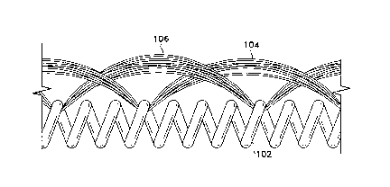
Figure 1 shows a length of the fibered coil
(100). It is made of several components: a helical coil
(102), a first fibrous element (104), and a second
fibrous element (106). The end of the coil may be sealed
to form a cap (108) .
The helical coil (102) is typically of a
radiopaque material such as tungsten, tantalum, gold
platinum, and alloys of those materials. Stainless
steels are also suitable. The use of various polymers,
such as polyethylene, polyurethane, and the like as the
coil material is also contemplated. The use of polymeric
materials typically involves the use of known radiopaque
fillers such as powdered tantalum, powdered tungsten,
barium sulfate, bismuth oxide, bismuth carbonate, or the
like. Preferred, however, is an alloy of platinum with a
_4_ 2170358
minor amount of tungsten. This alloy is very flexible
and yet the tungsten takes away a measure of ductility
from the resulting coil.
The coil may be from 0.2 to 100 cm in length or
S more. The diameter of the coil is from 0.004" to 0.015",
typically from 0.008" to 0.012". The wire making up the
coil is 0.0005" to 0.002" in diameter. The coil may be
wound to have a tight pitch, that is to say, that there
is no space between the adjacent turns of the coil, or it
may have some space between adjacent turns. Most
desirable, from the point of view of having a high
content of fiber, is a coil which is slightly stretched
in the manner and in the amount described below.
The first (104) and second (106) fibrous
elements typically are bundles of individual fibers (5 to
100 fibers per bundle), but may be individual fibers.
The fibers may be of a number of different thrombogenic
materials. Suitable synthetic fibers include
polyethylene terephthalate (e.g., DACRON*), polyesters,
especially polyamides (e. g., the Nylons), polyglycolic
acid, polylactic acid, and the like. Other less
desirable synthetic polymers, because of their decreased
thrombogenicity, include fluorocarbons (Teflon) and
polyaramids (Kevlar). Natural fibers such as silk and
cotton are also quite suitable.
The fibered coil (100) shown in Figure 1 is in
the general 'shape as found in the catheter lumen. The
coil (102) has been stretched to place the first fibrous
element (104) and second fibrous element (106) close
along the outer periphery of t.'he coil (102). This
stretching lessens the overall diameter of the fiber coil
(100) as seen by the catheter :lumen.
As may be seen more clearly in Figure 2, the
multiple fiber elements are alternately looped along the
coil. That is to say that the looping of the first fiber
* Trade-mark
~ ~~ ~~35
WO 96/00035 - 5 - PCT/US95/08075
element (104) through coil (102) alternates with the
looping of the second fiber element (106) through coil
(102). The fiber elements may be looped through the coil
(102) as shown in Figures 1 and 2 or they may be tied at
intersections with the coil (106) although, because of
the interference between the knot end catheter offered by
the knot, a mere looping is preferred. The end passage
of the fibers through the coil desirably involves a knot.
Only a pair of fibrous elements (104 and 106) are shown
in Figures 1 and 2; multiple such fiber elements may be
used, however. Additionally, it is quite desirable that
the spacing of the fibrous elements as they cross the
coil need not be equal.
As is portrayed in 'the side view found in
Figure 3, multiple filament numbers having a short coil
spacing (110), an intermediate. coil spacing (112), and a
long coil spacing (114). The:ae various fiber spacings
tend to increase the randomness of the fibered center of
the randomized coil after it is released from the
catheter. This benefit will be discussed in more detail
below.
A significant aspect: of this invention is shown
in Figure 4. That drawing, a cross-section view, shows
that the various fiber elements (in this example, 104 and
106) occupy a small radial sector of the coil's
circumference. Although, upon deployment, the various
fiber elements will shift toward each other to a modest
degree, the filaments must be placed in the same 90°
quadrant (105) to attain maximum benefit of the
invention. This quadrant is measured perpendicularly to
the axis of the stretched coil.
Finally, Figure 1 shows an end (108) on coil
(102). Such ends (108) are typically produced by heating
the end of the coil (102) to melt a small section of the
358
WO 96/00035 _ 6 _ PCT/US95/08075 ....
coil and form a closed end (7_08) . Figure 5A shows a
close-up of the end (108) and the coil (102).
Figure 5B shows an additional variation in
which the coil (102) encompae>ses a control wire (116) and
an end cap (118) having a hole therethrough. Use of such
a control wire (116) allows "ganging" of the coils or
placement of a number of coils "nose-to-tail" within the
catheter and therefore gives the attending surgeon the
choice of using one or more coils without reloading the
catheter.
Figure 6 shows the shape of the coil (102)
after it has been deployed from the catheter. The coil
(102) encompasses an interior region (124) which has
fiber passing through the region which is formed by
creation of a secondary diameter (126). This region
(124) of fibers provides for additional thrombogenicity
in the open region (124) among the secondary coil (126)
turns. This added and widely spaced fiber results in an
enhanced thrombus formation rate - typically a matter of
concern in using these device's for treatment of vascular
problems. We have found that by use of this procedure of
fiber attachment, upwards of .55% of the fibers found on
the coil are introduced into the open region (124),
preferably more than 75% and, most preferably, more than
85%.
The coils (102) discussed above are "preformed"
so as to allow the coil (102) to assume the secondary
diameter (126) shown in Figure' 6. The patent to Ritchart
et al. (U. S. Patent No. 4,994,.069), discussed above,
discusses 'a number of ways to preform such coils, e.g.,
by crimping the coil at various intervals. Another way
to preform the coils, particularly when using the
preferred platinum/tungsten alloy mentioned above is by
winding the coil on a mandrel into the secondary diameter
shown in Figure 6 and then modestly heat-treating the
PCT/US95/08075
.,.~ WO 96/00035 _ 7
thusly-wound coil. The coil will retain sufficient
flexibility to extend, in a .linear fashion, through a
catheter lumen.
This device may be deployed in the same manner
as are the coils described in the Ritchart et al or Chee
et al patents discussed abovf~. In general, a vascular
catheter is introduced into t:he bloodstream at a
convenient site, often the femoral artery in the groin,
and advanced to the site of concern. As has been noted
elsewhere, these sites ore often in the cranial arteries
but may be in any other site where occlusion is desired.
Guidewires are typically used to direct the catheter to
the desired site but blood flow is used to direct flow-
directed catheters. Once the distal end of the catheter
is at the site, the catheter lumen is cleared of
guidewires and the like. The inventive coil is then
introduced into the lumen, often with the help of a
cannula to preserve the shape of the elongated coil until
it enters the catheter lumen. A pusher, typically
similar in shape to a guidewire is then introduced into
the catheter lumen to push the inventive coil along the
interior of the catheter and .out its distal end. Once the
coil is safely in place, the ~~atheter is removed from the
body.
This invention has been described using
specific details to augment the explanation of that
invention. However, it is noi= our intent that the
specifics so used would be in any manner limiting to the
claimed invention. It is our intent that variations of
the invention which would be considered equivalent to one
having ordinary skill in this art be within the scope of
the claims which follow.