Note: Descriptions are shown in the official language in which they were submitted.
CA 02175867 2005-06-09
- 1 -
DEVICE FOR VAPOR STERILIZATION OF
ARTICLES HAVING LUMENS
BACKGROUND OF INVENTION
Field of Invention
The invention relates to the vapour sterilization of
articles such as medical instruments having long narrow
lumens therein, and more particularly, to a device for
delivering a gaseous antimicrobial directly into the lumen
of an article during the sterilization process.
Background Information
The need to sterilize articles such as medical
instruments arid others for use in the agriculture and
fermentation industries is well known. In recent years,
many methods of vapour sterilization have been developed.
While these methods offer the advantage of being generally
faster than sterilization by immersion in an antimicrobial
solution, they suffer from one major disadvantage, namely
the inability to sterilize the interior of a long narrow
tube in a short period of
217~8s~
- 2 -
time. Thus, with regard to medical instruments such as
endoscopes, the difficulty in sterilizing the lumen can
often negate the general advantage of using vapor
sterilization.
One way of overcoming the above disadvantage is set
forth in U.S. Patent Nos. 4,410,492 and 4,337,223. The
apparatus described therein comprises a sterilizing
chamber with means for introducing an antimicrobial gas
into the chamber and circulating the gas within the
chamber. Disposed within the chamber is a socket for
receiving the tubular end of a medical instrument. The
socket is connected to a valve and a recirculating pump
and the antimicrobial gas is recirculated from the
chamber through the lumen of the instrument. The
commercial apparatus employs ethylene oxide as the
antimicrobial and requires a sterilization times of
about 3 hours for flexible endoscopes and about 2 hours
for the shorter, rigid endoscopes. Ethylene oxide is a
known.toxic substance and the process thereby
experiences concomitant toxicity problems. , In addition,
the method and apparatus described in these references
cannot be used to sterilize an instrument within a
sterile pack since one end of the instrument must be
attached to the socket.
Thus there is a current need for an effective
method to sterilize medical instruments such as
endoscopes in a reasonably short period of time,
preferably in one hour or less. The method and device
of the present invention makes vapor sterilization of
such instruments practical by delivering vapor directly
to the interior of the lumen in the endoscope, whether
or not it is in a sterile pack.
2I ~~86~
- 3 -
SUMMARY OF THE INVENTION
The present invention comprises a method and device
for providing antimicrobial vapor directly into the long
narrow lumen of medical instruments and similar
articles. The device and method are intended for use
with solution vapor sterilization procedures. In these
procedures, the article is placed within a sterilization
chamber, the pressure in the chamber is reduced, and a
liquid solution of antimicrobial agent is introduced
into the chamber where it vaporizes. Alternatively, an
antimicrobial vapor may be introduced directly into the
chamber after the pressure therein has been reduced. In
either case, the instrument is sterilized by exposure to
the vapor or active species generated from it rather
than by direct contact with a liquid antimicrobial. The
procedure may further involve the use of heat or, e.g.,
low pressure gas plasma to enhance the antimicrobial
activity, reduce sterilization times, and/or remove
residual any antimcrobial agent from the instrument.
In its simplest form, the device of the present
invention comprises a vessel containing a small amount
of the antimicrobial solution, and a means for
connecting the vessel to the lumen of the instrument to
provide a source of antimicrobial vapor directly to the
lumen during the vapor sterilization process. The
device is placed on the instrument prior to disposing
the instrument in the sterilization chamber. As the
pressure in the chamber is reduced, the antimicrobial
solution contained in the vessel is vaporized and passes
from the vessel into the lumen of the instrument.
215867
- 4 -
With the use of the device and method of the
present invention, vapor sterilization times for
endoscopes can be reduced to one hour or less. In
addition, the method and the device may be used to
sterilize endoscopes in a sterile pack since the device
of the present invention may be attached to and packaged
with the endoscope before the endoscope is placed within
the sterilization chamber. Upon opening of the pack,
the device may be retrieved for re-use or preferably
discarded with the pack.
The device and method of the present invention
reduce sterilization time required for instruments
having long narrow lumens therein. Reduced
sterilization times are also achieved with the
instruments encased in a package designed to maintain
sterility after the removal from the sterilized chamber.
In addition, as antimicrobial vapor is provided directly
into the lumen of the instrument, lower concentrations
of antimicrobial solutions may be used in the
sterilizer, and this together with the reduced
sterilization times provides improved materials
compatibility with respect to both the instrument
components and the packaging or wrapping materials.
A device according to the inventions delivers an
antimicrobial vapor to a lumen of an article during
solution vapor sterilization. The device comprises a
first member which comprises a vessel having an inner
sealed chamber containing an antimicrobial solution and
a wall forming at least a portion of the chamber. A
connector connects the vessel to the article lumen. A
second member connects to the first member in moveable
relation thereto. The second member comprises an
CA 02175867 2006-04-03
- 5 -
opening member whereby movement of the second member in a
predetermined direction relative to the first member
moves the opening member toward the wall to open the wall
and place the chamber into fluid communication with the
article lumen.
A method for sterilizing an article lumen according
to the invention comprises the steps of: enclosing an
antimicrobial solution in a sealed chamber of a first
member, the sealed chamber having a thin wall; connecting
a second member in moveable relation to the first member,
the second member comprising an opening member;
connecting the wall to the article lumen so that the wall
is in fluid communication with the article lumen; and
then moving the second member in a predetermined
direction relative to the first member and thereby moving
the opening member toward the wall, opening the wall and
placing the chamber into fluid communication with the
article lumen; and isolating a user from the
antimicrobial solution during the process of opening the
chamber.
Preferably, the opening member opens the wall via
penetration thereof and comprises a spike having a first
end and a second end, and wherein the first end faces the
wall and comprises a sharpened tip. Preferably, a central
lumen extends coaxially through the spike and
communicates with the connector whereby the vessel is
placed into fluid communication with the article lumen
through the spike lumen when the spike penetrates the
wall.
Preferably, the first and second members
interconnect in telescoping relationship with each other.
A detent and surface on the opposing members preferably
limits the degree to which the first and second members
can telescope apart.
To ease the breaching of the wall a threaded
interconnection can be provided between the first and
CA 02175867 2006-04-03
- 5a -
second members wherein rotation of the first and second
members relative to each other moves them together to
breach the wall. A tactile detent can be provided to let
a user know when the members are fully rotated together.
Preferably, a guard is disposed between abutting
surfaces on the first and second members to prevent the
first and second members from moving together
sufficiently to breach the wall. The guard preferably
2~ ~~86'~
6 -
has a contrasting appearance to the first and second
members whereby the presence or absence of the guard can
easily be visually determined. In a preferred form,
the guard comprises a ring encircling the device between
the first and second members and is inelastic so that to
remove the ring from between the first and second
abutting surfaces it must be deformed beyond its elastic
limit.
BRIEF DESCRIPTION OF THE DRAWINGS
FIG. 1 is a perspective view of one embodiment of
the device, according to the present invention, attached
to the end of a tube;
FIG. 2 is a perspective view of another embodiment
of the device of the present invention, showing the end
of the device for making a connection to a tubular
member;
FIG. 2A is a perspective view of a variation of the
device of FIG. 2;
FIG. 3 is a plan view of another embodiment of a
device of the present invention;
FIG. 3A is a variation of the device of FIG. 3;
FIG. 4 is a plot of sterilization time verses
efficacy and showing enhanced efficacy of attaching an
HzOi device to a lumen prior to sterilization;
FIG. 5 is an exploded view of a further embodiment
of a device of the present invention;
' 21~586~
_,_
FIG. 6 is an exploded view in section of the device
of FIG. 5;
FIG. 7 is an end view of the opener of the device
of FIG. 5;
FIG. 8 is a plan view in section of the assembled
device of FIG. 5, prior to use;
FIG. 9 is a plan view in section of the assembled
device of FIG. 5, during use;
FIG. 10 is a perspective disassembly view of a
further embodiment of a device of the present invention;
FIG. 11 is a plan view in section of the assembled
device of FIG. 10, during use;
FIG. 12 is a close-up plan view of a distal portion
of a capsule portion of the device of FIG. 10; and
FIG. 13 is a sectional view taken along lines 13-13
of FIG. 12.
DETAILED DESCRIPTION OF THE INVENTION
The method and device of the present invention
relates to the sterilization of articles such as medical
instruments having a long narrow tube therein. The term
instruments as used herein applies to medical or
surgical devices such as endoscopes, catheters, tubing,
or similar instruments or articles having an internal
lumen which is preferably used in a sterile condition as
in, for example, the agricultural or fermentation
~I7586~
_8_
industries. The method and device of the present
application show particular advantages in the solution
vapor sterilization of lumens exceeding ten centimeters
in length and having an internal diameter of about 7
millimeters or less. As endoscopes typically have
lumens with internal diameters of 1 to 4 millimeters and
lengths of up to 1.5 meters or more for flexible
endoscopes and at least 45 centimeters for rigid
endoscopes, the method and device of the present
application have particular applicability to the
sterilization of these instruments. With the use of the
device of the present invention, antimicrobial vapor is
supplied directly to the lumen or interior of the tube
of the instrument during the vapor sterilization
process.
The antimicrobials used with the method and device
of the present invention include solutions of
glutaraldehyde, hydrogen peroxide, chlorine dioxide or
other antimicrobials in an inert solvent, the only
requirement being that the solution be liquid at
atmospheric pressure and a vapor at the temperature and
pressure of the sterilization process. Though the
higher concentration solutions of antimicrobials are
more effective, problems with materials compatibility
and shipping and handling may arise at very high
concentrations. For example, a 30% to 50% solution of
hydrogen peroxide in water is both very effective and
presents few shipping and handling problems, while
higher concentrations of up to 70% become increasingly
more difficult and dangerous to handle.
In solution vapor sterilization, the procedure
generally used is as follows: The article to be
2I~~8~~
- 9 -
sterilized is placed within the sterilization chamber,
the chamber is sealed, and a vacuum is drawn on the
chamber to reduce the pressure to less than about 50
torr, and preferably to 20 torr or less. An
antimicrobial solution is then injected into the chamber
where it vaporizes and contacts the exposed surfaces of
article. The time necessary for total kill of specific
microbial agents varies with the type and concentration
of antimicrobials present, and with the degree of
exposure to the microbial agent. Microbials disposed in
cracks, crevices or internal tubular structures are
somewhat protected from the antimicrobial agent and
require more time for total kill than microbials on the
external surface of the article. Heat or high frequency
radiation may be used to increase the effectiveness of
the antimicrobial and its penetration into remote areas
of the instrument.
The device of the present invention comprises a
vessel for containing a small amount of antimicrobial
solution, and a means for connecting the vessel directly
to the lumen or the end of the tube of the article to be
sterilized. When the article with device containing
antimicrobial solution is disposed in the sterilization
chamber and a vacuum drawn on the chamber, antimicrobial
vapor generated from the solution within the vessel
flows directly into the lumen.
The effectiveness of the method and device of the
present invention was demonstrated by the following
experiments:
50 inch (127 centimeters) lengths of Tygon tubing
having a 2 millimeter inside diameter were used to
2I~~86~
- 10 -
simulate an endoscope in the sterilization test. A
paper strip (2 mm x 13 mm) containing approximately 2.0
x 106 Bacillus subtilis (vat. globigii) spores was
placed in each tube equidistant from each end. A
syringe containing 0.05 milliliters of 10% by weight
hydrogen peroxide solution in water was provided for
each tube. Each of the samples was individually
packaged in a TYVEK"'/MYLAR~' envelope prior to
sterilization.
One third of the samples (three units) were placed
in the package with the syringe unattached to the end of
the tube. Another one-third of the samples were
packaged with the syringe attached. Individual samples
were placed within a 65 liter sterilization chamber and
sent through a hydrogen peroxide vapor sterilization
cycle wherein the pressure within the chamber was
reduced to 3 tort for the total exposure time minus 15
minutes, and 0.5 tort for the final 15 minutes of
exposure. No additional hydrogen peroxide was injected
into the chamber.
The remaining one-third of the samples, packaged
with the syringe attached to the end of the tube as
described above, were sent through a hydrogen peroxide
vapor sterilization cycle supplemented with high
frequency radiation plasma which is known to generate an
active species from the hydrogen peroxide. Again a 65
liter chamber was used, and the pressure within the
chamber was reduced to 3.0 tort for the total exposure
time minus 15 minutes and 0.5 tort for the final 15
minutes of exposure. Again, no additional hydrogen
peroxide was injected into the chamber. Plasma was
generated only during the final 15 minutes of exposure
217586'
- 11 -
at 2.05 MHz with 320 watts of power, pulsed 0.3
milliseconds on to 1.0 milliseconds off.
At the conclusion of the sterilization cycle, the
paper strip was removed from each tube and placed in a
glass vial containing 10 ml of a sterile pH 7.0
phosphate buffer solution. This solution contained 10
milligrams of TWEEN 80 to aid in removal of any spores
from the paper strip and 0.0066 milligram of catalase to
neutralize any remaining hydrogen peroxide. Five glass
beads were placed in the solution, and the solution was
vortexed for two minutes to completely macerate the
paper strip. Three decimal dilutions of the solution
were made with sterile pH 7.0 phosphate buffer, and the
original solution and the decimal dilutions were poured
into sterile glass Petri plates. A culture medium was
added and the plates were incubated for four days at
30'C. After incubation the number of viable organisms in
each plate was counted, and the number of spores on the
paper strip calculated by multiplying the spore count by
the appropriate dilution factor.
The results of the experiments are presented in
Table I below, and plotted in FIG. 4, where S/So
represents the ratio of the number of organisms
surviving the test to the initial number of organisms
which were placed on the paper strip prior to the test.
As shown by these data, no reduction in microbial
population was achieved in samples where the syringe was
not attached to the tubing, even after an exposure time
of 75 minutes. Attaching the syringe to the end of the
tube according to the method of the present invention
produced total kill in 35 minutes without low
temperature gas plasma, and in 25 minutes when the
21'~ 5 8 6'~
- 12 -
antimicrobial activity was enhanced by the use of low
temperature gas plasma.
Table I
Sterilization
Sample Time - Min. ~fficacv (S~S"1
A 3 5 8 . 6 x 10't
4 5 8 . 9 x 10't
75 1.1 x 10°
2 0 7 . 0 x 10't
25 5.8x10't
35 p
C 20 8.5 x 10⁻³
0
0
Sample A - Syringe unattached
Sample B - Syringe attached
Sample C - Syringe attached plus plasma
A preferred embodiment of the device to be used
in accordance with the teaching of the present invention
is shown in FIG. 1. The device indicated generally at
10 is shown attached to a tube 12. In the device
depicted in FIG. 1. the means for connecting the vessel
14 to the end of the tube comprises an expandable sheath
16, one end of which is securely attached to the vessel,
and the other end of which comprises an elastic ring 18
making a releasable attachment about the end of the
2I7~8 6'~
- 13 -
tube. The sheath 16 may be attached to the vessel in
any known manner and, as shown in FIG. 1, the sheath 16
is attached to the vessel by a second elastic ring 20
disposed over the lip 22 about opening 24 of vessel 14.
Though the vessel shown is cylindrical, the vessel may
comprise any three dimensional container preferably of
semi-rigid material, having an opening therein. The
vessel may be made of, e.g., polyethylene,
polypropylene, glass or any other material which is
nonreactive to the antimicrobial solution of vapor. The
sheath may also be formed of polyethylene, polypropylene
or other material which is relatively nonreactive to the
antimicrobial vapor. The elastic rings may be formed of
natural latex or butyl rubber which are relatively
resistant to the antimicrobial vapors; however,
resistivity is less critical when the device is
constructed for one time use. Disposed within the
vessel may be a substrate 26 comprising a woven or
nonwoven fabric or sponge for containing the liquid
antimicrobial solution. The vessel preferably has a
means 28 associated with the opening for attaching a
closure cap over the opening prior to use in order to
maintain the antimicrobial solution therein. As shown
in FIG. 1, means 28 comprises threads for a screw cap
fitting about the lip of the vessel.
Another embodiment of the device of the present
invention is depicted in FIG. 2 where the device is
indicated generally at 30. The means for connecting the
vessel 34 to the end of a tubular instrument comprises a
bushing 36 disposed within the open end of the vessel.
In the particular embodiment shown in FIG. 2, the
bushing comprises a series of rings 38 and 40 of
inwardly extending plastic flaps defining a flexible
2Z ~~86~
- 14 -
aperture 32 to receive the tubular instrument. The
flaps can be made of any flexible material which is non-
reactive to the antimicrobial solution or vapor, such as
polyethylene, and of sufficient thickness that the flaps
provide resistance to withdrawal of a tube inserted
through the aperture. Disposed within the vessel is a
substrate 42 containing the antimicrobial solution.
Preferably, the vessel 34 is provided with means 44 for
attaching a closure cap thereto prior to use. As shown
in FIG. 2, means 44 comprise threads for attaching a
screw cap (not shown) within the opening of the vessel.
FIG. 2A illustrates a variation in the design of the
device of FIG. 2 which utilizes the same basic vessel
and means for attachment to a tubular device. In the
device shown in FIG. 2A, end 45 of the vessel opposite
the open end is provided with aperture 46 for attaching
a disposable cartridge 47 containing a supply of
antimicrobial on a substrate such as a woven or nonwoven
fabric or sponge 48 as illustrated. The aperture 46 of
the vessel is designed in conjunction with neck 49 of
the cartridge to provide quick and easy attachment and
release of the cartridge and the vessel. In the
embodiment shown in FIG. 2A, aperture 46 is provided
with reverse threads for engaging the threads of the
neck 49 of the cartridge. In this variation of the
device it is not necessary for a substrate containing
the antimicrobial solution to be disposed within the
vessel since the antimicrobial solution is provided in
pre-measured aliquots in the cartridges. With the
device of FIG. 2A one achieves the convenience and
accuracy of disposable, pre-measured aliquots of
antimicrobial solution without the expense associated
with the device of FIG. 2.
2~ 7586'
- 15 -
The following table sets forth the effectiveness of
the devices depicted in FIGS. 1 and 2 in a sterilization
procedure described below.
Table II
Effect of Devices on Efficacy of Sterilization
Inside Tubes
Efficacv (S/S,~
No Device Device
Material I. D. Length FIG. 1 FIG. 2A
(cm) (cm)
Surgical Tygon 0.64 10 0 - -
0 . 64 2 0 4 . 4 x 10's - -
0 . 6 4 3 0 1.1 x 10'1 - -
0.64 45 8.8 x 10'1 0 0
Rubber Tubing 0.64 25 1.7 x 10'1 -
0.64 45 7.9 x 10'1 0 0
The efficacy is recorded in terms of the ratio of
the number of microorganisms surviving the test, S, to
the number of challenge organisms, So (approx. 1 x 10⁶)
on a paper strip disposed within the tube equidistant
from the ends. In the sterilization procedure, 100
microliters of 30% aqueous HZ02 solution was supplied in
each of the devices. The devices were attached to the
ends of tubes of the indicated length and 0.64 cm in
internal diameter. All of the tube samples were placed
within TYVEK~/MYLAR~ packaging prior to sterilization.
The packaged tubes were placed within the sterilizing
chamber and the pressure therein was reduced to about
0.1 torr in about 10 minutes. Additional 30% H202
21~~8~7
- 16 -
solution was injected into the chamber to achieve a
concentration of 2.0 milligrams HZO2 per liter of chamber
volume. Following injection of the HZO2, the tubes were
retained in the chamber an additional 50 minutes.
Injection of the HZ02 solution raised the pressure in
the chamber to about 6 torr and the pressure was again
reduced to about 0.1 torr. During the last 10 minutes
of exposure, low temperature gas plasma was generated in
the chamber at 300 watts. The challenge micro organisms
used in the test were Bacillus subtilis (var. globigii)
spores.
As shown in Table II above, when the tube length was
only 10 centimeters, sterilization was achieved without
the use of the device according to the present
invention. However, for tubing of 20 and 30 centimeters
in length, a device of the present invention would be
needed in order to achieve sterility within the exposure
time of the test. For tubes of 45 centimeters in
length, total kill was achieved during the 1 hour
exposure time of the test, using either of the devices
depicted in FIG. 1 and FIG. 2.
A further experiment used 7 mm medical grade Teflon
tubing 183 cm in length. The tubing was cut into three
pieces to obtain a 5 cm long center section which was
joined in the end sections by external tubing
connectors. In the experiment, approximately 1.0 x 10⁴
bacillus (var. globigii) spores were deposited in the
center section of the Teflon tubing. The tubing was
assembled and subjected to sterilization with hydrogen
peroxide vapor as described above at a concentration of
2.0 mg./liter of chamber volume. The chamber was
217~86v
evacuated to a pressure of 0.1 tort before the peroxide
was injected as an aqueous solution and allowed to
vaporize. After 20 minutes, a continuous gas plasma was
generated in the chamber at 300 watts, 13.5 MegaHertz
and the sterilization continued for an additional 5
minutes after which the vacuum was released with
sterile, filtered air, and the number of surviving
spores determined.
The experiment was first conducted without a device
of the present invention attached to the tubing, then
repeated with a device of FIG. 3 as described below
containing 100 ml of 30% hydrogen peroxide attached to
one end of the tubing. The experimental results of the
tests are presented in Table III below.
Table III
sterilization of 1 mm Tubinv
Efficacy jS/S~
Material I-D. Length No Device FIG. 1 Device
Teflon 1 mm 183 cm 1.9 x 10'1 0
The data of Table III demonstrate the efficacy of
the method of the present invention in sterilizing of
very long tubes having very small diameters used in
certain endoscopic procedures.
Additional embodiments of the device of the present
invention are depicted in FIGS. 3 and 3A. The device
shown in FIG. 3 indicated generally at 50, coagrises a
vessel 52 in the form of a pouch constructed of a
2I X5861
- 18 -
flexible material. The means for connecting the vessel
or pouch 52 to the end of an instrument tube comprises a
first drawstring 54, and preferably a second drawstring
62. These drawstrings are preferably arranged in the
configuration as shown in FIG. 2 to be drawn from
opposite sides of the pouch. The pouch is preferably
provided with an airtight seal to maintain the
antimicrobial solution therein prior to use, and
includes a means for creating an opening in the sealed
pouch so that it may be disposed over the end of a tube.
The seal may be created by sealing the ends 66 of the
pouch, and of the lumen as often the means for opening
the sealed pouch may comprise, for example, a line of
weakening at 68, preferably in combination with a notch
also shown generally at 68, to permit the pouch to be
opened by tearing off one end.
FIG. 3A shows a device indicated generally at 50A,
similar to device 50, but wherein the airtight seal and
the means for creating and opening the sealed pouch is a
line of fastening 64 similar to a "zip-lock" closure.
Optionally, opening flaps 70 may be provided on either
side of the pouch adjacent closure 64 of FIG. 3A, or the
line of weakening 68 of FIG. 3. These flaps are firmly
secured to the pouch. In use, after the sealed end 66
of the pouch of Fig. 3 has been removed along the line
of weakening 68, the flaps when pulled oppositely from
each other will distend the opening of the pouch for
disposal around the end of an instrument tube. The
flaps of Fig. 3A, when pulled in opposite directions,
can be used to open the zip-lock fastening, or if the
fastening is already opened, to distend the opening for
disposal around the end of an instrument tube. A
substrate 72 such as a woven or nonwoven fabric or
217~8fi7
- 19 -
sponge may be disposed within the pouch for containing
the antimicrobial solution.
In a preferred construction, the drawstrings are
provided with a locking means as illustrated. Though
many means for locking or catching a drawstring are
known in the art and may be used in conjunction with the
present invention, the locking means depicted at 56 at
FIG. 3 comprise a catch 60 for a serrated edge 58
provided on the drawstring. As shown in FIG. 3, the
catch, comprising an opening for disposing one end of
the drawstring therethrough, is located at the opposite
end of the drawstring. The catch, however, may be
provided by a flap, opening therein, attached to the
edge of the pouch, provided the other end of the
drawstring must also be attached to the pouch. When two
drawstrings are used, one or both drawstrings may be
provided with a locking means. By pulling the end 73 of
the drawstring, the flexible pouch is gathered and a
firm fastening may be made to a tube inserted within the
pouch.
Preferably, a highly concentrated solution of
hydrogen peroxide is used as the liquid antimicrobial in
the device of the present invention. However, in high
concentrations, hydrogen peroxide can quickly cause
damage to living tissue. A system for applying such
solution to an instrument lumen while reducing the
chances of accidental exposure of a user to the
antimicrobial solution is highly desirable. The
following embodiments provide such advantage.
FIG. 5 illustrates a further embodiment of a device
100 according to the invention. The device 100
~°~.7586~
- 20 -
comprises in gross a capsule 102, an opener 104, and a
safety ring 106 positioned between the capsule of 102
and the opener 104. Turning to FIG. 6, the capsule 102
comprises a cylindrical body 108 having a distal end 110
and a proximal end 112. At the proximal end 112, the
capsule body 108 expands radially to form a cup shaped
well 114. A membrane wall 116 is disposed within the
capsule body 108 adjacent to well 114.
A cap 118 of generally discoidal shape has a
distally projecting annular flange 120 which fits within
the well 114. The cap 118 is sonically welded to the
capsule 102 at the proximal end 112 to seal a quantity
of antimicrobial solution 122 within a chamber 124
defined between the cap 118, membrane wall 116 and
capsule body 108. During storage the antimicrobial
solution 122 may tend to diffuse through the capsule 102
and out of the chamber 124 thereby decreasing its
quantity and potency. The antimicrobial solution 122
thus preferably comprises 197 mg of 59% hydrogen
peroxide solution upon construction such that after a
reasonable storage period such as ten months, the
chamber 124 will retain approximately 100 mg of a 45%
hydrogen peroxide solution.
So that it may be more easily breached, a central
portion 126 of the membrane wall 116 has a slightly
thinner thickness than the remainder of the membrane
wall 116. Six radial ribs 128 extend from the capsule
body 108 towards, but not into, the membrane wall
central portion 126 to support the membrane wall 116
during the breaching process.
~175$6"~
- 21 -
At the capsule body distal end 110, an annular
flange 130 slopes outwardly and proximally, thus
providing a barbed appearance in cross-section. The
distal flange 130 preferably slopes in a gentle fashion,
such as a 17° slope from an imaginary coaxial centerline
132 of the device 100. A central annular flange 134
slopes outwardly and proximally from the capsule body
108 at a slightly more aggressive angle than the distal
flange 130. A pair of diametrically opposed slits 136
extend proximally in the capsule body 108 from its
distal end 110 to allow some flexibility in the capsule
body 108 and to thereby ease its entry into the opener
104.
The opener 104 comprises a cylindrical body 140
having a proximal end 142 facing the capsule 102 and a
distal end 144. A hollow spike 146, coaxially disposed
within the opener body 146, extends toward the membrane
wall 116 and terminates in a beveled and sharpened tip
148. Preferably, the tip 148 is beveled at a 30° angle
from the device center line 132. Also, a central lumen
150 extends coaxially through the spike 146.
Three equilaterally spaced posts 152 extend
outwardly radially from a fixed end of the spike 146 to
the opener body 140 and thereby support the spike 146
therein. Preferably, each of the posts 152 has a
distally facing fillet brace 154 for added support. A
circumferentially interrupted annular embossment 156
extends radially inwardly in a very shallow manner from
the opener body 140 (see also FIG. 7). When the capsule
102 is inserted into the opener 104 with the capsule
distal flange 130 beyond the opener embossment 156,
engagement therebetween prevents the capsule 102 from
2175867
- 22 -
being easily removed from the opener 104 while still
allowing a relative degree of movement between the
opener 104 and capsule 102 as will be more fully
described hereinafter.
A retaining ring 158 holds a mist-filter screen 160
within the opener body distal end 144. The mist-filter
screen 160 is round with a diameter exceeding that of
the opener body 140 whereby it is frictionally retained
within the opener body 140 by the retaining ring 158.
Preferably, the mist-filter screen 160 has a mesh
opening of 105 microns and is formed of polypropylene.
A plurality of axially aligned embossments 162 on an
outer surface of the retaining ring 158 ease insertion
and securely retain the mist-filter screen 16o and
retaining ring 158 within the opener body 140 (see also
FIGS. 8 and 9).
Alternatively, a series of detents (not shown), each
with a distally facing caroming surface and a proximally
facing radial surface could be provided within the
opener body 140, axially adjacent the posts 152. The
mist-filter screen 160 would thus have a diameter equal
to the inside diameter of the opener body 140 and be
held between the posts 152 and the proximally facing
surfaces of the detents. The screen could be easily
inserted through the opener distal end 144 and caromed
over the detent caroming surfaces into place between the
posts 152 and detents.
The safety ring 106 separates the opener 104 from
the capsule 102. With the safety ring 106 trapped
between the opener body proximal end 142 and the lip 115
on the capsule 102, the spike 146 is prevented from
~17~86'~
- 23 -
contacting the membrane wall 116 (see also FIG. 8). The
safety ring 106 is provided with a thin wall section 164
and a diametrically opposed pull tab 166 whereby manual
pressure applied to the pull tab 166 is sufficient to
deform the thin wall section 164 beyond its elastic
limit, preferably breaching the thin wall section 164,
thereby permitting removal of the safety ring 106 from
the device 100.
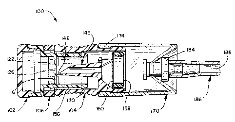
FIG. 8 illustrates the assembled device 100 prior to
use, with an adapter 170 affixed thereto. The adapter
170 comprises a cylindrical tubular body 172 formed of a
soft thermoplastic elastomer, such as Schafer, GmbH
THEKA-FLEX, S 2030 M. A shallow inwardly facing annular
flange 174 at a proximal end 176 of the adapter body 172
is received within a correspondingly shallow annular
groove about the opener body 140 to hold the adapter 170
to the device 100.
A truncated cone 178 extends inwardly, proximally,
from a distal end 180 of the adapter body 172 and
terminates in a central opening 182. A luer fitting 184
of an instrument to be sterilized 186 having a lumen 188
therein, is shown received within the opening 182.
Those of skill in the art will appreciate that the
dimensions of the cone 178 can be varied to accommodate
various types of instruments to be sterilized and that
other engaging means may be substituted therefor.
To use the device 100, an appropriately sized
adapter 170 is selected for the particular instrument
186 to be sterilized. The adapter 170 is attached to
the device 100 as shown in FIG. 8. The pull-tab 166 on
the safety ring 106 is grasped and pulled to separate
217586"
- 24 -
the safety ring thin wall section 164 and remove the
safety ring 106 from the device 100. To aid the user in
removing the safety ring 106 and in later rotating the
capsule 102 relative to the opener 104, the opener body
140 is provided with several textured finger
indentations 190 for easier grasping. After the safety
ring 106 is removed, the capsule 102 and opener 104 are
pushed together so that the spike 146 breaches the
membrane wall 116 as shown in FIG. 9. Preferably, the
capsule 102 is then rotated one full turn to ensure
proper breaching of the membrane wall 116. The
antimicrobial 122 is then free to leave the chamber 124
and flow into the instrument lumen 188.
In general practice, the device 100, with adapter
172 and instrument 186 attached as the membrane wall
breached 116 as shown in FIG. 9 are then placed into the
sterilization chamber (not shown ) of a solution vapor
sterilizer (also not shown). A vacuum applied to the
sterilization chamber causes the antimicrobial 122 to
vaporize and migrate into the instrument lumen 188 to
effect sterilization thereof.
FIGS. 10 to 13 illustrate a further embodiment of a
device 200 according to the invention. The device 200
is similar in nearly all respects to the device 100 with
the exception of the following differences.
Accordingly, portions of the device 200 which are
identical to the device 100 and were previously
described with respect thereto, will be designated with
like referenced numerals having a prime symbol (').
To reduce the force a user must exert to breach the
membrane wall 116', the capsule 102' threads into the
2175867
- 25
opener 104'. A raised embossment 202 surrounds the
capsule body 108' adjacent the lip 115'. A pair of
threads 204 formed in the embossment 202 receive,
respectfully, a pair of pins 206 which project into the
opener body 140'. Each thread 204 comprises a caroming
portion 208 and a circumferential portion 210.
The pins 206 enter the threads 204 through the
caroming portions 208 as the capsule 102' is rotated
relative to the opener 104', thereby pulling the capsule
102' axially into the opener 104'. The circumferential
portion 210 of the threads 204 allows the capsule 102'
to be rotated an additional one quarter turn after it is
fully received within the opener 104' to insure proper
breaching of the membrane wall 116'.
In the previous embodiment, the interaction of the
central flange 134 and the opener body 140 seals the
capsule 102 to the opener 104 to prevent antimicrobial
122 from leaking out of the device 100 between the
capsule 102 and opener 104. In the present embodiment,
an O-ring 212 about the capsule body 108' replaces the
central flange 136 and engages the opener body 140' to
seal the capsule 102' therein.
In the previous embodiment, the spike 146 is
provided with a simple bevelled tip 148 to penetrate the
membrane wall 116. In the present embodiment, the
bevelled tip 148 is replaced by a cutting tip 214 which
ie placed off of the central axles of the spike 146' and
which acts in a fashion similar to that of a can opener
to cut open the membrane wall 116. It will be
understood that the cutting tip 214 may take various
21'~586~
- 26 -
forms, however a sharp apex 216 and a sharp leading
cutting edge 218 improve its cutting ability.
Proper breaching of the membrane wall 116' is a
prerequisite to adequate sterilization. Accordingly,
operators of the devices 100 or 200 prefer some tactile,
audible, visual or other feedback that the device has
been operated properly. In the previous embodiment,
breaching of the membrane wall 116 tends to occur
suddenly, thus driving the capsule 102 and opener 104
together in a violent manner creating both an audible
and tactile snap. Also, the lip 115 will abut or
closely approach the capsule body proximal end 142 in
this position to provide a visual indication of proper
operation.
In the present embodiment, the threading interaction
between the capsule 102' and opener 104' breaches the
membrane wall 116' more gently than in the previous
embodiment. Thus, the user receives less tactile
feedback that the membrane wall 116' has been breached.
It may be desirable to provide such feedback in the form
of a snapping interaction between parts on the capsule
102' and opener 104' or perhaps to provide a visual
indication or other feedback that the opener 104' is
fully actuated.
FIGS. 12 and 13 illustrate one method of providing
such feedback. As each pins 206 travels its respective
thread circumferential portion 210, it encounters a
detest 220. The pins 206 cam over the detests 220 and
snap over a sharp trailing edge 222 thereon to become
trapped beyond the detests 220. Thus, the detests 220
provide both an audible and tactile feedback that the
21'~586~1
- 27 -
proper interaction has been achieved between the capsule
102' and opener 104'. Further, they prevent the capsule
102' and opener 104', and further prevent the capsule
102' from being easily backed out of the opener 104'.
Alignment marks (not shown) or other visual indicia mark
also be provided on the capsule 102' and opener 104' to
indicate full actuation.
Although the present invention has been described in
terms of specific devices for use in a preferred method
of vapor sterilization, it will be understood that
various modifications in the device and method will be
apparent to those skilled in the art and are within the
scope of this invention.