Note: Descriptions are shown in the official language in which they were submitted.
2178127
VARIABLE STIFFNESS COILS
Field of the Invention
This invention is an implantable vaso-occlusive
device. It is a helically wound coil having a central
section along its longitudinal axis which central section
is somewhat stiffer than at least one of its end regions.
Background of the Invention
Many commercially available vaso-occlusive
coils have capped ends which are formed by the simple
procedure of heating the coil end sufficiently to liquify
the composite metal into a round cap-like artifice.
Although the rounded cap provides a surface which is
relatively minimal when contacting the internal surface
of a delivery catheter, we believe that the tip could be
improved upon, at least from the aspect of coil delivery.
Some coils appear to have lost a measure of
flexibility in the region near the tip, perhaps because
of the heat necessary to melt the metal at the adjoining
tip. This short region of stiffness produces a leg which
presses against the lumen of the catheter, at least until
the tip clears the distal end of that catheter. The
energy stored in pushing the coil through the distal end
of the catheter causes the coil to jump forward and the
catheter to retract as the coil leaves the catheter. If
a very precise placement of the catheter tip is desired,
e.g., where a small-necked aneurysm is accessed, such a
lurching and slipping is particularly not desirable.
Out intent in this invention is to improve the
stiffness characteristics of the vaso-occlusive coil so
to enh~nce the ease with which the coi-ls advance through
2178127
- the catheter, improve the handling of the coil as it
exits the catheter, and improve the coil's ability to be
deployed gently as it leaves the catheter.
Finally, the flexible tip promotes the position
stability of the coil during placement. The "droop" of
the flexible distal section tends to engage the vessel
wall and press the trailing stiffer section of the coil
against the opposing wall. The resulting coil mass is
formed more quickly and more compactly.
Our solution to this problem is to assure that
at least one end of the vaso-occlusive coil is somewhat
more flexible than the adjoining midsection. The leading
end of the coil, i.e., the end of the coil which is
distally placed, is most important, although for
practicality's sake, it is desirable that both ends be so
constructed. In such a way, the coil may be introduced
from the catheter in either direction into the blood
system. There are several ways to increase the
flexibility of these end regions: vary the diameter of
the wire making up the coil, change the spacing of the
coil turns, vary the diameter of the coil, and change the
inherent properties of the material in the wire, such as
by annealing.
This technique is useful whether using coils
which have electrolytic or mechanical detachment links at
their ends, or when using coils having attached
thrombogenic fibers. The technique is especially useful
on coilæ having secondary shapes when those coils are
relaxed. There are other helical coil devices having
varying pitch spacing or the like.
For instance, U.S. No. Patent No. 485,652,
to Pfingst, issued November 8, 1892 describes a car
spring -- apparently a railroad car spring -- in which
the diameter of the rod making up the coil gradually
tapers. The inner diameter of the spring appears to be
21~8127
of constant diameter throughout. It is obviously quite
stiff.
U.S. No. Patent 4,553,545, to Mass et al.,
shows a intravascular prothesis made up of a helical
5 spring having a variable pitch. The device is intended
to hold a human body lumen open and consequently is
fairly stiff.
U.S. No. Patent 4,760,8249 shows a planar blank
intended for the manufacture of a coil spring. The coil
spring iæ suitable for a translllm~n~l implantation. The
device is either used as a stent to hold a vascular lumen
open or it may be used as a blood filter. The coil
spring filter may be used as a vena cava inferior filter
to prevent the formation of emboli and their passage into
15 the lung.
U.S. No. Patent 4,830,023, to de Toledo, shows
a medical guidewire having a coil tip. The device has a
degree of flexibility and a tip region of greater
flexibility. The coil making up the helically wound
2 0 spring is in two pieces: one having a greater pitch than
the other.
Similarly, U.S. No. Patent No. 5, 069,217, to
Fleischacker Jr., shows a coil of varying pitch soldered
to the end of a guidewire combination.
U.S. No. Patent No. 5,171,383, to Segae et al.
shows a guidewire having varying flexibility along the
axis. The flexibility is varied by changing the heat
treatment temperature along the length of the guidewire.
None of these publications show the concept
30 described herein in which at least a portion of the
center of the vaso-occlusive device i8 less flexible than
at least one of the ends.
SUMMARY OF THE INVENTION
This invention is an implantable vaso-occlusive
device. In general, it is a vaso-occlusive coil which,
2178127
viewed along its longitudinal axis, has a center section
which is somewhat stiffer than one or the other or both
of its end sections. This permits the vaso-occlu-sive
device to be deployed more gently from the catheter and
results in a procedure which places the coil with more
certainty at a specific point in a human body vascular
lumen or other site to be occluded.
The device is typically used in the human
vasculature to form emboli but may be used in any site in
the human body where an occlusion such as one produced by
the inventive device is needed.
The device may be made in a number of ways.
The wire forming the end section or sections of the vaso-
occlusive device may be of a smaller diameter. The wire
may be of different physical characteristics. Such
differences in physical characteristics may be produced
by annealing the wire. The end section or sections may
be made more flexible by changing the diameter of the
section as compared to the diameter in mid-section.
The device may be used with or without the
presence of ancillary fibers (e.g., Dacron) to enhance
the device's overall thrombogenicity. The device is
preferably made in such a way that it has both a primary
coil diameter and, once it is deployed from the catheter,
a self-forming secondary form.
BRIEF DESCRIPTION OF THE DRAWINGS
Figure 1 shows a side view of a generic linear
coil for the purpose of depicting the conventions used in
describing the inventive device.
Figure 2 is a close up of a section of a
generally linear coil also for the purpose of depicting
the conventions used in describing the inventive device.
Figure 3 shows a side view of a device for the
purpose of showing a secondary shape.
~ : ~178127
--5--
Figure 4 shows a partial side view of the
device made according to the invention in which the
spacing of helical turns of the coil is varied.
Figure 5 shows a side view of a variation of
the inventive device in which the diameter of the helical
coil is varied to provide differences in flexibility.
Figure 6 shows a variation of the invention in
which the diameter of the wire is varied in order to vary
the resulting flexibility of the inventive vaso-occlusive
device.
Figure 7 shows a variation of the invention in
which the diameter of the end section of the coil is
varied in order to vary the resulting flexibility of the
inventive vaso-occlusive device.
lS Figures 8A, 8B, and 8C show the procedure for
deploying a vaso-occlusive made according to the
invention and depict the way in which the device reacts
as it deployed into a vascular lumen.
DESCRIPTION OF THE II~Iv ~ ION
This invention is a helically wound vaso-
occlusive coil which may be introduced into the human
body, particularly into the human vasculature, uæing a
catheter. The inventive coil has at least one region
25 adjacent the end of the coil which has a greater
flexibility than the midsection of the coil.
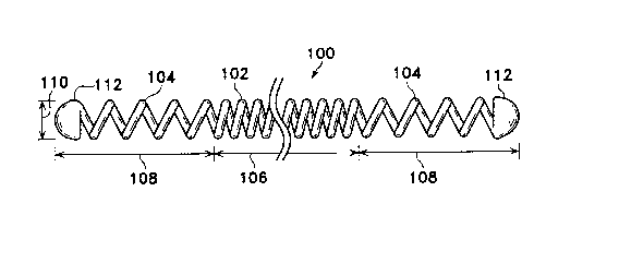
Figures 1-3 show a generally linear coil (100)
used to describe the conventions and terms used in
relation to this invention. The coil (100) depicted here
is made up of a central region (102) and two end regions
(104). Central or mid-region (102) has a length (106)
and end regions (104) similarly have lengths (108).
Although the two lengths of the end regions (104) are
shown to be equal to each other, the lengths need not be
equal. The coil (100) is helically wound from a wire.-
The diameter of the coil (100) is referred to as the
2 1 7 8 1 2 7
"primary~ diameter (110). The tips or caps (112) are
shown at the physical ends of the coils.
Figure 2 shows a close-up of a section of a
coil (100). Figure 2 shows the axis (114) of the coil
(100). The pitch angle ~ is shown as (116). That angle
is measured from the center of a line (120) of a coil
turn to a line perpendicular to the axis (116). As was
mentioned above, the pitch spacing or angle may be varied
in some aspects of this invention to produce a region of
higher flexibility.
Finally, Figure 3 shows a vaso-occlusive device
(122) made according to this invention having what we
term a "secondary shape n . In this instance the secondary
shape is a vortex-like shape. We term the shape
~secondary~ because it generically is formed by taking a
- wire which has been formed into a "primary" helical shape
(as seen in Figures 1 and 2 as (100)) and further forming
another shape which is not linear. There are numerous
secondary vaso-occlusive coil forms known in the art. A
selection of secondary shapes may be found in U.S. Patent
Nos. 4,994,069 (to Ritchart et al.), 5,382,259 (to Phelps
et al.), and in 5,304,194 (to Chee); the entirety of
which are incorporated by a notice. Specific secondary
shapes are not critical to this invention. In many
instances of use, it is not critical that the vaso-
occlusive device even have a secondary shape.
Nevertheless, for many procedures, particularly those
involving occlusion of a flowing vascular stream, a
secondary shape helps to assure effective embolization.
The wire making up the vascular device will typically be
of a metallic material, such as platinum, gold, rhodium,
rhenium, palladium, tungsten, and the like, as well as
alloys of these metals. These metals have significant
radiopacity and in their alloys may be tailored to
accomplish an appropriate blend of flexibility and
stiffness. They are also largely biologically inert. A
~ 2178127
highly desired metallic composition is a platinum alloy
containing a minor amount of tungsten.
The wire may, of course, be of other suitable
biocompatible materials, e.g., polymers, composites of
metals or alloys and polymers, etc. Polymeric wire
materials are often mixed with a radiopaque material,
e.g., barium sulfate, bismuth trioxide, bismuth
carbonate, powdered tungsten, powdered tantalum, or the
like, to promote their passive ability to be visualized
during fluoroscopy.
The diameter of the wire often used in this
invention will be in the range of 0.0005 and 0.005
inches. Larger diameter wire may be desirable for
certain very specific indications where occlusion is
needed at a high volume flow rate site. Such might
include repair an infant's Vein of Galen and treatment of
arteriovenous malformations (AVM's). Larger diameter
wire would be chosen because of its springiness.
Materials with higher innate springiness, e.g., platinum
alloys with high tungsten content, would also be
suitable.
The primary coil diameter (110 in Fig. lJ will
no~;n~lly be in the range of 0.008 to 0.025 inches; For
most neurovascular indications, a range of O.OlS to 0.018
inches is preferred. The axial length of the primary
shape will usually fall in the range of 0.5 to 100 cm,
more usually 2 to 40 cm. Depending upon usage, the coil
- may well have 10-75 turns per centimeter, preferably 10-
40 turns per centimeter. All of the ~ n~ionS here are
provided only as guidelines and are not critical to the
invention. However, only dimensions suitable for use in
occluding sites within the human body are included in the
scope of this invention.
Fig. 4 shows one variation of the inventive
vaso-occlusive device. A magnified, partial side view-of
a coil (130) is seen. In this view, the diameter of the
2178127
wire and the primary diameter (110) of the coil is
maintained to be approximately or substantially constant
in the end region (132) and in the center region~(134).
We say ~approximately or substantially constant" in that
one excellent way to produce the coils of this invention
involves winding the coil stock at nominally constant
pitch and simply stretching one or more of the ends to
produce the increased pitch spacing. The diameter of the
coil in the stretched region will obviously decrease when
such a step is taken.
For the coil wire diameters, wire compositions,
and pitch spacings with which we are familiar, an
increased pitch spacing of 25~ or more is sufficient to
provide the increased lateral flexibility to attain the
goals of the invention. The length of the end regions
(e.g., (104) in Fig. 1 and (132) in Fig. 4; others
discussed below) may be selected in several ways. For
instance, for most coils, an end region in length of at
least 1.5 times the diameter of the selected vascular
region is sufficient. This may translate into an end
region length of 2-3 mm in some cases. A length of 0.5
to 1.5 cm is typical. The total percentage of
comparative high flexibility would typically lie between
2.5 and 20% of the total primary axial length of the
coil.
The flexibility of this variation of the coil
and its brethren discussed below are all measured
perpendicular to the coil axis using a "rolling" 1-cm
moment arm. That is to say that a specific coil is
grasped at a point and the force is applied one
centimeter away. The force required to achieve a
specific deflection for the coil in the more flexible end
section is compared to the less flexible mid-section.
The ratio of these forces (i.e., force per unit
deflection in end section:: force per unit deflection in
mid-section) should be less than 1Ø Typically the
2178127
g
ratio will be between 0.35 and 0.95 with a preferred
range of 0. 4 to 0. 75, most preferably 0.6 to 0. 75.
Fig. 5 shows a further variation of the-
invention in which the enhanced flexibility of the end
5 portion is provided by a variation in the primary
diameter of the vaso-occlusive coil.
Fig. 5 shows the portion of a coil (138) having
an end section ( 140) and a primary diameter somewhat
larger than the primary diameter of the mid-section
(142). The diameter of end section (140) is sufficiently
larger than the diameter of mid-section (142) so that it
is able to meet the criteria mentioned above. That is to
say that the ratio of force needed for a unit deflection
of the end section is less than the unit of force needed
15 to deflect an isolated portion of the mid-section (142);
and preferably is of a ratio between 0.5 and 0. 95.
Figure 6 shows another variation of the
inventive vaso-occlusive helical coil (150) in which the
diameter of the wire making up mid-section (152) is
20 larger than the diameter of the wire making up at least
one end section ( 154) . This axially contiguous
relationship between two sections of the coil may be
carried out by providing the coil wire to the device for
rolling the primary coil in a series of different
25 diameters. The two sections may be brazed or soldered
together. Again, it is only necessary that the two
sections (152) be of different wire diameters, and hence
flexible, to conform to this variation of the invention.
As is the case with all of these variations, the
variation desirably has a secondary shape such as one of
those mentioned above.
Figure 7 shows a variation of the inventive
device in which the flexibility of the end section is
varied throughout the end section. The concept behind
3 5 this variation may be accomplished using any of the
procedures described above. Figure 7, however, shows a
2178127
-10--
coil (156) with a constant diameter wire but which has
been wound on a tapered mandrel. The central section
(157) of the coil (156) is of generally constant
diameter. The end section (158) portrayed has a
5 decreasing diameter as the end of the coil is approached.
Clearly, it is our intent that the flexibility
of the various sections of our inventive coil need not be
a constant, but may vary along the axis. The variation
may be linear or may vary at some other rate.
Figures 8A, 8B, and 8C depict a common
deployment method for the inventive vaso-occlusive
devices described here. It may be observed that these
procedures are not significantly different than those
described in the Ritchart et al. patent mentioned above.
The major difference in the procedure is the ability of
the end section of vaso-occlusive to quickly bend as it
exits the catheter and engage the lumen wall.
Specifically, Figure 8A shows the distal tip of a
catheter (160) which is within the lumen of an artery
(162). The distal or end section (164) of the vaso-
occlusive device is shown emanating from the distal tip
of catheter (160). The beginning of, or distal of the
mid-section (166) of the vaso-occlusive device is shown
proximally of the lumen. In Figure 8A, the distal end
25 portion (164) vaso-occlusive device is beginning to
"droop" toward the wall of the blood vessel (162). -
In Figure 8B, the end section (164) of the
vaso-occlusive device has proceeded farther out of the
catheter (166) and has engaged the wall of the blood
vessel (162). In Figure 8C, the end section (164) is
completely along the wall of vessel of (162) and the
secondary shape of the vaso-occlusive device is beginning
to form. The mid-section (166) extends rrom one vascular
wall to the other. As the vaso-occlusive device
35 continues to extend from the catheter, it will become
2178127
more convoluted and will form an occlusive site within
vessel (162).
Modification of the above-described variations
of carrying out the invention that would be apparent to
those of skill in the fields of medical device design
generally, and vaso-occlusive devices specifically, are
intended to be within the scope of the following claims.
- 30