Note: Descriptions are shown in the official language in which they were submitted.
WO9~11G027 ~ ~ 782C5 PCT/SE9~/01166
Method of selectin~ specLfic b~cteriophages
Technical Area of the rn~ention
The present invention concems a method for selecting a molecule, such as an antibody,
antigen7 peptide, protein or fragment thereof, which molecule is expressed together with a
phage coat protein on the phage's surface.
Back~round Qf the Invention
Monnrlr~n~3l antibodies were introduced in 1975 by George Kohler and Cesar Milstein The
concept comprises fusing immune B Iymphocytes from mice with a tumour cell line, for
instance a myelomalplasmacytoma. The resulting hybrid myeloma (= hybridoma) will posses
the following two disbnct properties: 1. produce specific antibodies; and 2. Iive infinitely in
cell culture. The first of these properties is inherited from the immune mouse cells, whereas
the second one comes from the tumour cell line. The hybridoma prepared as outlined above,
will produce so-called mrmnclr1n:~l antibodies of high specificity and in infinite amounts;
properties which makes them especially suitable for use in biomedical ~3rplj~tionc
Human therapy using mtm~rlon~l antibodies does however require human antibodies, among
other because an unwanted glycosylation appears on the mouse antibodiesl which renders
these antibodies directly unsuitable for human therapy (Borrebaeck et al., 1993). Human
m~norl~nAI antibodies have however shown themselves to be considerably much harder to
produce than the mouse anbibodies1 especially because human beings can not be immunised
due to ethical considerations. This means that the starting material, i.e. the immune B
Iymphocytes, has not been optimal. The main problem has been that the number of immune B
Iymphocytes has been very low in non-immunised individuals, which makes it extremely
difficult to select specific anbbodies from said B Iymphocytes.
In 1985 Smith (Smith, 1985) published a method which dramatically changed how antibodies
and especially human antibodies could be produced. Smith showed how small peptides could
be expressed together with a phage coat protein on a fil~m~ntoll~ phage (virus which infects
bacteria). As fil:~m~ntollc phages allow even foreign proteins to be expressed on some of
their own coat proteins, such as for instance protein 3 or prQtein 8, these phages are very well
suited for expression of even the relabvely big antibody fragments, such as for instance Fab
of Fv (McCaffery et al., 1990; Barbas et al., 1991; Huse 1991).
The method for placing the anbibody fragment on the phage surface is the following:
From a starting material which comprises B Iymphocytes, such as blood, Iymphoid tissue or
the like, the B Iymphocytes are separated and a gene library of the antibodies produced by
said B Iymphocytes is erected. The genes encoding the variable heavy and light antibody
WO 95/16027 2 ~ ~ 8 ~ ~ 5 PCT/SE9~/01166 ~
domains (VH and VL) are amplified through the so-called PCR-method (PCR=Polymerase
Chain Reaction), which was first described applied on antibodies by Larrick et al. ( 1989)
These amplified gene segments, which codes for all different antibody specificities found in
the starting material used, are thereafter cloned into a so-called phagemid vector with a
random combination of different VH/VL genes (Huse et al., 1989). The result of this cloning
is that all available sp~ ~ificiti~ can be immortalised in one single step and in a following
step they may be expressed on the surface of a fil~nn~nto~c phage together with for example
coat protein 3. Those phages which express an antibody fragment with the sought after
specificity can then be selected by taking advantage of the surface displayed antigen receptor,
i.e. the antibody fragment. In summary, it can be said that all antibody specificities in a
certain starting material can be directly immortalised by PCR ~mrlifir~ti~n and thereafter
expressed on the surface of a phage.
Theoretically this method gives access to the complete pool of antibodies foundin the
immune system. This pool consists of up to 1014 different antibody specificities and at a given
point of time in a human beings life approximately 108 109 different cr~rifi~iti~-c will be
aYailable. The selection of one (I) antibody specificity out of the pool of for instance 109 is a
very difficult task, in many cases impossible if there are not more than one or a few copies of
the wanted specificity.
Different modes of selection have been published, all of which depend on for instance
conventional affinity ~ , ' y of the phages or simply a panning procedure where the
phages are bound to an antigen covered plastic surface from which the specifically bound
phages, i.e. those containing a specific antibody fragment can be isolated. Antigen specific
panning and affinity .,l~ ,l,y will in the best of cases only reward a purification
factor of 1000 times, and in many cases only a factor of S0-100 times per step.
l)~finition gf th~ Iny~nticn
It has now been found that a surprisingly much simpler and more efficient selection of the
phages expressing antibodies or antibody fragments of wanted specificity on their surface
can be achieved by linking specific phage replication to the antigen recognition of said
antibodies or antibody fragments on the phage's surface.
Further, the selection according to this inYention, although especially suitable for selecting
hurnan antibodies, may be used for the selection of any molecule, which may be expressed on
the surface of a phage together with phage coat protein3.
~ WO 9S116027 2 1 7 8 ~ 0 5 PCT/SE9~/01166
Examples of such molecules are peptides, proteines, antigens, antibodies and fragments
thereof, and in this specification and the claims, the term "ligand 1" will be used to
J, .~ r said molecules.
Further, the terrn "ligand Il" will, in this specification and the claims, be used to ~l~nnrnin~f,-
any group or molecule, which can interact specifically, i.e.bind or be bound by said ligand I
on the surface of a phage. Examples of groups or molecules which may act as ligand II are
peptides, proteins or fragments thereof, organic molecules, hormones or fragments thereof.
Detailed description of the invçntion
The aim of the method according to the present invention is to make available an efficient
method of selection based on that specific recognition of a phage, through a ligand I on its
surface leads to an ability to replicate and multiply.
The present invention links Ic~,og~l;L;un of a ligand carried on the phage and the phage's
replication. This a direct mimicry of the humoral immune system theory of clone selection
where only antigen specific B Iymphocytes proliferate and L~ ClL~ in an antigen driven
process. Since ligand recognition is linked to phage replication this means that only the
specific phages replicate, i.e. multiply and this makes possible an easy selection of the phage
carrying a ligand even if this phage is surrounded by hundred thousands of non-specific
phages.
The method according to the present invention which comprises linking specific phage
replication and I~Lo~ll;LiL~ll of a ligand I on the phage surface, is achieved by
a.) letting a helper phage stock, which phages do not have gene3 but carry protein3 in their
coats, infect bacteria which carry a phagemid vector with a cloned ligand I;
b.) add a fusion protein comprising protein3 or a part thereof, and a ligand II specifically
interacting with said ligand I, so that ligand I and ligand II bind specifically to each other;
c.) let said specific phages, which carry ligand 1, ligand Il and protein3 on their surface infect
bacteria and thereby replicate and multiply.
WO 9S/16027 PCl'~SE9~/0116G
2~2~ ~
Any fjl~nn~nh-l~c phage may be used as helper phage by removal of gene3, because this
renders the phage non-infectious since protein3, expressed by gene3, is the protein which
binds to the pili of the bacterium and thereby mediates an infection of bacteria by phages. '~
Examples of fil~m~nt~ phages, which may be L~ u~ d into helper phages usable in this
invention are M13, fd and fl It is preferred to use a M 13 helper phage, which after the
removal of gene3 has been named Ml 3 MD~3.
The fusion protein may be a true fusion protein or a similarly linked molecule making
available a ~ " of protein3 and a ligand II. In this cl7~rifi~ti~n and the claims, the
term "fusion protein" is used in the meaning to encompass both genetically produced fusion
prot~ins and chemically linked molecules of protein3 and ligand II, and further, the term
"fusion protein" is also ment to encompass molecular structures constructed with a receptor-
ligand pair between protein3 and ligand Il. An example of such a receptor-ligand pair is
biotin-avidin, but other such receptor-ligand pairs are well-known in the art. Thus, the "fusion
protein" may be any ~,u~L;~ iull linking protein3 and ligand Il, directly or indirectly.
The ligand II in the fusion protein, for instance an antigen, will interact specifically with
those phages having a specific ligand 1, an antibody or anhbody fragment, on their surface
and these phages can now infect bacteria, such as E. coli, as ligand II is linked to protein3,
which mediates infection. Thereby, replication and mllltirlic:~ti~n can occur and the ligand
recognition is linked to specific phage rep1ication. All other phages which are non-specific for
the ligand II in the fusion protein do not receive the ability to infect are left behind as a
background during the selection process.
In order to produce a helper phage stock of a truncated infechous phage, such as for instance
M13MD~3, this is transfected into bacteria, such as E coli, which already contain gene3 on a
plasmid, for example a pUC 19 plasmid. The resulting extruded phage, will not contain gene3
but protein3 and can thus only infect bacteria, such as E. coli once. The thus produced helper
phage stock is now used to infect E. coli cont~uning phagemid vectors with cloned regivns
from different ligands, for example anhbodies. The result will be a new phage stock where
the phages express a ligand I on their surfæe linked to a truncated protein3 from the
phagemid vector. These phages cannot infect again and thus they can not replicate and
multiply
Apart from the plasmid pUC19, any other bacterial expression vector may be used for cloning
gene3 into the bacteria.
~ WO 95/16027 2 1 7 8 2 0 5 PCT/SE9~/01166
s
The method according to the invention, linking replication of a phage to specific recognition,
allows for the first time the use of starting materials for generation of antibodies, which
. includes only very few copies, because it makes possible the amplification of the specific
phage many million times. In this manner a method is created which gives access to the
wanted antibody crerifiriti~c after the same principle which the body uses for selecting its
antibody specific B cells.
It is especially preferred to use the process according to the present invention for selection of
human antibodies, by using said human antibody as ligand 1.
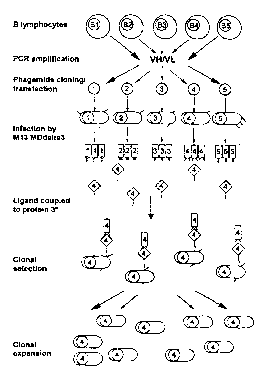
Description of the Fipnlres
Figure I gives a description of the principle for linking ligand iel~og..;~iun to phage
replication by selection of specific phages according to the present invention (* M13
MDdelta3 is a helper phage lacking gene 3, i.e. not itself infectious).
Figure 2 shows the result of an experiment where the specificity and selectivity of the
selection process is ,~ ,, .. ~1. r ~ rd
Workinp FY~mrl~c
PI~Jala~ivll of helper ~h~e stock . M131vlD~
The const~uction of a mutant phage M13MD~3 (devoid of gene 3) was performed by
digestion of the replicative form ~RF) DNA of M13KO7 (Viera, J. and Messing, J. 1987) with
BspHI and Xmnl removing the fragment between residue 1299 and 2646 (according to the
numberjng of Wezenbeek et al. (Wezenbeek, P. M. G. F., Huselbos, T. J. M. and
~..I..,..I"...k,l." G. G. 1980) In order to ~ UI~JU~ a fragment from residue 1299 to 1525
containing gene VIII and part of gene IX, this sequence was PCR amplified from M13 KO7
template using the primers 1299(BspHI): S'-ACTTCCTCATGAAAAAGTC-3' and
1525(Xmnl): S'-GGGAAATTATTCTTATCAGCTTGC-3'. Following digestion, the PCR
fragment was cloned in the 7.3 kb RF DNA originating from M13KO7. Helper phage stocks
were prepared as described (~oog. l,o,."., H. R., Griffiths, A. D., Johnson, K. S., Chiswell,
D. J., Hudson, P. and Winter, G. 1991), except for the use of TGI ~ r~.. ,... ~ with pUCI9
vector, containing gene 3 that produced an intact protein 3 from M13. The result phage thus
had the same proteins as the wild type but did not contain gene 3. This phage was able to
infect a male host cell once but any y~hc~qll~ntly extruded particles were non-infectious.
-
WO951160~7 PCTISl~9~/01166
2t 78~0~ --
Example I
Three different phage stocks, where each stock contain phages which express antibody
fragments specific for ~ ,.,Li~ hen egg Iysozyme (HEL), phenyloxæolon (phox) or
gpl20 0n the human ;."" ,.", :~J~ ric;~ ,~ y virus (LlC), were prepared separately. The three
different phagemids are transfected into XL 1 Blue bacteria and are cultivated with ampicil~in
selection. Thereafter these bacteria are infected with a helper phage, M13MDA3, which does
not itself contain gene3, thereby producing non-infectious phage stocks sincc protein 3 which
mediates infection is not included. The three different phage are prepared by centrifugation
and filtration and are mixed with different amounts of a fusion protein between a truncated
protein3 (only the 98 N-terminal amino acids) and HEL (dp3-HEL), whereupon it isincubated over night. The following day XL I Blue bacteria are infected with these three
phage stocks and Figure 2 shows that only the phages carrying the correct receptor on their
surface, i.e. the antibody fragment specific for HEL has been given the ability to replicate and
multip~y. The linking between ligand recognition and replication has increased the specific
phage titre from a background level of 102 up to more than 108 cfu/ml, which is a sp~cific
increase of more than a mil~ion times. Further,( as appears from the figure 2), the non
specific phages did not replicate at all, but stayed on the background titre of 10~ cfu/ml.
Examp1e ~
Three different phagemids, which express antibody fragments specific for hen egg Iysozyme
(HEL) or phenyloxæolon (phox) or gpl20 on the human imml-nm1~f~ nl~y virus (LTC),
were mixed in the relation 1:1500:1500. This mixture were transfected into -XLI Blue
bacteria and are cultivated with ampicillin selection. Thereafter these bacteria are infected
with a helper phage, M13MI~3, which does not itself contain gene3, thereby producing non-
infectious phages. The phages are prepared by ~, "", r, . ",l " .,, and filtration and are mixed
with 30 weight % of a fusion protein of a truncated protein3 ( only the 98 N-terminal amino
acids) and HEL (dp3-HEL), which is incubated over night. The following day XL I Blue
indicator bacteria are infected with this phage stock and are cultivated over night with
ampicillin selection. A little more than one hundred colonies are selected and are cultivated
further in a 96 hole cultivating plate where they arG infected by the wild type of the helper
phage M13 KO7, which carries the gene3. This results in the production of phages from
every colony, which can be detected using a phage-ELISA. Table I shows that the
c~n~ ntrAti~mfactorinthefirst antigenspecificstepis>lO~tlmesand<~ L~l~/101
times after the second selection step. This happens because the fusion protein (dp3-HEL)
links antigen recognition with specific replication of HEL specific phages, i.e phages
expressing the antibody fragment specific against HEL on their surface.
~ WO 9~116027 2 ~ 7 8 2 C 5 PCI-/SE9.1101166
References
Barbas, C. F., Kang. A. S., Lemer, R. A. & Benkovic, S. J. Proc. Natl. Acad Sci. (USA) 88,
7978 (1991).
Borrebaeck, C. A. K., Malmborg, A. & Ohlin, M. ImmunoL Today 14, 477 (1993)
Huse, W. D., Sastry, L., Iverson, S. A., Kang, S. A., Alting, M. M., Burton, D R, Benkovic,
S. J. & Lemer, R. A. Science 246, 1275 (1989).
Huse, W. D. In Antibody E~ ir.GGI i"~,, A PracticalApproach (Borrebaeck, C. A. K., ed.)
p. 103, W. H. Freeman and Co., New York (1991).
Larrick, J. W., Danielsson, L., Brenner, C. A., Abrahamsson, M., Fry, K. E. & Borrebaeck,
C. A. K. Biochem. Biophys. Res. Commun. 160,1250 (1989).
McCafferty, J., Grlffiths, A. D., Winter, G. & Chiswell, D. J. ~lature 348, 55Z (1990).
Smith, G.P. Science228, 1315 (1985).
Viera, J. and Messing, J. 1987. Production of single strand plasmid DNA. 1987. Meth.
EnzymoL 153:3-1 1
Wezenbeek, P. M. G. F., Huselbos, T. J. M. and ~t~h~ k rl ~, G. G. 1980. Nucleotide
sequence of fil:lnn~-n~ c b~ Gliu~ M13 DNA genome: Cul~ Jll with phage fd. Gene
11:129-148.
H~uy,~ ou~ll, H. R, Griffiths, A. D., Johnson, K. S., Chiswell, D. J., Hudson, P. and wintGer,
G. 1991. Multi-subunit proteins on the surface of fil ~ ntol~e phage: Methodologies for
displaying antibody (Fab) heavy and light chains. NucleicAcid. Res. 19:4133-4137.
WO 95116027 ~ 1 ~ 8 ~ a 5 PCTISE9Jlû1166 ~
Table 1
Clonal mixture Initial ratio Final ratio Enrichment factor
First round of enrichment
pEXrnide HEL/
pEXrnide Phox +
pEXrnide LTC : -
1/3 x 104 82/20 1.2 x 10⁵
1/3 x 105 49/59 2.5 x 10⁵
1/3 x 106 4/104 1.1 x 10⁵
1/3 x 107 0/108
Second round of enrichment
1/3 x 10~ 10315 6.1 x 10⁹
1/3 x 109 55/53 3.1 x 10⁹
1/3 x lol 16192 5.2 x 10⁹
113 x 101l 2/106 5.6 x 10⁹
1/3 x 10l2 0/108