Language selection

Search

Patent 2179085 Summary

Third-party information liability

Some of the information on this Web page has been provided by external sources. The Government of Canada is not responsible for the accuracy, reliability or currency of the information supplied by external sources. Users wishing to rely upon this information should consult directly with the source of the information. Content provided by external sources is not subject to official languages, privacy and accessibility requirements.

Claims and Abstract availability

Any discrepancies in the text and image of the Claims and Abstract are due to differing posting times. Text of the Claims and Abstract are posted:

  • At the time the application is open to public inspection;
  • At the time of issue of the patent (grant).
(12) Patent: (11) CA 2179085
(54) English Title: SURGICAL PINS
(54) French Title: BROCHES CHIRURGICALES
Status: Term Expired - Post Grant Beyond Limit
Bibliographic Data
(51) International Patent Classification (IPC):
  • A61B 17/03 (2006.01)
  • A61B 17/00 (2006.01)
  • A61B 17/064 (2006.01)
(72) Inventors :
  • DAY, RALPH PAUL (United Kingdom)
  • ANDERSON, KENNETH ROBERT (United Kingdom)
(73) Owners :
  • ETHICON, INC.
(71) Applicants :
  • ETHICON, INC. (United States of America)
(74) Agent: NORTON ROSE FULBRIGHT CANADA LLP/S.E.N.C.R.L., S.R.L.
(74) Associate agent:
(45) Issued: 2006-12-05
(22) Filed Date: 1996-06-13
(41) Open to Public Inspection: 1996-12-16
Examination requested: 2003-06-09
Availability of licence: N/A
Dedicated to the Public: N/A
(25) Language of filing: English

Patent Cooperation Treaty (PCT): No

(30) Application Priority Data:
Application No. Country/Territory Date
9512128.1 (United Kingdom) 1995-06-15

Abstracts

English Abstract

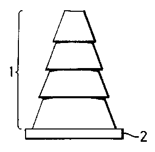
A pin for attaching a surgical membrane barrier to tissue structure of a patient comprises a pin body 1 formed from a sequence of four truncated cones which define a serrated profile which enables the pin to engage the membrane barrier and to be pushed-fitted into the tissue structure. The head portion 2 of the pin serves to engage the membrane barrier and retain it in position on the underlying tissue structure. The pin is made from a resorbable polymer, such as PDS Polydioxanone and is of substantially solid construction throughout. In an alternative embodiment, a circularly cylindrical extension is provided between the largest truncated cone and the pin head. The arrangement serves to maintain a spacing between the membrane barrier and the underlying tissue.


French Abstract

Broche pour attacher une barrière de membrane chirurgicale à une structure de tissu d'un patient comprenant un corps de broche 1 formé d'une séquence de quatre cônes tronqués qui définissent un profil dentelé qui permet à la broche d'enclencher la barrière de membrane et d'être poussé-engagé dans la structure de tissu. La portion de tête 2 de la broche sert à enclencher la barrière de membrane et à la garder en position sur la structure de tissu sous-jacente. La broche est faite d'un polymère résorbable, tel le polydioxanone PDS et d'une construction substantiellement solide. Dans un mode de réalisation alternatif, une extension cylindrique circulaire est prévue entre le cône tronqué le plus grand et la tête de broche. Les arrangements servent à maintenir un espace entre la barrière de membrane et le tissu sous-jacent.

Claims

Note: Claims are shown in the official language in which they were submitted.

-7-
The embodiments of the invention in which an exclusive property or privilege
is
claimed are defined as follows:
1. A pin for attaching a surgical membrane barrier to tissue structure of a
patient,
the pin being formed from a resorbable polymer and being of substantially
solid
construction throughout and having a profile which enables the pin to engage
the
membrane barrier and to be push-fitted into the tissue structure, in
combination with
said surgical membrane barrier for use therewith.
2. A pin as claimed in claim 1, wherein said surgical membrane barrier is
formed
from a resorbable polymer.
3. A pin as claimed in claim 1 or claim 2, wherein the profile is tapered.
4. A pin as claimed in any one of claims 1 to 3, wherein the profile is
serrated.
5. A pin as claimed in any one of claims 1 to 4, wherein the profile is
circumferentially continuous.
6. A pin as claimed in claim 5, wherein the profile is defined by a sequence
of
substantially coaxial truncated cones.
7. A pin as claimed in claim 6, wherein said sequence comprises four truncated
cones.
8. A pin as claimed in claim 6 or claim 7, wherein the radial cross-section of
said
cones is substantially circular.
9. A pin as claimed in any one of claims 6 to 8, wherein the angle of each
cone is
in the range 67.5 degrees to 75 degrees.
10. A pin as claimed in claim 9, wherein said angle is substantially 70.6
degrees.

-8-
11. A pin as claimed in any one of claims 1 to 10, wherein the profile extends
axially over a length of between 2mm and 10mm.
12. A pin as claimed in claim 11, wherein said length is substantially 4.0mm.
13. A pin as claimed in any one of claims 1 to 12, comprising a head and a
spacer
portion between said head and said profile.
14. A pin as claimed in claim 13, wherein said spacer portion has an axial
length of
between 2mm and 5mm.
15. A pin as claimed in claim 14, wherein said axial length of said spacer
portion is
substantially 2.0mm.
16. A pin as claimed in any one of claims 1 to 15 and having a substantially
circular
radial cross-section.
17. A pin as claimed in claim 16 and having a minimum diameter of between
0.5mm and 2.0mm.
18. A pin as claimed in claim 17, and having a minimum diameter of
substantially
0.5mm.
19. A pin as claimed in any one of claims 16 to 18, and having a maximum
diameter of between 2mm and 10mm.
20. A pin as claimed in claim 18 and having a maximum diameter of
substantially
3.0mm.
21. A pin as claimed in any one of claims 1 to 20 and made from a polymer of p-
dioxanone or poly[oxy(1-oxoethylene)oxyethylene].

-9-
22. A pin as claimed in claim 21 and made from PDS Polydioxanone (R.T.M.).
23. A pin as claimed in any one of claims 1 to 20 and made from a copolymer of
glycolide with L(-) lactide.
24. A pin as claimed in claim 23 and made from Vicryl Polyglactin 910
(R.T.M.).
25. A method of manufacturing a pin for attaching a surgical membrane barrier
to
tissue structure of a patient, the pin being of substantially solid
construction throughout
and having a profile which enables the pin to engage the membrane barrier and
to be
push-fitted into the tissue structure, wherein the pin is formed from a
resorbable
material.

Description

Note: Descriptions are shown in the official language in which they were submitted.


2 ~ 19085
SURGICAL PINS
The present invention relates to pins for attaching
a surgical membrane barrier to tissue structure of a
patient.
It is known that, after performing certain surgical
procedures on a patient, it can be desirable to place a
membrane barrier on the site of the operation. Such
membrane barriers exclude cell types such as epithelial
elements, thereby promoting "productive" cell lines with the
aim of encouraging qualitative and quantitative regeneration
of functional tissue.
There are many clinical situations wherein a secondary
means of fixing and retaining the appropriate membrane
barrier to the underlying tissue structure is essential if
the tissue regenerative process is to be encouraged.
It has long been recognised that secondary surgical
interventions undertaken for the sole purpose of removing
non-absorbable "scaffolding" structures (placed at the time
of primary surgery) are undesirable. Apart from the
additional discomfort and inconvenience to the patient, the
secondary intervention can prejudice the quality and speed
of the delicate biological regenerative process initiated
soon after the placement of the membrane barrier.
The current systems available for providing membrane
support are constructed from highly engineered medical grade
titanium alloys. Apart from the disadvantages of requiring
secondary surgery, as mentioned above, such titanium alloy
systems are extremely expensive and require expensive
manipulating instruments, such as drivers, graspers and
surgical mallets. In addition, there is considerable
difficulty with handling and manipulating such systems.
Furthermore, the size constraints of existing designs can
render it difficult to obtain adequate purchase in spongy
cancellous bone.
It has therefore been considered desirable to provide
a tissue tacking system made from a resorbable material,
thereby precluding the need for secondary surgery, and to

217985
2
provide a system which overcomes the other disadvantages of
conventional systems mentioned above.
Bone pins have therefore been made from synthetic
resorbable polymers so as to overcome the need for secondary
surgery. However, such bone pins have relied upon screw
threading for retention, and this presents a major inherent
problem in that they are unable to resist the high torque
forces produced when attempting to fix them into dense bone.
It would therefore be desirable to provide a
resorbable surgical pin which overcomes this disadvantage.
Pins made from synthetic resorbable polymers are
known. These are of hollow construction to allow the pins
to become infiltrated by, incorporated by, and ultimately
resorbed by host tissue. However, this construction
necessarily complicates the manufacturing process and is not
feasible for pins having small dimensions. Furthermore,
such a hollow construction is not necessary for facilitating
the processes of guided tissue and/or bone regeneration.
In accordance with a first aspect of the present
invention there is provided a pin for attaching a surgical
membrane barrier to tissue structure of a patient, the pin
being formed from a resorbable polymer and being of
substantially solid construction throughout and having a
profile which enables the pin to engage the membrane barrier
and to be push-fitted into the tissue structure, in
combination with a said surgical membrane barrier for use
therewith.
In accordance with a second aspect of the present
invention there is provided the use of a resorbable polymer
to manufacture a pin therefrom for attaching a surgical
membrane barrier to tissue structure of a patient, the pin
being of substantially solid construction throughout and
having a profile which enables the pin to engage the
membrane barrier and to be push-fitted into the tissue
structure.
The profile is advantageously provided by a serrated
edge to the pin, such as provided by a sequence of truncated
cones.

2179Q~5
3
A suitable material for the resorbable pin is a
p o 1 y m a r o f p - d i o x a n o n a o r
poly[oxy(1-oxoethylene)oxyethylene), such as PDS
Polydioxanone (R.T.M.), although a copolymer of glycolide
with L (-) lactide, such as Vicryl Polyglactin 910 (R.T.M.)
may alternatively be used. The chemical structures of these
compounds are described in GB 1 540 053 and GB 1 416 196
respectively.
Preferred embodiments of the present invention will
now be described with reference to the accompanying
drawings, wherein:
Figure 1 shows an axial cross-section of a membrane
pin in accordance with the present invention; and
Figure 2 shows an axial cross-section of a membrane
pin incorporating a membrane support, also in accordance
with the present invention.
With reference to Figure 1, the membrane barrier
support pin comprises a pin body 1 of substantially solid
construction throughout in the form of a sequence of
truncated circular cones which are arranged so as to provide
a serrated profile which can frictionally grip the sides of
a pre-drilled hole in underlying body tissue, such as bone.
The angle of each truncated cone is 70.6°. The minimum
diameters of the truncated cones are respectively 0.5mm,
0.7mm, 0.9mm and l.imm, the maximum diameters being
respectively l.2mm, l.4mm, l.6mm and l.8mm. The larger end
of the pin body is provided with a circularly cylindrical
head 2 having a diameter of 3.Omm. This head serves to
secure a resorbable membrane barrier in position on
underlying body tissue.
An alternative embodiment is shown in Figure 2,
wherein, between the largest truncated cone and the head 2
there is provided circularly cylindrical extension having a
length of 2mm and a diameter of l.8mm. This serves to
maintain an adequate spacing beneath the membrane barrier to
allow consolidation and subsequent organisation of the blood
clot, which is believed to be fundamental to the osteogenic
process, since the osteocyte progenitor cells are contained

2 ~ 7905
4
therein. As with the embodiment shown in Figure 1, the head
2 serves to retain the membrane barrier above the underlying
body tissue.
In each case the pin is made from PDS Polydioxanone.
This material advantageously provides residual intrinsic
strength for up to six weeks after implantation.
The pins may be conveniently inserted by compaction
into a pre-drilled lmm diameter channel in underlying
tissue. Such channels are advantageously countersunk and
made using a tungsten carbide tapered fissure bur running at
low speed and continuously irrigated with sterile saline.
Pre-clinical, in-vivo experiments have additionally
shown that membranes made from PDS Polydioxanone serve to
promote osteogenesis, with the quality of the resultant bone
being well-organised and vascularised, in marked contrast to
the poorly-organised woven-type bone which has been observed
following the use of resorbable barrier membranes of
different construction and materials. It is therefore
considered appropriate to manufacture the pin and the
membrane barrier from the same polymeric material.
Membrane barrier pins in accordance with the present
invention can conveniently be manufactured using compression
or injection moulding techniques and can be conveniently
manufactured in the form of a group of four such pins
attached to a small removable stalk of PDS Polydioxanone to
facilitate ease of handling by the clinician.
It will be appreciated that such membrane support pins
have an extremely wide area of potential application, e.g:
1. Ora 1 Surctery
Implantology . a membrane support for guided
regeneration.
Periodontology . a membrane support for guided
bone and tissue regeneration.
2. Maxillo-Facial / Cranio-Facial Surgery
Membrane Support to facilitate guided bone regeneration
for bony defects, such as mandibular cyst cavities;

~ 179035
congenital defects, such as cleft palates;
maxillary sinus augmentation lifting;
orbital floor repairs (severe comminuted fractures).
5 3. Orthopaedic Surctery
Facilitation of guided bone or other tissue regeneration
for many defects, both as a consequence of trauma
surgery and elective surgery, e.g. non-union of
fractures, such as of long bones; regeneration of large
bony defects, such as in post-ablative surgery.
4. ENT Surgery
Membrane support to facilitate nasal septum
reconstruction.
5. General/Plastic Surgery
Means for support of suture/tape slings used for tissue
support, augmentation etc.
Although the present invention has been described with
reference to preferred embodiments, it will be appreciated
that numerous modifications and/or variations are
contemplated by the present invention. For example,
although specific dimensions of the membrane support pin
have been given above, the size can of course be varied to
satisfy the demands of the particular clinical application.
In particular: the angle of each truncated cone could take
any value within the range 67.5° to 75°; the profile could
extend over any length between 2mm and lOmm; the spacer
portion, if such is provided, could have any axial length
within the range 2mm to 5mm; the minimum diameter of the pin
could have any value between 0.5mm and 2.Omm; and the
maximum diameter of the pin could have any value between 2mm
and lOmm.
Although the membrane barrier pins of the present
invention have been described in relation to the attaching
of resorbable membrane barriers, it will be appreciated that
advantages of the invention may still be gained even if a

217905
6
non-resorbable membrane barrier is attached using such pins.
Furthermore, although the pins are preferably made from PDS
Polydioxanone, it will be appreciated that any suitable
resorbable polymer could be used, the choice of material
being dependent on the strength retention profile required
of the particular clinical application.

Representative Drawing
A single figure which represents the drawing illustrating the invention.
Administrative Status

2024-08-01:As part of the Next Generation Patents (NGP) transition, the Canadian Patents Database (CPD) now contains a more detailed Event History, which replicates the Event Log of our new back-office solution.

Please note that "Inactive:" events refers to events no longer in use in our new back-office solution.

For a clearer understanding of the status of the application/patent presented on this page, the site Disclaimer , as well as the definitions for Patent , Event History , Maintenance Fee  and Payment History  should be consulted.

Event History

Description Date
Inactive: Expired (new Act pat) 2016-06-13
Grant by Issuance 2006-12-05
Inactive: Cover page published 2006-12-04
Inactive: Final fee received 2006-09-15
Pre-grant 2006-09-15
Letter Sent 2006-04-13
Notice of Allowance is Issued 2006-04-13
Inactive: IPC removed 2006-04-10
Inactive: IPC from MCD 2006-03-12
Inactive: Approved for allowance (AFA) 2006-01-11
Amendment Received - Voluntary Amendment 2005-12-15
Inactive: S.30(2) Rules - Examiner requisition 2005-06-20
Inactive: Application prosecuted on TS as of Log entry date 2003-07-17
Letter Sent 2003-07-17
Inactive: Status info is complete as of Log entry date 2003-07-17
All Requirements for Examination Determined Compliant 2003-06-09
Request for Examination Requirements Determined Compliant 2003-06-09
Amendment Received - Voluntary Amendment 2003-06-09
Application Published (Open to Public Inspection) 1996-12-16

Abandonment History

There is no abandonment history.

Maintenance Fee

The last payment was received on 2006-06-09

Note : If the full payment has not been received on or before the date indicated, a further fee may be required which may be one of the following

  • the reinstatement fee;
  • the late payment fee; or
  • additional fee to reverse deemed expiry.

Patent fees are adjusted on the 1st of January every year. The amounts above are the current amounts if received by December 31 of the current year.
Please refer to the CIPO Patent Fees web page to see all current fee amounts.

Owners on Record

Note: Records showing the ownership history in alphabetical order.

Current Owners on Record
ETHICON, INC.
Past Owners on Record
KENNETH ROBERT ANDERSON
RALPH PAUL DAY
Past Owners that do not appear in the "Owners on Record" listing will appear in other documentation within the application.
Documents

To view selected files, please enter reCAPTCHA code :


To view images, click a link in the Document Description column (Temporarily unavailable). To download the documents, select one or more checkboxes in the first column and then click the "Download Selected in PDF format (Zip Archive)" or the "Download Selected as Single PDF" button.

List of published and non-published patent-specific documents on the CPD .

If you have any difficulty accessing content, you can call the Client Service Centre at 1-866-997-1936 or send them an e-mail at CIPO Client Service Centre.

({010=All Documents, 020=As Filed, 030=As Open to Public Inspection, 040=At Issuance, 050=Examination, 060=Incoming Correspondence, 070=Miscellaneous, 080=Outgoing Correspondence, 090=Payment})


Document
Description 
Date
(yyyy-mm-dd) 
Number of pages   Size of Image (KB) 
Representative drawing 1998-03-08 1 2
Description 1996-06-12 6 264
Abstract 1996-06-12 1 22
Claims 1996-06-12 3 82
Drawings 1996-06-12 1 9
Representative drawing 2005-05-17 1 2
Claims 2005-12-14 3 74
Reminder of maintenance fee due 1998-02-15 1 111
Reminder - Request for Examination 2003-02-16 1 112
Acknowledgement of Request for Examination 2003-07-16 1 173
Commissioner's Notice - Application Found Allowable 2006-04-12 1 162
Correspondence 2006-09-14 1 38