Note: Descriptions are shown in the official language in which they were submitted.
~ 1 7q66s
Page 2 of 11
Shawn Cohen, M D. and John Chen, M.D.
June 21, 1996
Title:
Angled Hollow Cannula with Mobile Fixation Flange
Field of the Invention:
The present invention relates to an apparatus to deliver an
instrument or material or energy source into a closed chamber; and one
possible application thereof in the treatment of living organisms, more
specifically, the human eye.
Background to the Invention:
The human eye is a fluid-filled globular structure that converts an
optical image into neural signals for cortical processing and
interpretation. The transparent fluid media of the eye is divided by the
lens into an anterior chamber and the vitreous cavity. The vitreous is a
gel-like substance made up of collagen fibrils in a stroma of highly
hydrophillic substances called glycosaminoglycans (GAG). The GAG
component absorbs water avidly, causing the vitreous to swell, and this
provides the vitreous with its shock-absorbing or resistive effect to
compression. The vitreous is loosely attached around its periphery at the
front of the eye and more firmly at the margin of the optic nerve head, as
it enters the back of the eye. In the vitreous, there are floating cells
which have the capacity to form scar tissue. Vitreous liquefaction and
2l19665
Page 3 of 11
Shawn Cohen, M.D. and John Chen, M.D.
June 21, 1996
degradation with increasing age, a process called syneresis, or trauma
causes it to collapse in on itself. The vitreous may then pull on its
adhesions to the retina, the light-sensitive part of the eye, to cause a
detachment and resultant loss of vision. In other instances, such as
diabetes, new blood vessels that proliferate in response to a lack of
proper circulation, do so using the vitreous surface as a scaffold on which
to grow. Vitreous traction, as a result of syneresis or trauma, may then
result in shearing of these friable vessels. Hemorrhage into the vitreous
induces a significant loss of vision. Organization of this vitreous blood
into a scar may induce a retinal detachment as the scar condenses and
pulls on the retina. In order for the retina to return to its original
flattened position, the vitreous and its scar tissue must carefully be
removed in a process called a vitrectomy.
Access to the vitreous, for the purpose of a vitrectomy, is usually
through the anterior part of the eye so that the extremely delicate and
tissue-thin retina, which lines the back of the eye, is avoided. The current
standard of care is to employ the "pars plana" approach. In this approach,
as opposed to previous approaches in which very large incisions are
employed, three tiny (0.9-1.0 mm) incisions are created, one for
intraocular infusion, one for the fiber optic light probe and one for the
cutting and suction probe. Accessory instruments can be used in place of
the cutting and suction probe, eg. forceps or a laser source. The infusion
cannula reinflates and maintains the eye structure after the intraocular
incisions are made and also serves to tamponade and flatten the retina.
21 7q665
Page 4 of 11
Shawn Cohen, M.D. and John Chen, M.D.
June 21, 1996
Currently, infusion cannulae are straight-tipped cannulae and are
inserted into pars plana incision ports. The position of these cannulae is
not stable and thus they need to be sutured in place to secure their
positions during surgery. Recently, a new technique of pars plana incision
ports was invented (to be published in 2 months time) in which these
incisions are self-sealing and do not require closure with a suture at the
end of the procedure. However, with these new self-sealing incisions, the
older, standard, straight infusion cannulae are not suitable because of
poor positioning and instability. Also, the need to suture these cannulae in
place defeats the purpose of the newer incision technique (sutureless
surgery).
2 1 19665
Page 5 of 11
Shawn Cohen, M.D. and John Chen, M.D.
June 21, 1996
Objects and Statement of the Invention:
It is therefore an object of the present invention to provide an
apparatus for the delivery of an instrument or material or energy source
into a closed chamber, which overcomes some or all of the aforementioned
problems.
As embodied and broadly described herein, the invention provides an
apparatus for the delivery of an instrument or material or energy source
into a closed chamber, said apparatus comprising:
- a luer-lock or other system or adaptor for attachment to an
input source (eg. infusion line);
- a flexible tubing system through which substrates or energy
sources or instruments may pass;
- an angled rigid tip securely attached to the tube and placed on
the opposite end of the tube to the luer-lock or source
attach ment;
- a flexible flange with "memory" or an inherent property to
return to a prespecified orientation when stresses placed upon
it are removed, said flange being capable of riding freely on
the angled rigid tip;
- the above being either constructed entirely from materials
capable of being sterilized in high temperatures for reuse or
constructed with the intent to be discarded after each use.
~ l ~q66~
Page 6 of 11
Shawn Cohen, M.D. and John Chen, M.D.
June 21, 1996
In a preferred embodiment, an incision is made in the sclera 5.0 to
5.5 mm away from the limbus. A tunnel is made from this point within the
sclera (the fibrous outer shell of the eye) parallel to the eye surface for
2.0 mm and then a trocar is used to penetrate the globe. The tip of the
apparatus, with its flange retracted away from the wound, is introduced
under the scleral flap and is directed parallel to the eye surface towards
the puncture site. Next, the tip is rotated into position so that the
intraocular portion is perpendicular to the surface of the eye and the
extraocular portion is parallel to the surface of the eye. The flange is then
advanced over the cannula and either sutured in place or slipped under the
flap of sclera. In the latter scenario, the cannula is secured naturally in
place as the memory in the flange returns the wings to a outstretched
position, fixating it and the cannula tip in place. Through this cannula, the
surgeon may feed a light source through the tube and into the eye and/or
an infusion source of a balanced salt solution. In this way, it may be
possible to merge the light and infusion sources into a single cannula.
In a variant, the present invention may be used, in a larger
dimensional scale, in concert with other endoscopic surgical systems,
such as a system used for intraabdominal surgery.
The present invention offers several advantages over other
apparatuses known in the art. Th0 problem of stability of the intraocular
cannula is now significantly reduced since the cannula is fixed into place
by the flexible flange and by nature of its entraocular component being
2 1 7~6~5
Page 7 of 11
Shawn Cohen, M.D. and John Chen, M.D.
June 21, 1996
parallel to rather than perpendicular to the ocular surface. The risk of
damage to intraocular structures is significantly reduced as the
intraocular portion of the probe is perpendicular to the ocular surface
instead of following its natural tendency to point forwards towards the
crystalline lens of the eye.
Brief Description of the Drawings:
The following is a description by way of preferred embodiment,
reference being made to the following drawings, in which:
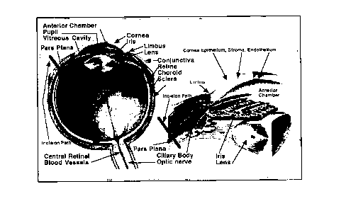
FIG. 1 is a cross-sectional schematic of the human eye;
FIG. 2 is a photograph of the invention in its resting mode;
FIG. 3 is a scale diagrammatic representation of the invention with
specific characteristics of some of the unit's parts.
2~7q665
Page 8 of 11
Shawn Cohen, M.D. and John Chen, M.D.
June 21, 1996
Description of a Preferred Embodiment:
For background reference, figure 1 provides a basic cross-sectional
view of the human eye illustrating various components thereof. The outer
shell of the eye consists anteriorly of a transparent cornea and more
posteriorly of a three-layered wall. This wall, from outside to in, includes
the fibrous sclera, the nourishing and blood-rich choroid layer and the
light-sensitive retina that lines the inner aspect of the eye. Between the
anterior border of the retina and the ciliary body is an area of relatively
safe entry into the eye and is called the pars plana. The pars plana
corresponds to the incision site for the placement of a vitrectomy cannula
in a pars-plana vitrectomy procedure.
Referring now to figure 2, there is provided a means of connecting
the apparatus to the input source, eg. an infusion source of balanced salt
solution. While the preferred embodiment is described using a luer-lock
system, for example there may be provided an adaptor that allows the
apparatus to receive a fiber optic light source or some other input.
Referring now to figures 2 and 3, the tubing diameter, length and
material used in the preferred embodiment includes but should not be
limited to tubing used for a standard 1 9-gauge Surflo winged infusion set
(P-216-1 1 9).
The preferred embodiment includes but is not limited to a 1 9-gauge
217q6~5
Page 9 of 11
Shawn Cohen, M.D. and John Chen, M.D.
June 21, 1996
metal hollow-core cannula. Said cannula may be constructed with either a
flattened end as shown or a bevelled end that may allow the outflow from
the cannula to be directed away from the bevelled surface, rather than in
line with or parallel to the intraocular cannula axis.
Said cannula is attached to said tubing by means of heat shrinkable
tubing or suitable heat-resistant and, preferably waterproof, glue.
The preferred embodiment includes but should not be limited to an
angled tip of 90~ and should not be limited to the dimensions specified. a
variant includes an angled tip of adjustable angle, analogous to the
bending angle on some soda straws or by some other technique created by
one skilled in the art. A variant includes a length of 5 mm for the
extraocular portion and 3.5 mm for the angled intraocular portion.
Disposed along the cannula tip is a winged flange that may be glided
freely over the cannula. A variant of the flexible flange dimensions and
material used in the preferred embodiment includes but should not be
limited to 3.2 x 2.5 mm. The wings of the flange may contain one or more
alternative fixation points which may include suture or button holes.
The preferred embodiment also includes a variant in which the
cross-sectional geometric shape of the angled tip of the cannula may be
varied to produce, for example, an ellipse. Such a shape would have the
advantage of producing a flatter surface and thus less elevation and
217qS6~
Page 10 of 11
Shawn Cohen, M.D. and John Chen, M.D.
June 21, 1996
distortion of the scleral tunnel into the eye.
The above description of a preferred embodiment should not be
interpreted in any limiting manner since variations and refinements are
possible which are within the spirit and scope of the present invention. It
should be understood that cannulae of various constructions known to
those skilled in the art which provide for the same or similar function as
the invention could also be included within the scope of the invention.
