Note: Descriptions are shown in the official language in which they were submitted.
Li'O 95/t9.t30 ~ ~ ~ ~ ~ j ~ PCT/US95100.l00
MACROENCAPS'ULATED SECRETORY CELLS
Field of the Invention
The present invention relates to macroencapsulation of secretory cells in a
hydrophilic gel material, therapeutic methods employing the macroencapsulated
secretory
cells, and preserving the secretory cells by macroencapsulation.
Background of the Invention
Secretory cells are cells that are characterized by secreting biological
products,
such as, but not limited to, hormones (e.g., insulin), growth factors,
cytokines, and so
forth. Their role in biological processes is well known, and need not be set
forth here.
A number of diseases and pathological conditions are related to a failure of
the secretory
cells to work properly, such as a deficient production of the secretory
products, e.g.
hypothyroidism and cretin dwarfism, hoth due to thyroid hormone deficiency,
hypophysial
dwarfism due to pituitary growth hormone deficiency, Lesch-Hyhan Syndrome due
to
hypoxanthine-guanine phosphoribosyltransferase deficiency, fulminant hepatic
failure due
to the hepatotrophic factor deficiency, extracellular matrix disease due to
chondrocyte
deficiency, and insulin dependant diahetes due to insulin deficiency.
One approach to treat such conditions is to transplant the secretory cells
into the
patient. The transplanted material, in order to be clinically safe and
effective, must (I)
be non-immunogenic, non-thrombogenic, bio-stable, and completely non-toxic to
cells and
tissues of the host, (2) maintain cell viability for an extended period of
time, (3) permit
free passage of nutrients, secretagogies (a substance that stimulates
secretion), and cell
products, (4) facilitate surgical implantation and cell reseeding, and (5) be
easily fixed in
place and, likewise, removed.
Pancreatic islet transplantation to treat insulin-dependant diabetes has been
the
subject of renewed interest due to technological advances in isolating islets
of Langerhans.
By way of background, the human pancreas contains islets of Langerhans
(hereinafter
. "pancreatic islets") that are scattered throughout the exocrine pancreas
with some
concentrations near the pancreatic ducts. The pancreatic islets, taken
together, can be
" 30 thought of as a single endocrine organ occupying around 1 % of the volume
of the
pancreas. Within the pancreas, small islets (up to 160 tem diameter) tend to
distribute
throughout the exocrine tissue. These small islets represent 75% of the islets
in number
wo ssit9aso 2 ~ ~ ~ ~. ~ ( rCT~s9siooaoo
2
but only about 15% in volume. Islets greater than 250Pm diameter constitute
only 15%
of the total number of islets but 60% of the volume. These islets are
localized near larger
ducts and blood vessels, and are not surrounded by acinar tissue. A human
pancreas may a
contain over I million islets, and each islet typically consists of several
thousand cells.
Each islet is comprised of a central core of insulin producing beta cells (B-
cells) and a
surrounding mantle of glucagon containing alpha cells (A-cells), somatostatin
secreting
delta cells (D-cells) and pancreatic polypeptide containing cells (PP-cells).
Insulin
producing B-cells makeup the majority of the cells, and comprise up to about
80% of the
islets in a human.
The clinical applications of pancreatic islet transplantation have been
limited by
the inability to prevent islet allograft-xenograft rejection, i.e., a
rejection of the
transplanted pancreatic islets due to the host's immune system attacking the
transplanted
pancreatic islets. To counteract the rejection, the pancreatic islets have
been transplanted
in combination with the administration of immunosuppressive agents.
Immunosuppressive therapy, however, has proved to be a double-edged sword;
while reducing the risk of rejection, it impairs the body's overall
immunological defenses.
Various methods of protecting the transplanted tissue from the host immune
response have
been explored by many investigators. As discussed below, although temporary
success
has been reported (See Lacy, Diabetes Reviews 1 (1):76 (1993), effective long-
term
methods have yet to he achieved.
The five major approaches to protecting the transplanted tissue from the
host's
immune response all involve attempts to isolate the transplanted tissue from
the host's
immune system. The immunoisolation techniques used to date include:
extravascular
diffusion chambers, intravascular diffusion chambers, intravascular
ultrafiltration
chambers, microencapsulation, and macroencapsulation. All of these methods
have
failed, however, due to one or more of the following problems; a host fibrotic
response
to the implant material, instability of the implant material, limited nutrient
diffusion
across semi-permeable membranes, secretagogue and product permeability, and
diffusion
lag-time across semi-permeable membrane barriers.
For example, a microencapsulation procedure for enclosing viable cells,
tissues, "
and other labile biological membranes within a semipermeable membrane was
developed
by Lim in 1978. (Lim, Research report to Damon Corporation (1978)). Lim used
WO 95/19430 2 ~ g ~ ~ 5 6 PCTNS95/00400
3
microcapsules of alginate and poly L-lysine to encapsulate the islets of
Langerhans. In
1980, the first successful in vivo application of this novel technique in
diahetes research
was reported ((Lim, et al., Science 210:908 (1980)). The implantation of these
microencapsulated islets of Langerhans resulted in sustaining a euglycemic
state in
- 5 diabetic animals. Other investigators, however, repeating these
experiments, found the
alginate to cause a tissue reaction a.nd were unable to reproduce Lim et al's
results
(Lamherti, et al., Applied Biochemistry and Biotechnology 10:101 (1984);
Dupuy, et al.,
Jour. Biomed. Material and Res. 22:1061 (1988); Weber, et al., Transplantation
49:396
(1990j; and Soon-Shiong, et al., Transplantation Proceedings 22:754 (1990)).
The water
solubility of these polymers is now considered to be responsible for the
limited stability
and biocompatibility of these microcapsules in vivo ((Dupuy, et al. supra,
Weber, et al.
supra, Soon-Shiong, et al., supra, and Smidsrod, Faraday Discussion of
Chemical
Society 57:263 (1974)).
Recently, Iwata et al., (Iwata, et al. Jour. Biomedical Material and Res.
26:967
(1992)) utilized agarose for microencapsulation of allogeneic pancreatic
islets and
discovered that it could he used as a imedium for the preparation of
microbeads. In their
study, 1500-2000 islets were micioencapsulated individually in 5°f
agarose and implanted
into streptozotocin-induced diabetic mice. The graft survived for a long
period of time,
and the recipients maintained normoglycemia indefinitely.
Their method, however, suffers from a number of drawbacks. It is cumbersome
and inaccurate. For example, many beads remain partially coated and several
hundred
beads of empty agarose form. Addiitional time is thus required to separate
encapsulated
islets from empty beads. Moreover-, most of the implanted microbeads gather in
the
pelvic cavity, and a large number of islets are required in completely coated
individual
beads to achieve normoglycemia. Furthermore, the transplanted beads are
difficult to
retrieve, tend to be fragile, and will easily release islets upon slight
damage.
A macroencapsulation procedure has also been tested. Macrocapsules of various
' different materials, such as poly-2-hydroxyethyl-methacrylate, poly
vinylchloride-co-acrylic acid, and ca:llulose acetate were made for the
immunoisolation
of islets of Langerhans. (See Altman, et al., Diabetes 35:625 (1986); Altman,
et al.,
Transplantation American Society ofArtificial Internal Organs 30:382 (1984);
Ronel, et
al., Jour. Biomedical Material Research 17:855 (1983); Klomp, et al., Jour.
Biomedical
WO 95/19430 ~ ~ g ~ 15 6 PCT1US95100.100
4
Material Research 17:865-871 (1983)). 1n all these studies, only a transitory
normalization of glycemia was achieved.
Archer et al., Journal of Surgical Research, 28:77 (1980), used acrylic ,
copolymer hollow fiber to temporarily prevent rejection of islet xenografts.
They
reported long-term survival of dispersed neonatal murine pancreatic grafts in
hollow fibers
which were transplanted into diabetic hamsters. Recently Lacy et al., Science
254:1782-
1784 (1991) confirmed their results, but found the euglycemic state to be a
transient
phase. They found that when the islets are injected into the fiber, they
aggregate within
the hollow tube and result in necrosis in the central portion of the islet
masses. The
central necrosis precluded prolongation of the graft. To solve this problem,
they used
alginate to disperse the islets in the fiber. Using this method they were able
to achieve
long-term graft survival. However, this experiment has not been extensively
repeated.
Therefore, the membrane's function as an islet transplantation medium in
humans is
questionable.
Thus, there exists a need for achieving secretory cell transplantation, and in
particular, pancreatic islet allograft and xenograft survival without the use
of chronic
immunosuppressive agents.
The inventors have surprisingly discovered that macroencapsulating secretory
cells
in a hydrophilic gel material results in a functional, non-immunogenic,
macrobead that
can be transplanted into animals and can be stored for long lengths of time.
The
macroencapsulation of the secretory cells of the present invention provides a
more
effective and manageable technique for secretory cell transplantation. The
macroencapulation technique can also be used to macroencapsulate other
biological agents,
such as enzymes, microorganisms, trophic agents including recombinantly
produced
trophic agents, cytotoxic agents, and chemotherapeutic agents. The
macroencapsulated
biological agents can be administered to treat conditions known to respond to
the
biological agent.
CA 02181156 2001-05-18
Summary of the Invention
In accordance with one aspect of the invention there is provided a method of
producing an agarose coated, agarose-collagen secretory cell bead comprising;
(a)
suspending secretory cells in a solution containing collagen, (b) adding
agarose to
said suspended secretory cells of step (a) to form secretory cells suspended
in a
mixture of agarose and collagen, (c) forming a collagen-agarose semisolid bead
from
said suspended secretory cells of step (b), (d) treating said collagen-agarose
semisolid
bead of step (c) to polymerize collagen contained in said semisolid bead,
whereby a
solid collagen-agarose bead is formed, (e) coating said solid bead of step (d)
with
agarose to obtain an agarose coated, agarose-collagen secretory cell bead.
In accordance with another aspect of the invention there is provided a method
of producing an agarose coated, gelfoam secretory cell bead comprising; (a)
suspending secretory cells on gelfoam, (b) rolling said gelfoam containing
said
suspended secretory cells into a sphere, (c) coating said sphere with agarose
to obtain
an agarose coated, gelfoam secretory cell bead.
In accordance with another aspect of the invention there is provided a method
of producing an agarose coated, agarose secretory cell bead comprising; (a)
suspending secretory cells in agarose, (b) forming a bead from said suspended
secretory cells of step (a), (c) incubating said bead of step (b) in
humidified air, (d)
coating said bead of step (c) with agarose to form an agarose coated, agarose
secretory cell bead.
In accordance with still another aspect of the invention there is provided an
agarose coated, agarose-collagen secretory cell bead.
In accordance with yet another aspect of the invention there is provided
secretory cell beads selected from the group consisting of agarose coated,
agarose-
collagen secretory cell beads; agarose coated, gelfoam secretory cell beads;
and
agarose coated, agarose secretory cell beads for use in treating a patient
having a
condition caused by an impaired functioning of secretory cells.
CA 02181156 2001-05-18
Sa
In accordance with still another aspect of the invention there is
provided use of secretory cell beads selected from the group consisting of
agarose
coated, agarose-collagen secretory cell beads; agarose coated, gelfoam
secretory cell
beads; and agarose coated, agarose secretory cell beads, in the manufacture of
a
biological agent for the treatment of a patient having a condition caused by
an
impaired functioning of secretory cells.
In accordance with a further aspect of the invention there is provided a
method
for preserving secretory cells, comprising: (a) forming beads selected from
the group
consisting of agarose coated, agarose-collagen secretory cell beads; agarose
coated,
gelfoam secretory cell beads; and agarose coated, agarose secretary cell
beads; and (b)
incubating said secretory cell beads.
In a particular embodiment the present invention seeks to provide a secretory
cell macrobead that can be transplanted into animals to treat conditions
caused by an
impaired functioning of the host's secretory cells.
In a further particular embodiment this invention seeks to provide a secretory
cell macrobead that can be stored for long lengths of time.
In accomplishing these and other particular embodiments, there has been
provided, in accordance with one aspect of the present invention a method of
producing an agarose coated, agarose-collagen secretory cell macrobead; an
agarose
coated, gelfoam secretory cell macrobead; and an agarose coated, agarose
secretory
cell macrobead.
w CA 02181156 2001-05-18
Sb
In another aspect of the invention, there is provided a method of treating a
patient having a condition characterized by an insufficiency in a secretory
cell
product, comprising transplanting into said patient a therapeutically
effective amount
of secretory cell macrobeads selected from the group consisting of agarose
coated,
agarose-collagen secretory cell macrobeads; agarose coated, gelfoam secretory
cell
macrobeads; and agarose coated, agarose secretory cell macrobeads.
In yet a further aspect of the invention, there is provided a method for
preserving secretory cells, comprising forming macrobeads selected from the
group
consisting of agarose coated, agarose-collagen secretory cell macrobeads;
agarose
coated, gelfoam secretory cell macrobeads; and agarose coated, agarose
secretory cell
macrobeads; and incubating said secretory cell macrobeads.
Other objects, features and advantages of the present invention will become
apparent from the following detailed description. It should be understood,
however,
that the detailed description and the specific examples, while indicating
preferred
embodiments of the invention, are given by way of illustration only, since
various
changes and modifications within the spirit and scope of the invention will
become
apparent to those skilled in the art from this detailed description.
Brief Description of the Drawings
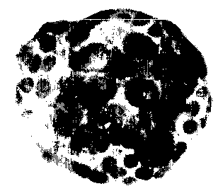
Figures 1 and 2 show agarose coated, collagen-agarose pancreatic islet
macrobeads.
W095119430 ~ ~ ~ ~ ~ J~ PCTlU595/00400
C
6
Figure 3 shows the glucose levels,of diabetic mice transplanted with agarose
coated, collagen-agarose pancreatic islet macrobeads.
Figure 4 shows the glucose levels of diabetic mice transplanted with agarose
coated, gelfoam pancreatic islet macrobeads.
Figure 5 shows the glucose levels of diabetic mice transplanted with agarose
coated, agarose macrobeads.
Figure 6 shows the glucose levels of diabetic mice transplanted with free
pancreatic islets.
Figure 7 shows the glucose levels of diabetic mice transplanted with agarose
coated, collagen-agarose mac>~obeads and free pancreatic islets.
Figure 8 demonstrates a glucose tolerance test for normal mice, streptozotocin-
induced diabetic mice, streptozotocin-induced diabetic mice receiving
allotransplant in
the kidney capsule "(KCT mice)", and streptozotocin-induced diabetic mice
receiving
agarose coated, agarose-collagen pancreatic islet macrobeads; agarose coated,
gelfoam
pancreatic islet macrobeads; and agarose coated, agarose pancreatic islet
macrobeads
(collectively referred to as "head mice").
Detailed Description of the Invention
The present invention relates to macroencapsulation of biological agents, and
preferably, secretory cells in a hydrophilic gel material, therapeutic methods
employing
the macroencapsulated biological agents, and preferably, secretory cells, and
preserving
the biological agents, preferably secretory cells by macroencapsulation. The
hydrophilic
gel material comprises agarose, and combinations of collagen-agarose and
gelatin
sponge-agarose. Gelatin sponge will hereinafter be referred to as gelfoam.
The term biological agent denotes a living organism and its products, e.g.
proteins, enzymes, hormones, polypeptides, serum, antibodies, and antibiotics
and also
genetically engineered cells. Biological agents include enzymes, e.g., glucose
oxidase,
lactase complex, microorganisms, e.g., Klebsiella aerogenes for removal of
ammonia and
urea, trophic agents, including recombinantly produced trophic agents, e.g,
recombinantly
produced growth hormone, and cytotoxic agents.
The term secretory cell includes a pancreatic islet, aithough technically, a
pancreatic islet is not a secretory cell, but mostly a cluster of secretory
cells scattered
WO 95/I9430 PCT/US95/00400
7
throughout the pancreas and comprising its endocrine portion. In humans, they
are
composed of at least four different types of secretory cells: alpha cells
which secrete the
hyperglycemic factor, glucagon; beta cells which are the most abundant (70% -
80%)
and secrete insulin; delta cells which secrete somatostatin, and polypeptide
cells which
secrete polypeptide hormone.
As explained previously, transplanted material must be compatible with the
host.
Agarose has a long history of use in biological research, and its quality is
well-controlted.
Collagen is the most abundant protein in mammals, provides firm mechanical
support and
serves as the hiological space for cell replication, differentiation,
organogenesis,
individual growth and wound repair. Collagen also has good biocompatibility.
Gelfoam
is non-immunogenic and has been used extensively in surgical procedures. It is
also
well-tolerated by secretory cells.
The biological agents, and preferably, secretory cells, are first isolated
using
procedures well known in the art. In. a preferred embodiment, pancreatic
islets are
cultured at either 4°C, 24°C, or at 37°C before they are
macroencapsulated. This method
allows one to select only surviving islets after the isolation trauma. Also,
the islets
become less immunogenic resulting in the protection of macrobeads form
fibrosis.
In one embodiment of the invention, a biological agent, preferably pancreatic
islets, and more preferably about 50,000-700,000 pancreatic islets, are
suspended in an
aqueous solution of collagen, preferably about 0.5%-2~ atellocollagen
solution.
Atellocollagen is obtained by treating collagen with pepsin, which removes
antigenic
telopeptides, responsible for intermolecular cross linkage of collagen. About
0.5%-5%
of agarose, preferably about 1 % , is then added to the suspended pancreatic
islets to form
pancreatic islets suspended in a mixture .of collagen and agarose. The mixture
containing
the pancreatic islets is then transformed into a semisolid bead using
techniques well
known in the art, preferably by dropping the mixture onto mineral oil or a
Teflon~ sheet.
The semisolid bead is then transferred to an antibiotic medium, washed, and
then
incubated under standard conditions to polymerize the collagen, preferably at
37°C in a
humidified 5% CO, atmosphere, whereby a solid collagen-agarose macrobead is
formed.
In another embodiment of the invention, a biological agent, preferably
pancreatic
islets, and more preferably about 50,000-700,000 pancreatic islets, are spread
onto the
' CA 02181156 2000-12-21
g
surface (3-5 cm) of a gelatin sponge. The gelatin sponge is then rolled into a
sphere.
Agarose, 3%-5%, is poured onto the sphere to form a head.
In yet another embodiment of the invention, a biological agents, preferably
pancreatic islets, and more preferably about 50,000-700,000 pancreatic islets,
are placed in
an agarose solution ranging from about 0.5%-S% agarose, preferably about 1%
agarose:
The mixture is then transformed into a macrobead by contacting the mixture to
mineral oil
or Teflon. The bead is then transferred to an antibiotic medium, washed, and
incubated
overnight, preferably at 37°C in a humidified 5% C02 atmosphere.
In all the aforementioned embodiments, the macrobeads are uniformly coated
with
agarose, preferably by rolling the bead 3-4 times in a Teflon spoon containing
about 500-
2,000 ul of 5% -10% agarose. Similarly, the term biological agent macrobeads,
as used
herein, denotes macroencapsulated biological agents in the form of a bead.
The macrobeads may be used as a vehicle to deliver the biological agent to the
body
where the agent will perform its known function. More than one type of
biological agent
may be encapsulated in one bead. For example, a macrobead can contain multiple
enzymes,
such as hemoglobin and glucose oxidase. Such a bead can be administered to
remove
bilirubin. These beads can be used either for oral administration of digestive
enzymes
(lactase complex) or for selective removal of undesirable amino acids from the
body.
Encapsulation of the enzymes will also prevent the degradation of the enzyme
in the
lumen. Furthermore, recombinant gene products can be safely delivered using
encapsulation as the medium. K. aero~enes gene, for example, can be
macroencapsulated
in macrobeads for urea and ammonia removal. Where the biological agent is
immunogenic
to the host, the macrobead allows the administration of the biological agent
without the use
of immunosuppressant or with decreased amounts of immunosuppressant.
The secretory macrobeads may be used to treat conditions caused by an impaired
functioning of the secretory cells of the subject, e.g. insulin dependant
diabetes, growth
factor deficiency disorder, and hormonal disorders, by transplanting the
secretory cell
macrobeads into the subject. The macrobeads may be inserted into the
appropriate location
for that particular treatment. For example, macrobeads containing hepatocytes
can be
implanted into the abdominal cavity to treat diseases related to liver non-
function. A
preferred application is transplanting 5-10 pancreatic islet macrobeads, each
containing
WO 95/19430 ~ ? 81 15 6 . PCTlUS95100400
9
50,000-700,000 pancreatic islets, into a patient to treat insulin-dependant
diabetes. The
macroheads can he inserted into the peritoneal cavity.
The secretory cell macroheads are transplanted into a patient in an amount
sufficient to treat the condition. An amount adequate to accomplish this is
defined as a
~ S "therapeutically effective amount" or "efficacious amount". Amounts
effective for this
use will depend upon the severity of the condition, the general state of the
patient, the
route of administration, the placement of macrobeads, and whether the
secretory cell
macroheads are being administered in combination with other drugs.
The secretory macrobeads can he used for allogeneic and xenogeneic
transplantation in combination with immunosuppressants or preferably, without
immunosuppressants. In a preferred emh~odiment, patients having chronic or
acute insulin
dependant diabetes are treated by xenotransplantating animal pancreatic
islets, e.g.
porcine, bovine, marine, rat, picin, or a:ny other suitable species into the
patient without
the use of immunosuppressants. The secretory cell macrobeads can also be
administered
1S in combination with other therapeutic agt;nts, e.g. the commonly used
triple drug therapy
(cyclosporine, azathioprine, and hydrocortisone), rapamycin, deoxyspergualin
or
antibodies, to treat the condition.
The macroheads can also he used as a means to store the biological agents, and
preferably secretory cells, for extended periods of time. To maintain the
viability of the
biological agents, and preferably secretary cells, the biological agents, and
preferably
secretary cell macrobeads are incubated until they are transplanted in the
animal.
When the secretary cells are pancreatic islets, the pancreatic islet
macrobeads are
incubated at a temperature of 24°C or 37°C.
2S EXAMPLES
Example I
Pancreatic islets were isolated from rats by a modification of the method
disclosed
in Gotoh et al., Transplantation 40:437 1;1985).
Collagenase solution (collagenase Type XI, Sigma Chemical, St. Louis, MO;
lmg/ml containing 2mglml of Sigma, Type V, bovine serum albumin and lmg/ml
CaClz)
was injected into the pancreas via the common bile duct. (Gotoh et al.,
Transplantation
W 0 95119430 PCTIUS95100.100
40:437 (1985), Supra). The pancreas was removed and collected in a flask
maintained on
ice. Once pancreata from 4 rats had been collected, the flask was placed in a
waterhath,
at 38°C, for 30 minutes. The resulting digested tissue was washed 4
times in cold (8°C)
HBSS (Hank's Balanced Salts Solution).
5 Undigested tissue, large lymph nodes, and other extraneous material were
removed
by repeated mobilization of the tissue, followed by removal of the
supernatant. Purified
islets were isolated on a discontinuous Ficoll gradient, consisting of 25%, 23
% , 21 %, and
11 % Ficoll layers, prepared in Euro-Collins solution (Frescenius AG, Gluchen
Steinweg,
Homburg V.D.H.) and centrifuged at 2000 r.p.m. for 16 minutes. The islets were
10 collected from the interface between 1 I % and 21 % and the interface
between 21 % and
2396 Ficoll layers. Islets from each fraction were pooled and washed four
times in HBSS
solution containing 10% fetal calf serum.
The pooled islets cells were then transferred to petri dishes containing RPMI
complete medium, i.e., cold RPMI 1640 medium (GIBCO, Grand Island, NY),
supplemented with 25 tnM HEPES, heat-inactivated fetal bovine serum (10%), and
antibiotic-antimycotic solution (lmlll00m1) which contains: 100ug/ml of
penicillin,
100ug/ml of streptomycin sulfate, and 25ug1m1 of amphotericin B. Any remaining
non-islet acinar, vascular, ductular, or lymph node tissue was identified with
the aid of
a dissecting microscope, and carefully removed with a fine-tip sterile
pipette. Final purity
was assessed by staining the islet preparation with diphenylthiocarhazone.
After isolation, the islets were incubated in bacteriological plastic dishes (
100 mm)
containing lOml of RPMI medium, at 37°C, in a humidified atmosphere
having 5% CO,;
for 4 days. The medium was changed every day, and the islets were then either
directly
transplanted or macroencapsulated.
Example II
A. Preparation of Agarose Coated,
aoarnce Collagen Pancreatic islet Macrobeads _ _
1000 pancreatic islets obtained by the method of Example I were washed four
times in RPMI complete medium as described in Example I, less fetal calf
serum. The
pancreatic islets were then added to a tube containing SOIeI of 1 %
atelocollagen solution
in phosphate buffered saline, to suspend the pancreatic islets. 100u1 of 1 %
low viscosity
WO 95/19430 PCT/U595/00400
agarose (Sigma Type XII) solution, prepared eittrer in RPMI or in MEM (minimal
essential medium), maintained at 60°C, was then added to the collagen-
pancreatic islet
suspension. The contents of the tube were then transferred immediately, as a
single large
drop; either onto sterilized mineral oil, maintained at room temperature, or
onto a Teflon~
sheet. After one minute, the drop became a semisolid macrobead which was then
transferred to RPMI antibiotic medium, at 37°C. The maccobeads were
washed three
times with the same medium to remove all oil. Finally, they were rinsed twice
with
complete medium (37°C) and incubated overnight, at 37°C, in a
humidified atmosphere
having 5% C02. During this period, the: collagen polymerized and the
pancreatic islets
rested on the collagen fiber.
The next day, the solid macroheads were transferred to a Teflon~ spoon which
contained approximately lml of 5% agarose in RPMI or in MEM medium. The solid
macrobeads were then rolled in this solution 2-3 times in order to uniformly
coat them.
Before the agarose solidified, the macrobeads were transferred to mineral oil
in a Teflon~
IS dish to obtain smooth-surfaced macrobc;ads. After 60 seconds, the
macrobeads were
removed from mineral oil and washed 3 times with RPMI antibiotic medium, and
then
two times with RPMI complete medium. They were then incubated overnight, at
37°C,
in a humidified atmosphere having 5 % CO,. Agarose coated, agarose-collagen
pancreatic
islet macrobeads are shown in Figures 1 & 2.
B- Preparation of Agarose Coated,
. .la in Singe I~creati~lcl r Nfacrobe~de
A small piece of gelatin sponge (gelfoam), 3mmR was first soaked in RPMI
complete medium. The medium was squeezed out and the gelfoam was allowed to
rest
for 1 minute. One thousand pancreatic islets, prepared according to Example I,
were
washed four times with RPMI antibiotic medium. They were then suspended in 101
of
RPMI antibiotic medium. They were transferred by a fine-tipped plastic pipette
and
spread onto the surface of the gelfoam. After 20 seconds, the gelfoam was
rolled into
a small sphere. SOId of 5 % agarose was poured onto the surface of the sphere
to create
an pancreatic islet macrobead.
In order to uniformly cover the macrobead with S% agarose, 5001 of 5% agarose
was added to the macrobead in a Teflon's spoon and was rolled 3-4 times.
Before the
agarose solidified, the macrobead was transferred to mineral oil, and the dish
was rotated
W 0 95119x30 PC'T/US95I00.100
12
to obtain a smooth surface on the macrohead. The macrohead was washed 3-4
times in
RPMI antibiotic medium and then rinsed 2 times with RPMI complete medium. It
was
incubated overnight before being used for transplantation.
C. Preparation of Agarose Coated,
A_garos. Pancreatic Islet Macrobeads
One thousand pancreatic islets obtained by the method of Example 1 were first
washed 4 times in RPMI antibiotic medium. The pancreatic islets were
transferred to a
tube containing SOwI RPMI antibiotic medium and suspended thereon. 100u1 of 1
%a
agarose solution was then added to the tube. The entire contents of the tube
was
immediately transferred, as a single large drop, to either sterilized mineral
oil or a teflon
sheet. After I minute, the drop solidified to a macrobead. The macrobead was
transferced to RPMI antibiotic medium, maintained at 37°C. The oil was
then removed
by washing the macrobead 3 times with the same medium, and then by rinsing 2
times
with RPMI complete medium. The beads were incubated overnight at 37°C
in a
humidified atmosphere having 5% CO,.
The next day, these beads were transferred onto a Teflon~ spoon containing lml
of 5 % agarose in either RPMI or in MEM medium To uniformly coat the
macrobeads
with agarose, the heads were then gently rolled in agarose 2-3 times. They
were then
transferred to mineral oil, in a teflon dish, before the agarose solidified.
After 60 seconds,
the beads were removed from the mineral oil and washed 3 times in RPMI
antibiotic
medium and 2 times in RPMI complete medium. The beads were then incubated
overnight.
Example 1II -Transplantation of the Pancreatic Islet
Macrobeads Into Mice
A. cipsPnt Mice & Donor ats
The mice used were male C57BLI6 and BALBIc strains. Recipient mice were
made diabetic by a single i.v. injection of streptozotocin (170-200mg/kg).
Non-fasting plasma glucose levels were determined before the induction of
diabetes. All blood sugar levels in the recipient mice were monitored via tail
vein blood
samples with an ExacTech Pen Sensor. Only those mice with serum glucose level
> 400mgldl on the day of transplantation were used.
WO 95119430 G~ 181 15 6 P~T~s9~lOOdDO
13
Wistar Furth rats were used as donors for xenotransplantation.
B. Xencitransplantation of Pancreatic islet Macrobeads
Into the Peritoneal Cavitv
At the time of xenotransplantation, pancreatic islet macroheads of Example
II(A),
II(B), and II(C), respectively, were transferred gently to separate plates
containing RPMI
antibiotic medium. To remove all serum proteins, the medium was changed three
times.
Diabetic recipient mice were anesthetized with avertin. A midline incision was
made to
introduce a single pancreatic islet macrohead into the free peritoneal cavity.
A two-layer
closure of the incision was done with an absorbent suture. Control mice
received either
an empty macrobead i.p. (intraperitoneally), free pancreatic islets i.p., or
an empty
macrobead together with free donor pancreatic islets.
After transplantation, each recipient's blood glucose was checked daily or
every
other day until it reached the normal range; thereafter blood glucose was
checked only
2-3 times every week. Transplants were considered technically successful if
the serum
glucose was < 200mg/dl and remained there for consecutive bleedings. A
transplant was
considered to have been rejected if the serum glucose concentration rose above
200mg/dl
after a period of transient normoglycemia. Transplants were considered to have
failed or
to have become 'primary nonfunctional' if the blood glucose never became
normal (i.e.,
consistently remained > 200mg/dl).
C. In r peri oneal Gh!cose Toler nce Test
Approximately 70-84 days post-implantation, glucose tolerance tests were
performed. Glucose solution (l.Og/kg body weight) was intraperitoneally
injected into
mice who had been fasting for 6 hours (gam-3pm). Both pre- and post-injection
(0, 30,
60,and 120 minutes), blood samples were taken to determine plasma glucose
levels using
the ExacTech Pen Sensors.
For comparison, glucose tolerance tests were performed on normal C57BL/6 and
BALB/c mice, on streptozotocin induced C57BL/6 and BALBIc mice in which no
pancreatic islets had been transplanted, and on streptozocin-induced diabetic
BALB/c mice
in which free pancreatic islets had beE:n transplanted into the kidney capsule
("KCT"
mice).
W0 95119.130 PCTIIIS95l()0400
~~~1,15~
14
Control experiments were conducted to ensure that the euglycemic state in
diahetic
mice was being achieved via the macroencapsulated pancreatic islets and not
the
macrobeads themselves. Empty agarose coated, agarose-collagen macrobeads and
agarose
coated, gelfoam macrobeads were, therefore, prepared in the same manner as the
beads
of ExaupIes II(A) and (B).
D. Results of the Intraperitoneal Xenotransplantation
~d GluCncP Tnierance Test _
Upon implantation of pancreatic islet macrobeads, the changes observed in the
non-fasting plasma glucose level of STZ-diabetic streptozotocin induced
C57BLI6 mice
are shown in Figures 3 & 4. The recipients of agarose coated, agarose-collagen
pancreatic
islet macrobeads and agarose coated, gelfoam pancreatic islet macrobeads
maintained a
normoglycemic state for more than 60 days and, during this period, the body
weight of
these mice increased an average of 3 grams. When agarose covered, agarose
pancreatic
islet macrobeads were transplanted, 2 of 6 animals became normoglycemic after
21-33
days post transplantation (Figure 5) and remained euglycemic thereafter. All
other
transplanted animals failed to achieve a euglycemic state. Empty macrobeads
(n=6) did
not affect blood glucose.
When free pancreatic islets were transplanted intraperitoneally, 6 of 7
transplanted
animals became normoglycemic 1 day after transplantation; however, they
maintained this
state for only 3-10 days (Figure 6). When free pancreatic islets were
transplanted with
empty beads made of agarose coated, agarose-collagen macrobeads or agarose
coated,
gelfoam macrobeads, all the animals became normoglycemic within 24 hours and
remained so for more than 12 days (Figure 7). Subsequently, all animals became
hyperglycemic. Animals which contained empty macrobeads excited no tissue
reaction for
the 90 days they were followed.
The results obtained afrer performing the Glucose Tolerance Tests are
presented
in Figure 8. In normal BALBIc and C57BL16 mice and "KCT" mice, plasma glucose
peaked at 30 minutes and returned to baseline levels by 120 minutes.
Similar results were obtained when macroencapsulated pancreatic islets and
non-encapsulated pancreatic islets transplanted in the kidney capsule were
tested.
WO 95!19430 PCT/US95/00400
The results of these experiments demonstrate that the agarose coated, agarose-
collagen islet macroheads; agarose coated, agarose-gelfoam pancreatic islet
macrobeads;
and agarose coated, agarose islet macrobeads display the properties required
for a hybrid
artificial organ. Although all three types successfully secrete insulin,
agarose coated,
5 agarose-collagen and agarose coated, agarose-gelfoam macrobeads are more
suitable as
biohyhrid artificial organs due to the uniformity of results obtained in the
minimum
number of transplanted animals. Moreover, all the three types of beads showed
no
adverse effects. The macroheads remained free in the peritoneum showing
neither tissue
reaction, nor any adhesion to any organ. Thus, these hiohyhrid pancreatic
islets perform
10 their function as efficiently in the macroencapsulated heads as in their
natural habitat, the
pancreas.
In all of the mice, plasma glucose peaked at 30 minutes and returned to
baseline
levels by 120 minutes.
15 Example IV
Extended Storm I .sfe of Pancreatic islntc
Macroencapsulated beads prepaired according to Examples I(A), (B), and (C),
which were incubated for 4 weeks at 37C in-complete RPMI medium, were tested
for
their long-term preservation properties in vivo and in vitro. It was found
that the
macroencapsulated pancreatic islets which were incubated for 4 weeks were
functionally
similar to those which were incubated for I day.
This example demonstrates that tlhe method of macroencapsulation according to
the
present invention can be used for secrel:ory cell preservation, and
preferably, pancreatic
islet preservation.