Note: Descriptions are shown in the official language in which they were submitted.
2182371
WO 95/20675 PCTlUS95/00961
1
A METHOD FOR SEPARATING RARE CELLS
FROM A POPULATION OF CELLS
This invention relates to a method for separating
rare cells from circulating peripheral blood. More
particularly, it relates to the isolation of fetal nucleated
erythrocytes from maternal cells in a blood sample of a
pregnant woman.
BACKGROUND OF THE INVENTION
Fetal tissue, and in particular fetal DNA, is
routinely used in prenatal diagnosis and other medical
procedures which require an accurate assessment of the genome
of the fetus. Currently, the fetal tissue is obtained by the
use of amniocentesis, chorionic villus sampling (CVS),
fetoscopy, or cordocentesis, as described in Thompson and
Thompson Genetics in Medicine, 5th Edition, W.B. Saunders Co.,
Philadelphia, 1991.
In amniocentesis, a sample of amniotic fluid, which
contains fetal cells, is transabdominally removed from the
mother, with a needle and syringe. Amniocentesis has inherent
associated risks. The major risk is induction of miscarriage
which is estimated to occur at 1 in 200 amniocenteses. Other
risks include maternal infection and physical damage to the
fetus. In CVS, fetal trophoblast tissue is aspirated from the
villous area of the chorion transcervically or
transabdominally. The rate of fetal loss by this method may be
as high as 1 in 100. Cordocentesis provides a method of
obtaining fetal blood directly from the umbilical cord with
ultrasonic guidance. Each of these invasive methods carries
risks to both the mother and the fetus.
Accordingly, it would be desirable to have a non-
invasive method for obtaining fetal tissue or fetal DNA. It
would also be desirable to have a method for isolating and
enriching the fetal tissue from maternal tissue that is rapid
2
.. ~~ 2182371
and reliable in order to facilitate screening and pre-natal
diagnosis in clinical laboratories. It would also be
desirable to have a method of isolating and enriching rare
cells from a population of blood cells. Surprisingly, the
methods of the present invention accomplish these and other
related needs.
SUI~lARY OF T8E INVENTION
Methods for the enrichment and isolation of rare
cells, including fetal nucleated red blood cells, are
described.
In one aspect of the invention, there is provided
a method for isolating fetal nucleated red blood cells from
maternal blood in a blood sample, the method comprising the
steps of centrifuging the blood sample in a first
centrifugation vessel to obtain a red blood cell fraction;
transferring the red blood cell fraction to an upper
portion of a second centrifugation vessel, the second
centrifugation vessel containing a density gradient medium
consisting of a colloid dispersed in a meltable gel,
wherein the colloid is capable of maintaining the red blood
cell fraction in a substantially unaggregated state;
hemolysis of maternal red blood cells in the red blood cell
fraction to obtain an enriched fetal red blood cell
fraction, wherein the hemolysis occurs in a water soluble
medium disposed on the upper portion of the density
gradient medium; melting the gel; and centrifuging the
enriched fetal red blood cell fraction through the density
gradient medium to obtain a fraction enriched in fetal
nucleated red-blood cells.
In another aspect of the invention, there is
provided a method for obtaining fetal nucleated red blood
B
2a ~ 2 ~ g 2 3 7 1
cells from maternal blood in a blood sample, the method
comprises:
a) centrifuging the blood sample in a first
centrifugation vessel to obtain a red blood cell
fraction;
b) transferring the red blood cell fraction to an
upper portion of a second centrifugation vessel,
the second centrifugation vessel containing a
density gradient medium consisting of a colloid
dispersed in a meltable gel, wherein the colloid
is capable of maintaining the red blood cell
fraction in a substantially unaggregated state;
c) hemolyzing the maternal red blood cells in the
red blood cell fraction to obtain an enriched
fetal red blood cell fraction, wherein the
hemolysis occurs in a water soluble medium
disposed on the upper portion of the density
gradient medium;
d) melting the gel; and
e) centrifuging the enriched fetal red blood cell
fraction through the density gradient medium to
obtain a fraction enriched in fetal nucleated red
blood cells.
B
WO 95/20675 218 2 3'~ 1 pCT~7S95/00961
In another aspect of the invention, a kit for the
isolation of fetal nucleated red blood cells is provided, which
comprises a hemolyzing agent, a hemolysis inhibitor, a
hemolysis termination reagent comprising a high concentration
of hemolysis inhibitor; and the density gradient medium of the
invention.
BRIEF DESCRIPTION OF THE DRAWINGS
Figure 1 shows a centrifuge tube of the invention for
initial separation of the red blood cell fraction.
Figure 2 shows another embodiment of a centrifuge
tube of the invention for initial separation of the red blood
cell fraction.
Figure 3 comprises Figures 3A, 3B, 3C, 3D, and 3E and
shows the mean cell volume versus the fraction number for blood
samples subjected to varying first centrifuge spin speeds.
Figure 4 comprises Figures 4A, 4B, 4C, and 4D and
shows the mean cell volume versus the fraction number for blood
samples with and without chlorpromazine and with and without
high speed centrifugation. More specifically, Figure 4A shows
the mean cell volume of fractions from a low speed sample with
chlorpromazine, Figure 4B from a low speed sample without
chlorpromazine, Figure 4C from a high speed sample with
chlorpromazine, and Figure 4D from a high speed sample without
chlorpromazine.
Figure 5 comprises Figures 5A through 5H, and shows
the mean cell volume versus the fraction number for umbilical
cord blood samples without chlorpromazine (Figures 5A, 5C, 5E,
and 5G), and with chlorpromazine (Figures 5B, 5D, 5F, and 5H).
Figure 6 comprises Figure 6A and 6B, and is a
histogram showing the mean cell volume and the mean cell
hemoglobin concentration of umbilical cord blood and maternal
blood samples in isotonic (Figure 6A) and hypotonic (Figure 6B)
conditions.
DETAILED DESCRIPTION OF SPECIFIC EMBODIMENTS
Unless defined otherwise, all technical and
scientific terms used herein have the same meaning as commonly
WO 95/20675 ~~ PC'TlUS95/00961
4 _
understood by one of ordinary skill in the art to which this
invention belongs. Although any methods and materials similar
or equivalent to those described herein can be used in the
practice or testing of the present invention, the preferred
methods and materials are described. For purposes of the
present invention, the following terms are defined below.
As used herein, "erythrocytes" or "red blood cells"
or "RBC" include adult and fetal red blood cells, and may be
nucleated or non-nucleated.
As used herein, "gelatin" means a heterogenous
mixture of water soluble proteins of high,average molecular
weight, typically derived from collagen by hydrolytic action.
Suitable forms of gelatin are commercially available, such as
from Knox, Sigma Chemical Company, and Aldrich Chemical
Company.
As used herein, "tonicity" is the measure of the
concentration of a solution relative to cells. For example, an
isotonic solution (relative to a blood cell) is one in which
the concentrations of solids and salts are similar to those
found in nature, such that the cell neither gains nor loses
significant amounts of water by osmosis. A hypotonic medium is
one in which the salts and solids are of a lower concentration
than the cell, such that the cell gains water through osmosis.
A hypertonic solution is one in which the salts and solids are
of a higher concentration than the cell, such that the cell
loses water through osmosis.
Adult red blood cells have an average life span of
120 days. During the 120 days, the cells accumulate
irreversible changes, for example in hemoglobin glycosylation.
Loss of water without change in solid mass leads to a steadily
increasing density with RBC age, as described in United States
Patent No. 4,835,097 and in Borun, J. Clin. Invest. (1957) 36:
676-679.
Fetal blood cells are rare cells circulating in the
maternal blood stream. Fetal cells are believed to "leak" into
the maternal blood stream through the placenta. Estimates of
the frequency of this rare event vary, but have been reported
as approximately 1 in 10° to i in 1011 cells. Holzgreve, W. et
WO 95120675 ~ ~ 1 8 2 3 7 1 PCT~'i; S9i/00961
al., Lancet ;.990) _:1220. Duri.~.g the early period of
gestation, :etal red blood cells nay be nucleated. '~hus,
unlike non-nucleated fetal erythrocytes, they contain fetal DNA
and may be used for genetic analysis of the fetus without the
necessity of invasive procedures.
Methods for isolation of blood cells have been
described which use density gradients containing cell
aggregating or clumping agents such as methylcellulose,
Isopaque'", dextran and Ficoll'", as described in Boyum, Scand.
J. Clin. Lab. Tnvest (1968) 21 (Supp1.97) 31 - 50, and in
Bhat, N. M. Immunol. Meth (1993) 158:277-28p. Isopaque"' is
a sodium N-methyl-3,5,-diacetamino-2,4,6-triiodobenzoate, as
described in Boyum, suara. Ficoll'" (Accurate Chemical and
Scientific Corporation, westbury NY) is a synthetic high
polymer made by the copolymerization of sucrose and
epichlorohydrin. The molecules have a branched structure with
a high content of hydroxyl groups giving solubility in aqueous
media. Many of these agents are freely diffusible. These
agents cause erythrocyte clumping, and thus provide methods for
isolating leukocytes from red blood cells. However, under
these cell-aggregating conditions, fetal nucleated red blood
cells may become physically trapped within a clump of
aggregated maternal red blood cells, and therefore will
sediment ~rith maternal erythrocytes, as the average density of
the clump deterTM es its sedimentation characteristics.
Perccll density gradients have been described in
Rennie et al Clinica Chemica Acta (1979) 98:119-125, and in
Vincent_and Nadeau, Anal. Biochem. (1984) 141: 322-328. In the
Rennie study, an isotonic Percoll density gradient was used to
age-fractionate erythrocytes. Leukocytes (white blood cells)
were removed prior to the centrifugation process, as they co-
fract~onated with erytt~.rocytes in isotonic gradient conditions.
Thus, removal cf leukocytes for use in the Rennie method
required an additional time-consuming step.
Initial attempts to characterize petal cells
exploited the fact that :paternal cells contain no Y-
chrcnosomes, and thus cells containing :-specific DNA should be
B
" WO 95/20675 218 2 3 ~ 1 pCT~s95~00961
6
of fetal origin. However, this technique is not available
where the fetus is female and thus has limited practicality.
Fetal RBC's differ from maternal RBC's in various
ways, including the chemical structure of the hemoglobin
contained, the presence and activity of various enzymes such as
carbonic anhydrase, and their cell surface antigens. The
general size and hemoglobin content of fetal and maternal cells
is also different. Thus, when RBC age and lose water and
become more dense, the youngest of maternal cells, and the
youngest fetal cells, i.e. nucleated fetal RBC's, may have very
different densities. Saunders A.M. Clinical Chemistry (1991)
157: 1531.
Attempts to isolate fetal red blood cells from
maternal blood are described in U.S. Patent No. 4,416,778.
These techniques are cumbersome, time-consuming, expensive, and
difficult to adapt to large scale screening or clinical testing
applications.
More recent techniques have focussed on biochemical
differences between the maternal and fetal cells, for example,
cell surface antigens. Bianchi et al (PCT International
Application No. PCT/US90/06623) describes a method for
enriching fetal nucleated red blood cells from a peripheral
blood sample by the use of an antibody which binds an antigen
present~on the cell surface of the fetal cells. By
appropriately labelling the antibody, the fetal cell/antibody
complexes may be sorted from the maternal cells using flow
cytometry such as fluorescence-activated cell sorting (FACS),
or by using magnetic active cell separation (MACS).
Similarly, Ganshert-Ahlert et al, Am. J. Obstet.
Gynecol. (1992) 1350-1355 and PCT Publication WO 9323754,
describes a complicated method of enriching for fetal nucleated
erythrocytes using a triple density gradient on whole maternal
blood, followed by use of the transferrin receptor to enrich
fetal nucleated red blood cells. A flow cytometry or magnetic
separation step is then required to identify the labelled
cells. As noted in the Ganshert-Ahlert reference, the use of
the transferrin receptor still does not provide a reliable
identification of fetal cells in a circulating maternal cell
WO 95/20673 - PCTlLTS95100961
2182371
population. Further, his enrichment protocol recuires
expensive reagents and lengthy laboratory procedures, and is
thus unacceptable in many commercial or large-scale screening
and diagnostic applications.
The present invention provides a fast, economical and
reliable method of enriching rare cells from a population of
blood cells, and more specifically provides a method of
enriching fetal nucleated red blood cells from a maternal blood
cell population.
In one embodiment of the invention, the method of
isolating fetal nucleated red blood cells from a maternal
population comprises the steps of centrifuging the blood sample
in a first centrifugation vessel to obtain a red blood cell
fraction; transferring the red blood cell fraction to an upper
portion of a second centrifugation vessel, the second
centrifugation vessel having a density gradient medium
consisting of a colloid dispersed in a meltable gel, wherein
the colloid is capable of maintaining the red blood cells in a
substantially unaggregated state; hemolyzing maternal
erythrocytes in the red blood cell fraction to obtain an
enriched fetal erythrocyte fraction; melting the gel; and
centrifuging the enriched fetal erythrocyte fraction through
the density gradient medium to obtain a fraction enriched in
fetal nucleated erythrocytes.
The first centrifuge step provides an initial
enrichment which separates the low density red blood cell
fraction and all the white blood cells from.the more dense red
blood cells, and from the serum, and serum proteins.
Preferably, the first centrifuge tube of the invention is made
of soft plastic, in order to facilitate the movement of the
blood cells through the tube.
Plastic hourglass shaped tubes for use in the
present invention are preferably supported within the
centrifuge, to prevent excessive defor:,iity or collapse of the
tube at the narrow central channel portions. Support may be
provided by any suitable means. For example, a solid removable
B
WO 95/20675 ~ 2 1 B 2 3 7 1 Port: s9smo96 ~
8
support cast may be wrapped around the tube. In a preferred
embodiment of the invention, the tube is supported in a liquid
suDDOrt medium within a larger vessel, such as a test tube
The level of
liquid is at least high enough to cover the narrow portion of
the tube. Preferably, the weight of the volume of the liquid
support medium displaced by the sample tube is approximately
equivalent to the weight of 'the volume of the sample tube and
its contents. A preferred liquid support medium for use in the
invention is water.
A preferred centrifuge tube of the present invention
is shown in Figure 1. The tube (2) of Figure 1 is hourglass
shaped, comprising a narrowed central channel (4), together
with larger upper (6) and lower (8) chambers. The tube is
housed within an outer vessel (10), which contains a liquid
support medium (12), for example water, at a level sufficient
to immerse the narrowed portion of the hourglass shaped tube,
and preferably at a level equal to that of the sample during
2o centrifugation. The tube may be precalibrated, such that for a
blood sample (i3) of a given volume, and at a set centrifuge
spin speed and time, the desired fraction is isolated in the
narrow channel of the tube, which widens the fraction band,
thus greatly facilitating the harvesting of the desired red
blood cell fraction.
Another tube of the present invention is that shown
in Figure 2, wherein the tube (2) has a sealed narrow lower end
(14), together with a wide upper portion (6). The tube is
immersed in an outer vessel (10) which contains water (12) at a
level sufficient to immerse the entire narrow lower portion of
the tube. A particularly suitable tube for centrifuging small
blood samples, for some applications of the invention is a
plastic transfer pipet (Sigma Chemical Co., St. Louis MO, or
Samco, San Fernando, CA), with the narrow, bottom portion heat
sealed, and a transverse opening cut into the bulb portion of
the tube.
The centrifugation medium in the first centrifuge
step is preferably made slightly hypotonic by the addition of
WO 95/20675 2 ~ g 2 3 7 ~ PCT/US95/00961
9
water in an amount sufficient to increase the comparative
density of fetal and maternal erythrocytes, and to increase the
movement of the cells relative to each other, but not of
sufficient hypotonicity to provoke cell lysis. Preferably,
water is added in an amount between 20 and 30 % of the whole
blood volume. More preferably, water is added in an amount
approximately equal to 25% of the whole blood volume. In some
applications, an anti-coagulant may be present in the blood, or
may be added prior to the first centrifugation.
A further addition prior to centrifugation in some
applications is a small portion of a high density aqueous
medium calculated to raise the density of the plasma from 1.025
to 1.035 gm/ml. In one aspect of the invention, compounds
which permit red blood cell deformation are added to the blood
sample in the first centrifuge tube, in order to provide
additional cell deformity and increased movement of the cells
relative to each other. Suitable red blood cell deforming
compounds are known to those of skill in the art. A preferred
red cell deforming compound is chlorpromazine (2-chloro-N,N-
dimethyl-lOH-phenothiazine-10-propanamine) as described in
Hartmann and Glaser, Bioscience Reports (1991) 11:4 213 - 221.
The first centrifugation step of the present
invention comprises a series of increasing spin speeds. The
speeds may be adjusted manually during the course of the
centrifugation step, or preferably, may be pre-programmed into
a suitable automated centrifuge.
The first centrifugation is preferably conducted at
plurality of increasing speeds, rather than a single high speed
spin. This gradual approach provides a finer density
separation by density than may be achieved in single high speed
bulk separation steps.
In the first centrifugation step, the whole blood
fraction is initially spun at low speed to bring cells away
from the plasma, thus providing an initial contribution to cell
separation. The tube is then spun at one or more intermediate
speeds to permit movement of the cells relative to each other,
and to achieve equilibrium density of the cells relative to
each other. At the highest speeds, the cells are also packed
WO 95/20675 218 2 3 71 pCT~S95/00961
in their equilibrium density positions to create a blood cell
stack and to facilitate recovery of the red blood cell layer
after centrifugation.
In a preferred embodiment of the present invention,
5 the first spin occurs at less than 200 g for five minutes,
followed by a spin in the range of 2500 - 3000 g for fifteen
minutes, with a high speed spin at approximately 14,000 g for
five minutes. One of skill in the art would recognize that
optimization of centrifugation speeds and durations depends on
10 factors including the volume of blood sample, the type, shape,
and height-to-width ratio of the centrifuge tube, the tonicity
of the medium and the density modified plasma, and the presence
or absence of blood cell deforming compounds. Optimization of
these conditions is within the purview of the skilled artisan.
After the first centrifugation step, a fraction
containing the red blood cells is obtained. This fraction also
includes the white blood cells. The top of the tube contains
the plasma fraction. The nucleated red blood cells, which are
more dense than plasma but less dense than other red blood
cells, will fractionate at the top of the red blood cell stack
found just below the plasma and will be variably mixed with
white blood cells. The use of a precalibrated first centrifuge
tube permits easy extraction of the relevant fraction from the
narrow portion of the first tube, thus minimizing inclusion of
other blood fractions, including serum and plasma from the
first centrifugation step.
The fraction containing the red blood cells and white
blood cells may be hemolyzed to differentially disrupt the
maternal red blood cells. Differential hemolysis of the
maternal red blood cells permits the destruction of a
significant number of the remaining maternal red blood cells
while preserving the majority of the fetal-origin cells, Boyer
S.N. et al, Blood (1976) 47(6): 883 - 897. The differential
hemolysis may occur ~n any suitable reaction vessel. In a
preferred embodiment, the differential hemolysis of the
maternal red blood cells occurs in an upper portion of the
second centrifugation vessel, such that the hemolysis reaction
may be stopped by centrifuging the reaction products, i.e. the
WO 95/20675 ~ 18 2 j ~ ~_ PC'TlUS95/00961
11
preserved red blood cells, into the density gradient medium,
thus removing the red blood cells from the hemolysis reagents.
The differential hemolysis according to the invention
utilizes the fact that red blood cells may be disrupted in
solutions containing hemolyzing agents such as NH4- and HC03-
ions. The cell disruption may be decelerated by inhibitors of
the enzyme carbonic anhydrase. Carbonic anhydrase levels are
at least five fold higher in adult erythrocytes than in fetal
erythrocytes. Thus, the rate of NH4-HC03 mediated hemolysis is
slower for fetal red blood cells, including fetal nucleated red
blood cells, than for adult red blood cells, particularly in
the presence of carbonic anhydrase inhibitors. Preferred
carbonic anhydrase inhibitors for use in the invention include
acetazolamide, ethoxzolamide (6-ethoxyzolamide, Sigma Chemical
Co.) and methoxzolamide.
Differential hemolysis results in a population of
white blood cells together with red blood cells enriched for
fetal red blood cells. According to the present invention, the
level of enrichment of fetal cells after the hemolysis is at
least one thousand fold. The enriched fetal red blood cell
fraction is then centrifuged through the density gradient
medium in order to harvest the fraction enriched for fetal
nucleated red blood cells, and to remove red blood cell
fragments resulting from the hemolysis reaction and the
majority of white blood cells. According to the present
invention, the fetal nucleated red blood cells present in an
initial sample of 20 ml of peripheral blood may be reduced into
a 20 microliter sample, thus providing easy identification and
analysis on a microscope slide, or by polymerase chain
reaction.
The second centrifugation step of the present
invention utilizes a density gradient medium. After hemolysis,
the nucleated red blood cells are expected to equilibrate in a
density gradient at approximately the same density as
granulocytes, a component of the white blood cell fraction, as
described in PCT Application No. WO 9323754. However, in the
present invention, the tonicity and density of the gradient
medium allows separation and enrichment of the fetal nucleated
WO 95120675 2 ~ g 2 3 ~ ~. PCTlUS95/00961
12
erythrocytes from the white blood cell components of the
sample.
The density gradient medium for use in the present
invention is comprised of a colloid dispersed in a meltable
gel. The colloid imparts the required density to the gradient
medium. Thus, by altering the concentration of the colloid,
the density of the medium may be correspondingly altered. The
particulate nature of the colloid enables immobilization of
separate layers of density without diffusion of one layer into
another while in the gel state. Further, the colloid is
capable of maintaining the blood cells in a substantially
unaggregated state. As used herein, substantially unaggregated
means that the cells are able to move relative to each other
according to their densities and the tonicity of the medium,
and do not form clumps which trap cells such that the trapped
cells are unable to freely migrate through the density gradient
medium in accordance with their densities. A preferred colloid
which imparts the density to the medium for use in the
invention is polyvinyl-pyrrolidone coated silica, for example,
Percoll'", manufactured by Pharmacia, and available from Sigma
Chemical Co.
The density gradient medium for use in enriching
fetal nucleated erythrocytes according to the invention is
hypertonic. Under hypertonic conditions, red blood cells
shrink and thus become more dense. Under these conditions,
white blood cells maintain a constant density. Thus, by
selectively shrinking the erythrocytes in a hypertonic medium,
the density of these cells increases and they equilibrate
within the gradient at a different density from the white blood
cells.
The medium may be made hypertonic by the addition of
salts to the centrifugation mixture. Suitable salts for use in
the invention include sodium chloride, potassium chloride, or
lithium chloride, or any mixture thereof. Commercially ,
available balanced salt solution mixtures may also be used,
such as Dulbecco's phosphate buffered saline (PBS), Hanks
balanced salt solution, Earl's balanced salt solution and the
like.
" ._, WO 95/20675 ~' G 1 8 2 3 7 1 PCT/fS95/00961
Gels :.or uSe in the present invention are meltable
gels. As used herein, "meltable" includes any gel capable of
transition between a gel state and a sol state. As used
herein, "melt" describes the transition from the gel state to
the sol state, which may be accomplished by any suitable means,
including the application of heat, light, electric current,
magnetic or physical disruption, chemical compounds, and the
like. In a preferred embodiment of the invention, the meltable
gels are converted from the gel state to the sol state by the
application of heat. In this embodiment, the gels are
preferably in the gel state at room temperature, but are
capable of being converted to the sol state at a temperature
low enough to maintain the integrity of any cellular components
which are in association with the gel. In a most preferred
embodiment, the density gradient medium comprising the meltable
gel is in the gel state at room temperature, may be converted
from the gel state to the sol state at 37~C, and thereafter
remains in the sol state at room temperature for a period of
sufficient duration to carry out the methods of the present
invention.
In another embodiment of the invention, the meltable
gel may be converted from the gel state to the sol state by the
application of a chemical compound. For example, carrageenan
(Sigma Chemical Company, St. Louis MO) or aliginic acid (Kelco,
San Diego, CA) form a gel cross-linked with multivalent
cations. Application of a chelating agent, such as EDTA,
destroys the cross-linkage of the gel, and melts the gel into
the sol state. Chemical chelating agents are known to those of
skill in the art and are described in, for example, the Merck
Index, 11th Edition.
Non-limiting examples of meltable gels for use in the
invention include agar, agarose, low melting point agarose,
alginic acid, carrageenan, pectin, or gelatin. A preferred gel
for use in the invention is gelatin. It will be appreciated by
~S the person of skill in the art that combinations of these gels
may also be used. Preferably, when in the sol form the gel is
reasonably transparent, so that the separated fractions may be
seen for the purpose cf harvesting.
s
WO 95/20675 218 2 3 71 PCT/US95I00961
14
Methods of preparation of the colloid/gel density
gradient medium may vary depending on the time for which the
gradient is to be stored, the nature of the cells to be
separated, and the temperature at which the gel is melted. For
example, a gel that has a relatively high melting temperature
is typically prepared in a lower concentration than a gel with
a low melting temperature.
In a preferred embodiment of the present invention,
the density gradient medium is supplied in a second centrifuge
tube as a prepackaged unit. Thus, the density gradient may be
stored for lengthy periods of time, which eliminates the
preparation step in the laboratory. In use, the enriched red
blood cell fraction obtained from the first centrifugation step
may be transferred directly to the upper portion of the second
centrifuge tube, and the hemolysis reaction may take place in
that position. The gel may then be melted, and centrifuged
such that the reaction products of the hemolysis reaction i.e.
the preserved cells are driven into the melted gel. The
hypertonicity of the density gradient medium serves to
decelerate the hemolysis reaction.
In this embodiment of the invention, the prepackaged
density gradient medium may be supplied in kit form together
with any one or more of the following additional compounds:
hemolysis reagents, red blood cell deforming compounds such as
chlorpromazine, precalibrated first step centrifuge tubes, and
reagents for control experiments.
In another embodiment of the present invention, the
hemolysis reaction may occur in a separate reaction vessel, and
the hemolysis reaction may be stopped by the application of
chemical compounds, as.described above.
The density gradient may optionally include
preservatives, which may be in any form suitable for
incorporation into the density gradient, such as solid or
liquid preservatives. Non-limiting examples of suitable
preservatives include azide, propyl p-hydroxybenzoate, and
methyl p-hydroxybenzoate.
Methods for demonstrating successful separation of
centrifuged samples and enrichment of fetal nucleated red blood
WO 95/20675 ~ 18 2 3 ~ 1 pCT/US95100961
cells are known to those of skill in the art and include actual
harvesting of nucleated red blood cells and counting on a solid
support such as a prepared slide or a hemocytometer,
fluorescent in situ hybridization, and measuring a surrogate
5 for density of each red blood cell or each red blood cell
fraction.
In the first method, the enriched cell population may
be transferred to a solid support and stained with stains
specific for fetal or nuclear material. These methods are
10 known to the person of skill in the art. For example, the
Kleihouer-Betke adult hemoglobin extraction as described in
Kleihouer E. et al, Clinicia Wochenschr. (1957) 35: 637, and
Betke, K. Bibl. Haem. (1968) 29:1085, may be used to extract
hemoglobin from any remaining maternal red blood cells, and
15 thus preserve fetal hemoglobin. Then, the cells may be
examined for fetal hemoglobin. Similarly, the cells may be
examined for the presence of a nucleus, using a nuclear stain
known to those of skill in the art. Nuclear stains may
recognize chromatin, nuclear proteins, DNA or other nuclear
components. Non-limiting examples of nuclear stains include
methylene blue, hematoxylin, propidinium iodide, and thionin.
In the latter method, the mean cell volume (MCV) of
red blood cells in a subsample or fraction of all red blood
cells is such a surrogate. Similarly, the mean cell hemoglobin
concentration (MCHC) is another surrogate for measuring the
density of red blood cells. Both measurements are available on
hemalogs or routine automated hematology devices, e.g. Miles-
Technicon's "H" series, including H~1"', H~2'", and H~3'".
By the use of the methods and compositions of the
present invention, a population of fetal nucleated erythrocytes
may be enriched by a factor of one thousand fold or more, from
a starting volume of 20 ml, as exemplified in Table 1. In
Table 1, a starting number of fetal nucleated red blood cells
expected to be found in a 20 ml sample of maternal peripheral
blood ~~as calculated based on estimated "leak" values of
between 1 in 108 and 1 in 101- cells.
WO 95120675 218 2 3 71 PCT~S95I00961
16
TABLE 1
Enrichment Step Volume Expected Observed
NRBC
NRBC
Starting Volume 20 ml 20 - 100
After first centrifuge step 0.5 ml 20 - 100
After second centrifuge step 0.02 ml 20 - 100 20
(20 ~.1)
In some embodiments of the invention, it may be
preferred to omit the first centrifuge step. In this
embodiment, the hemolysis reaction occurs on the whole blood
sample rather than the enriched red blood cell fraction
obtained after the first centrifuge step described herein. Due
to the resulting neccessity of a large volume of the hemolysis
reaction, it is preferred to perform the hemolysis in a
separate reaction vessel, rather than on an upper surface of
the density gradient medium. Where the hemolysis occurs in a
separate reaction vessel, the hemolysis may be terminated by
the addition of hemolysis termination reagents, including high
concentration of inhibitors.
In another embodiment of the present invention, rare
cells may be separated from a population of blood cells.
Detection of the presence or absence of the rare cells may be
used for diagnosis or differential diagnosis of disease
conditions in which the rare cells are present. Alternatively,
the rare cells may be isolated according to the methods of the
invention for use in diagnosis or therapy.
In this embodiment of the invention, the method
comprises the steps of centrifuging the blood sample in a first
centrifugation vessel to obtain a fraction containing the rare
cells; transferring the rare cell fraction to an upper portion
of a second centrifugation vessel, the second centrifugation
vessel having a density gradient medium consisting of a colloid
dispersed in a meltable gel, wherein the colloid is capable of
maintaining the rare cells in a substantially unaggregated
state; melting the gel; and centrifuging the rare cell
WO 95120675 218 '2 3 ~ 1 p~/Ug9g/00961
17
fraction through the density gradient medium to obtain a
fraction enriched in rare cells.
Rare cells may be any cell which exhibits or can be
made to exhibit differential density gradient characteristics
such as increased or decreased density or altered shape.
Thus, these characteristics of rare cells may be manipulated by
varying the osmolarity of the environment or the cellular
content. Non-limiting examples of rare cells which may be
isolated according to the invention include erythrocytes
infected with viruses or other infectious agents, erythrocytic
infestations including parasitic infestations and trypanosomes,
cancerous cells, or abnormally shaped cells such as sickled
cells. Known erythrocyte infestations include those associated
with malaria parasites Plasmodium vivax and Plasmodium
falciparum as described in Ihalamulla R.L. et al. Trans. Royal.
Soc. of Tropical Medicine and Hygiene (1987) 81: 25 - 28. Rare
cells may include aberrantly shaped cells such as may be found
in the thalassemias, sickle cell anemia, and a variety of other
hematologic disorders. Rare cells also include adult nucleated
red blood cells, which may occur in some disease states, for
example, myeloproliferative disorders such as myelofibrosis and
polycythemis vera (Vaquez' disease).
Enriched rare cells, including fetal nucleated red
blood cells may be used in a variety of ways which are apparent
to the skilled artisan. For example, the DNA of fetal
nucleated red blood cells may be used in the polymerase chain
reaction with appropriate primers to detect the presence or
absence of a medical condition, such as a particular disease
allele. The cells may be used to create secondary or stable
cell lines.
The present invention will be further illustrated by
Examples 1 through 9, which are intended to be exemplary and
not scope limiting.
WO 95/20675 j 2 1 8 2 3 7 1 P~~~-S9~~OO961
18
F t TML' Tw
..XP~R_.._N.hL EXAMPLES
Example 1 - Preparation of sample for ~~rst centr~fuae step
A first centrifuge tube was prepared as follows. A
polyethylene (PE) transfer pipet, E & K r50020 (E & K
Scientific, Saratoga, CA) having a narrow (1 mm) stem was
sealed at the end farthest from the bulb by heat melting the
polyethylene until the opening is closed. The bulb was cut
transversely to provide a wide opening.
To fill the tube with sample, the sample was placed
in the remainder of the cut bulb still attached to the stem.
The filled capillary PE was placed into a 10 m1 test tube
containing 9.5 ml of water, and then placed into a Cent a IEC
centrifuge model 7. The entire assembly was centrifuged at low
g force (138g) for 5 minutes. This commenced the process of
cell separation and dislodged the air block in the capillary.
After the initial centrifugation at 138 g for 5
minutes, the red blood cells were loosely packed at the lower
end of the tube. The tube was further centrifuged at 2800 G
for 15 minutes, 7000 G for fifteen minutes, and 14,000 G for 5
minutes. The red blood cell stack in the capillary was then
cut with a scalpel into 10 equal fragments, each containing red
blood cells. The cells from each fragment were resuspended in
a medium containing salt and proteins to mimic plasma (0.9%
NaCl, 6% bovine serum albumin (BSA)). The cells were prepared
for microscopic slide examination to identify fetal nucleated
red blood cells or were analyzed for MCV and MCHC.
Example 2- Preparation of the Colloid/Gel Densitv Gradient
Medium -
10 grams of Knox' gelatin were layered aver 50 ml of
deionized water and permitted to soak in and swell. The
swollen granules of gelatin were then heated to 55°C until they
melted and fused. This was used as the 20% gelatin stock
solution. The stock solution may be used immediately or may
be stored as a gel at 4°C and melted before use.
A stock saline solution was prepared from NaCl
(4.96g), KC1 (0.76g), LiCl (0.21g), P7a-,HPO: (0.67g), and KH,P04
(0.25g), in ~0 ml cf deionized water. The stock saline
WO 95/20675 ~ ~ g 2 3 7 ~ PC"TIUS95100961
19
solution had a pH of 6.8, density of 1.085 g/ml, and an
osmolarity of 4389.2 mOsm.
Varying density gradient medium solutions of Percoll
were made according to the formula:
Vo = V D - (MS DS) - MG DG - (1-MS-MG)
Do - 1
where D - desired density
Vo - volume of Percoll added
V - final volume of working solution
Do - density of Percoll stock solution
MS - proportion of stock saline added,
calculated from TN/TS
TN - desired tonicity of final solution
TS - measured tonicity of stock salt mix
DS - density of stock saline solution
CG - concentration of stock gelatin as multiple
of desired gelatin
MG - proportion of stock gelatin added
calculated from 1/CG
DG - density of stock gelatin, and
where DO = 1.129 g /ml, DS = 1.047 g/ml, TS = 4389 mOsm, DG is
1.052 g/ml, and CG is lOx final of 2%.
Density gradient medium solutions (V = 100 ml) having
densities of 1.110 , 1.095, 1.080, and 1.065 g/ml, and having
tonicities of 300 (isotonic), 360 (slightly hypertonic), and
500 (strongly hype rtonic) mOsm were prepared according the
above relationship as follows:
WO 95120675 21 g 2 3 ~ ~. PCTlUS95100961
TABLE 2
DESIRED REQUIRED
VALUES VOLUMES
D TN V V V Vwater
5 1.065 300 43.86 6.83 10 39.3
1.080 300 55.49 6.83 10 27.67
1.095 300 67.12 6.83 10 16.04
1.110 300 78.75 6.83 10 4.41
1.065 360 43.36 8.2 10 38.43
10 1.080 360 54.99 8.2 10 26.8
1.095 360 66.62 8.2 10 15.17
1.110 360 X 78.25 8.2 10 3.54
1.065 500 42.2 11.39 10 36.4
1.080 500 53.83 11.39 10 24.77
15 1.095 500 65.46 11.39 10 13.14
1.110 500 77.09 11.39 10 1.51
The solutions were stable at room temperature for
20 about 1 hour, after which they began to gel.
Gradients were made within a glass 13 x 100 mm test
tube (total volume 9.5 ml) by adding one ml of each 300 mOsm
density gradient solution one layer at a time, starting with
the most dense and following in descending order.
After each layer was added, the tubes were chilled in
ice water. The solutions set in 15-20 minutes, at which time
the next solution was added to produce very sharp interfaces.
Each tube was sealed and stored at 4°C until use.
Example 3
Using one blood sample the following procedures were
performed. Half the blood was first run on a hypertonic
density gradient 360 m osmolar, with densities of 1.065, 1.080,
1.095, and 1.100 to separate the red blood cells from the white
blood cells. Red blood cells were isolated from the interface
at 1.095 / 1.110 or from the bottom of the tube. The red blood
WO 95/20675 PC"T/US95/00961
2182371
21
cells were then harvested, washed in isotonic salt solution and
made hypotonic by addition of a 25% volume of water.
The other half of the blood sample was made hypotonic
by addition of one quarter volume of water to the whole blood
without enrichment of the red blood cells from the white cells.
Chlorpromazine was added to a portion of the
hypotonic whole blood sample, to a final concentration of 1.0
mM.
Table 3 lists the g forces which were sequentially
applied to the various treatments of the blood sample (thus
Samples 1 -5), together with the treatment details of each
sample:
Table 3
Sample Sample Sample Sample Sample
1 c 3 4 5
Treatment Red blood Whole Red blood Whole Hypotonic
Details cell blood cell blood whole blood,
fraction fraction with
chlorpromazine
Centrifuge
Speed:
170 x g ,/ ,
1800 x Q J ,/ ,/ J I J
700o x g
laooo x g ~ ~ ~ r
Ten (or in some cases 9 or 11) fractions were
collected from each tube as described in Example 1, and the
mean cell volume of each fraction of the sample was calculated
using the Miles H~1TM automated hematology device. Mean cell
volumes for each of the fractions are provided in Table 4 and
in Figure 3.
WO 95120675 ~ 18 2 3 71 PCTIUS95100961
22
Table 4
Fraction Sample Sample Sample Sample Sample
Number 1 2 3 4 5
1 -- 123.1 120.1 -- 129.9
2 122.2 117.6 117.4 119.8 126.4
3 119.7 116.2 116.8 118.2 122.0
4 117.2 113.9 115.5 116.3 120.2
5 115.9 112.1 115.2 115.6 120.2
6 113.8 112.3 113.9 115.9 119.3
7 111.9 110.4 114.1 114.7 117.8
8 109.1 107.8 112.7 113.7 117.0
9 107.4 107.2 110.6 112.1 115.4
10 103.6 102.0 108.6 107.4 112.9
11 108.8
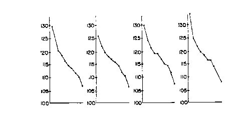
Figure 3 is a graphical representation of the results
of Table 4 with the mean cell volume being plotted against the
fraction number for Sample 1 (Figure 3A), Sample 2 (Figure 3B),
Sample 3 (Figure 3C), Sample 4 (Figure 3D), and Sample 5
(Figure 3E).
As can be seen from Table 4 and Figure 3, the slope
of mean cell volume of the ten fractions is steepest at higher
speed centrifugation (higher g forces) i.e. in Samples 1 and 2.
This steep slope represents a more efficient separation of
dense cells from the less dense cells. Thus, the low density
(high mean cell volume) cells which include the fetal nucleated
red blood cells fractionate in the uppermost fractions of the
tube. Generally, the mean cell volume of the hypotonic
fraction (Sample 5) is increased due to the increased cellular
water absorption from the hypotonic medium.
Examt~le 4
Using the whole blood samples of Example 3, a
comparison of low speed centrifugation with and without
chlorpromazine, and high speed centrifugation with and without
chlorpromazine was made.
WO 95/20675 ~ 18 2 3 ~ I. pCT~S95/00961
23
In sample 6, chlorpromazine was added to a final
concentration of 1.0 mM, and centrifuged to a final speed of
2800 g for fifteen minutes. Sample 7 contained no
chlorpromazine, and was centrifuged at a final speed of 2800 g
for fifteen minutes. In sample 8, chlorpromazine was added to
a final concentration of 1.0 mM and was centrifuged at a final
speed of 14,000 g for five minutes. Sample 9 contained no
chlorpromazine, and was centrifuged to a final speed of 14,000
g for five minutes. Fractions were collected as described in
Example 2, and assessed for mean cell volume. The results are
shown in Table 5, and Figure 4.
Table 5
Fraction Sample Sample Sample Sample
6 7 g g
1 129.6 126.1 129.9 132.8
2 -- 122.3 124.1 124.8
3 120.5 ~ 119.4 120.8 121.5
4 118.6 112.6 118.2 119.4
5 116.8 ~ 117.1 118.1 117.6
6 114.9 115.7 116.5 115.9
7 113.8 114.1 114.8 116.1
8 ~ 111.6 ~ 112.2
114.1 112.6
110.6 110.4 110.9 109.5
10 107.2 106.4 107.4 107.5
Figure 4 is a graphical representation of the results
of Table 5 with the mean cell volume being plotted against the
fraction number for Sample 6 (Figure 4A), Sample 7 (Figure 4B),
Sample 8 (Figure 4C), and Sample 9 (Figure 4D).
A comparison of samples 6 and 7 in Figure 4 shows a
steeper curve for Sample 6, which indicates the improved
density separation by the use of chlorpromazine at Lower g
forces. This improvement was not observed in Sample 8 relative
to Sample 9, that is at higher g forces, where there may be
greater cell shear or packing.
WO 95/20675 3 ~ 1. PCT/US95100961
24
Example 5
Four samples of umbilical cord blood were taken from
newborn children. Each sample was divided into two portions,
one portion having 25% water added (Samples 10, 12, 14, and
16), and the other portion additionally containing
chlorpromazine to a final concentration of 1.0 Mm (Samples li,
13, 15, and 17, respectively). Each sample was centrifuged
according to the procedure described above to a final speed of
14,000 g for five minutes. Curves of the mean cell volume were
compared as indicated above. Each fraction was also
microscopically examined for the presence of nucleated red
blood cells.
A general improvement in slope was again observed.
Table 5 provides the mean cell volume for each fraction of each
sample, with the number of nucleated red blood cells observed
(if any) in that fraction in parentheses.
Table 6
Fract. Samp. Sample Samp. Sample Samp. Sample Sample Samp.
10 11 12 13 14 15 16 17
1 157 157.5 148.4 151.5 150.6 151.8 143.0 141.3
(51) (65) (129) (119) (>450) (>600) (>250) (>300)
2 153.6 153.9 141.5 142.2 145.4 147.1 128.2 135.8
(3) (1) (25) (20) (5) (3)
3 145.1 142.2 136.5 153.6 139.4 122.1 126.6
(4) (4) (1)
4 146.5 149.1 133.5 135.1 140.2 133.8 122.1 125.4
(2) (1)
5 144.5 146.8 132.2 141.6 132.6 121.7 129.9
(1)
.
6 140. 134.1 126.3 128.3 135.8 130.5 122.0 124.9
0 ~
7 143.2 138.9 122.9 129.2 132.4 128.3 118.0 120.0
I ~
8 134.6 137.4 122.2 133.7 127.8 136.6 119.4 124.0
~ ~ ~ ~ ~ ~
136.2 123.8 125.2 119.9
'.34.3 I ~ 119.1
~ ~
125.1
~
117.5
10 I 127.0 113.6 113.4 108.1
~ ~ ~ 113.3
121.7
~
110.8
~
112.4
~
As is seen from Table 6, nucleated red blood cells
are present more often in fractions farther from the top in
absence of chlorpromazine. In each pair of tubes, the movement
WO 95/20675 21 g ~ 3 7 ~ PCT/US95/00961
of nucleated red blood cells toward-the highest fractions was
improved when chlorpromazine was used.
The mean cell volume data of Table 6 is graphically
presented in Figure 5. The mean cell volume is plotted
5 relative to fraction number for Sample 10 (Figure 5A), Sample
11 (Figure 5B), Sample 12 (Figure 5C), Sample 13 (Figure 5D),
Sample 14 (Figure 5E), Sample 15 (Figure 5F), Sample 16 (Figure
5G), and Sample 17 (Figure 5H).
10 Example 6
Using whole blood only, the mean cell volume and mean
cell hemoglobin concentration of 13 samples was examined before
and after a standard addition of water, 25% of volume, to each
sample. The results are shown in histogram form in Figure 6.
15 In Figure 6, each "X" represents an umbilical cord blood
sample, each "O" represents a maternal peripheral blood sample
at 12-19 weeks gestation, and "." represents a maternal
peripheral blood sample obtained at delivery (40 weeks). The
upper panel of Figure 6 (Figure 6A) shows the distribution of
20 isotonic whole blood samples, while the lower panel (Figure 6B)
shows the distribution in samples made hypotonic.
It can be seen from Figure 6 that the mean cell
volume of umbilical cord blood samples is well separated from
the maternal blood sample mean cell volume, both before and
25 after rendering samples hypotonic. Additionally, there is a
marked improvement of separation between maternal cells and
cord cells in MCHC measurements after rendering samples
hypotonic. Thus, the cell densities as observed by the MCHC
measurement, are as great as differences in MCV. However, in
non-isotonic conditions, the hemoglobin in each cell does not
change, but the larger fetal cells will swell or shrink more
than the smaller maternal cells. Thus, a better contrast is
observed between MCHC in cord blood samples and maternal
samples when the medium is non-isotonic, and facilitates
enrichment and isolation of the fetal nucleated erythrocytes in
density gradients.
WO 95/20675 PCT/US95/00961
21823'1
26
Example 7. -
A hemolyzing agent mixture of ammonium chloride and
sodium bicarbonate at approximately 300 m osmolar salt strength
was prepared. Maternal (m) and umbilical cord (c) blood
samples were exposed to either a physiological salt solution as
a diluent, or the hemolyzing mixture, either in the presence or
absence of 30 ~.1 of the carbonic anhydrase inhibitor
acetazolamide (at a final concentration of 1 mM), sodium
fluoride (at a final concentration of 150 ~Cm), or azide (1%).
The number of intact cells in the final volume of a 15x
dilution of blood in the lysis mixture was determined after 7
and after 17 minutes. The results are provided in Tables 7 and
8.
WO 95/20675 ~ 18 2 3 ~ I. PCTIUS95/00961
~7
Table 7
7 min. 17 min.
Sample Source Diluent Lyse Inhibitor count count
1 M 280 ~,1 - - 3292 3175
2 C 280 - - 4089 3922
3 M 30 250 - 228 183
4 C 30 250 - 760 752
5 M 2 5 .~w~~amiae 4 8 3 213
0
6 C 2 5 a~u,io~,m,a~312 8 2 4 7 6
0
7 M 2 5 ~oa~~ n~m;a~2 2 5 218
0
8 C ~ 2 5 ~od~um Ouonde4 8 5 4 9 0
0
9 M 250 azide 210 177
10 C 250 azide 384 390
13 M 2 5 .~i.m;a~ 3 8 9 18 5
0 at t=0
14 C 250 azide at 2301 427
t=7 min.
15 M 30 ~C1 250 - 2704 2657
lOxPBS
16 C 30 ~C1 250 - 4208 3958
lOxPBS
As can be seen from Table 7, cord blood is more
resistant to hemolysis when acetazolamide is present in the
mixture, while fluoride and azide have little protective
effect.
As can be seen from samples 15 and 16, hemolysis is
prevented (or cell count is preserved) in a hypertonic medium
(samples 15 and 16). This results from a equalization between
WO 95/20675 j ~ ~. PCT/US95100961
28
the extracellular concentration of salt and the intracellular
concentration of salt driven by the carbonic anhydrase mediated
hemolysis reaction. Thus, the intracellular and extracellular
salt concentrations remain stable relative to each other, thus
preventing hemolysis.
Table 8
7 min. 17 min.
Sample Source Diluent Lyse Inhibitor count count
i
i
1 M ~ 280 - - 2921 2963
~,1 I
2 C 280 - - 4346 4225
3 M 30 250 - 214 ~ 216
4 C 30 250 - 595 658
5 M - 2 5 .cwzor.m~a~ 2 8 6 18 8
0
6 C - 2 5 .~a.ro~m~e 2 6 5 2 0 0
0 0 5
7 M 3 0 ~ 2 5 .~wro~.m~a~ 2 8 5 3 0 3
1 0
lOxPBS
8 ~ C .m. 2 5 .~~a~ 219 0 ~ 2 2
0 0 9
9 M 30 ~C1 250 - 2476 2456
lOxNaCl
10 C 30 ~.1 250 - 3433 3371
lOxNaCl
11 M 20 ~C1 260 - 1744 1824
lOxNaCl
12 C 20 ~cl 270 - 2856 2646
lOxNaCl
13 M 10 ~cl 270 - 211 169
lOxNaCl
14 C 10 ~.1 - 607 ~ 419
~
lOxNaCl
WO 95/20675 21 g 2 3 7 ~ PCTIUS95/00961
29
As can be seen from Table 8, salt concentrations
above 400 m Osm will prevent lysis of both adult and cord blood
samples.
Example 8
A blood sample taken from a non-pregnant adult
individual was supplemented with umbilical cord blood in
amounts of 25%, 12.5%, 8.3%, and 6.25%. The fetal nucleated
red blood cells were isolated according to the method of
Examples 1 and 2. The final volume obtained after the
centrifugation step was 20 ~cl. The 20 ul volume was divided
into 3.5 ~,1 aliquots. For each aliquot, the number of nucleated
red blood cells was determined over a standard, constant path
on the Wright stained microscope slide of the specimen. The
total number of NRBC recovered from each 20 ul volume is shown
in Table 9.
TABLE 9
Sample NRBC Recovery
(# NRBC) i
25% Cord Blood 27
12.5% Cord Blood 15
8.3% Cord Blood 8
6.25% Cord Blood 6
The data in Table 9 indicate a linear relationship
between % of cord blood spiked into a normal blood sample, and
the amount of nucleated red blood cells recovered from the
sample, indicating the successful enrichment and identification
of rare cells according to the methods of the invention.
Example 9
A hemolyzing agent mixture as in Example 7 was added
in equal volume to blood samples. In separate tubes of the
experiment either maternal or cord blood was tested. In a
series of such tubes the carbonic anhydrase inhibitor 6-
ethoxyzolamide was added to the final concentration indicated
WO 95/20675 ~ ~ ~. PCT/US95/00961
'' 0
J
in Table 10. Each tube was assessed as having either visible
hemolysis or no hemolysis.
TABLE 10
6-Ethoxyzolamide 1.6 mg/21 ml Stock
Blood Dilution 1:1
Blood Inhib. PBS Lyse Visible Lyse Time
Adult 100 ~cl 5 ~cl 5 ~cl 90 ~cl 8' S0"
Cord 100 ul 5 ul 5 ~1 90 ul not lysedat 17 min.
Adult 100 ~cl 4 ~.1 6 ~cl 90 ~cl 7'
Cord 100 ~cl 4 ~.tl 6 ~tl 90 ~tl not lysedat 17 min.
Adult 100 ~cl 3 ~.1 7 ~,1 90 ul 7'
Cord 100 ul 3 ui 7 ul 90 ~cl not lysedat 17 min.
~
Adult 100 ~cl 2 ~cl 8 ul 90 ul 6' 30"
I
Cord 100 ~cl 2 ~cl 8 ul 90 ~cl lysis 17'
at
Adult 100 ~.1 1 ~cl 9 ~cl 90 ~cl 6' 30"
Cord 100 ~cl 1 ul ~ 9 ~cl 90 ~cl lysis 17'
at
TABLE 11
No Inhibitors
Dilution Visible Lyse Time
Maternal 1:15 2'15"
Maternal 1:7 2'40"
Maternal 1:3 3'10"
Maternal 1:1 4'20"
Cord ~ 1:15 4'10"
Cord 1:7 6'40"
Cord 1:3 9'50"
Cord ~ 1:1 ~ >20"
Table 11 shows the differential hemolysis in varying
blood sample dilutions for both maternal and cord blood in the
absence of a hemolysis inhibitor.
WO 95/20675 ~ ~ 1 8 2 3 7 ~ P~~2595/00961
31
It can be seen from Tables 10 and 11 that without
inhibitor there is rapid hemolysis of both maternal and cord
blood samples. In the presence of excess inhibitor neither
maternal nor cord blood samples will hemolyze. Further, there
is a range of concentration of inhibitor in which maternal
sample is hemolyzed and cord blood is stable for an extended
period of time.
The foregoing description of the preferred
embodiments of the present invention has been presented for
purposes of illustration and description. They are not
intended to be exhaustive or to limit the invention to the
precise form disclosed, and many modifications and variations
are possible in light of the above teaching, and are intended
to be within the scope of the invention.
Although the foregoing invention has been described
in some detail by way of illustration and example, for purposes
of clarity of understanding, it will be obvious that certain
changes and modifications may be practiced within the scope of
the appended claims.