Note: Descriptions are shown in the official language in which they were submitted.
21 83398
MAGNETIC RESONANCE IMAGING OF CEREBRAL BLOOD FLOW
AND DIAGNOSIS OF DIABETES
Field of The Invention
The present invention relates to magnetic resonance imaging of
cerebral blood flow and diagnosis of diabetes. More
particularly, the present invention relates to a functional
magnetic resonance imaging method which specifies the brain
activation sites with changes in cerebral blood flow as an
indicator, and a novel method of diabetes diagnosis using said
functional magnetic resonance imaging method.
Description of Related Art
For human sense and movement, homeostasis, and various higher
functions such as emotion, memory, language and thinking, there
are often control mechanisms for each specific site or region in
the brain. Such localization of functions in the brain had been
confirmed by observing in detail changes in behavior of patients
suffering from regional damage to the brain caused by trauma or
cerebral blood vessel impediment, or states of epileptic attack,
and by estimating the function of the damaged region. In
addition, the results of local electric stimulus experiments to
cerebral cortex had provided important evidence of localization of
various functions in the brain.
On the other hand, the recent rapid progress made in the areas
of electronic engineering technology and image processing
technology has urged achievement of new brain tissue imaging
methods such as computer tomography (CT), positron emmision
tomography (PET) and magnetic resonance imaging (MRI) and
apparatuses for the application of these new methods. These
apparatuses and methods have made it possible to easily diagnose
focal regions which have so far been confirmed only through
au~optic or operative findings, and furthermore, it is now
21 83398
possible to study localization of brain functions of sound
subjects without applying any of medicament.
Among others, MRI is widely applied in the clinical medicine
because of the capability of clearly depicting the slightest
tissue of human brain in vivo. More recently, a method known as
the functional MRI (hereinafter sometimes abbreviated as "fMRI")
was developed, which images actual activation regions of the
brain, and is attracting the general attention as a means for
research on cerebral and neural mechanisms and a new means for
brain diagnosis.
This fMRI utilizes the findings that the local blood flow is
increased at the activated brain regions where neurons being
excited. In the area of MRI technology including fMRI, various
contrivances are being made in electromagnetic pulse system for
irradiation onto the brain, of which ones commonly known include
the inversion recovery (IR) method, the gradient-echo (GE) method
and the spin-echo (SE) method.
From among these new methods, the GE method has developed as a
method for measuring the blood flow rate and the oxigenation
status of hemoglobin in blood. More specifically, oxi-hemoglobin
transports oxygen to brain and other tissues of the entire body.
Oxygen fed from the lung into blood is sent in the form of oxi-
hemoglobin through arteries to the brain and other tissues.
Hemoglobin having cut off oxygen transforms into deoxi-hemoglobin
and goes back to the heart through veins. Because of the
properties as a paramagnetic substance, the deoxi-hemoglobin
disturbs the static magnetic field, thereby impairing an MRI
signal.
The conventional GE method, T2* -weighted image with long echo
time has been used to image oxigen consumption. In this method,
however, the signal intensity is affected by the fluctuation of
cerebral blood flow, which maikes it difficult to specify the
activation site. To overcome this difficulty, a method
21 833~8
determining presence of only deoxi-hemoglobin, excluding the
influence of fluctuation of the blood flow, was developed to
improve conventional GE method, and is now accepted as a more
accurate method to analyze brain functions.
The improved GE method free from the effect of blood flow
however requires in practice prior administration of a contrast
medium or use of a high magnetic-field pulse not allowed for
medical purposes, because of the very low sensitivity. There is
another problem of unavailability of a clear image since the MRI
signal is strongly affected by a disturbance of the static
magnetic field.
Summary of The Invention
The present invention has an object to provide an improved
method of fMRI which permit clear imaging even the slightest
changes in brain functions by determining the change in blood flow
itself, but not in blood oxigenation level.
Another object of the present invention is to provide a method
for diagnosis of diabetes using an excited state of a specific
brain site based on fMRI as an indicator.
The present invention provides, in a method of magnetic
resonance imaging of cerebral blood flow using pulse sequence of
rapid gradient-echo method, wherein the pulse is RF pulse having
slice gradient, read gradient and phase-encode gradient, the
improvement comprises giving the RF pulse with relatively high
flip angle of between 45 to 60 degrees, and adding an additional
gradient to each of the slice gradient, read gradient and phase-
encode gradient, thereby diffusing and refusing proton spin in
the cerebral blood flow.
The present invention provides also a diabetes diagnosing
method which comprises imaging the cerebral blood flow after
administration of insulin by the above-described method, and
detecting the increase in blood flow in any one of the
21 83398
hippocampus, paraventricular nucleus of the hypothalamus,
dorsomedial nucleus of the hypothalamus and ventromedial nucleus
of the hypothalamus.
Brief Description of The Drawings
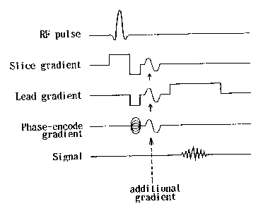
Fig. 1 is a schematic view illustrating the imaging pulse
sequense in the conventional GE method; Fig. 2 is a schematic view
illustrating the imaging pulse sequense in the method of the
present invention.
Fig. 3 illustrates an MRI representing changes in cerebral
blood flow of a diabetic model animal after administration of
insulin.
Detailed Description of The Invention
The fMRI of the present invention comprises a partial
improvement of the rapid gradient-echo method (RGE method) in
which changes in blood flow are emphasized for imaging the
activated region(s) of the brain. The ordinary RGE method uses,
for example, an imaging pulse sequense, of which the consititution
of one pulse is schematically shown in Fig. 1. In the method of
the present invention, in contrast, an additional gradient for
diffusing and refusing proton spin is added to the pulse sequence
as shown in Fig. 2.
While, in the RGE method, the ordinary RF pulse intensity is
represented by a flip angle of between 5 and 30 degrees, in the
method of the present invention, the RF pulse intensity is set at
a flip angle of between 45 and 60 degrees.
By adopting the additional gradient and the RF pulse intensity
as described above, it is possible to exclude the influence of a
disturbance of static magnetic field caused by the change in
concentration of deoxi-hemoglobin, and thus to clearly image an
activated region(s) with the change in blood flow as an
indicator. Furthermore, an image can be aquired with a higher
21 83398
time resolution (about 5 seconds), and a simultaneous combination
with a T2*-weighted image, a Tl-weighted image or a liquid
diffusion weighted image is also available.
The MRI method in the present invention makes it possible to
clearly image a very slight change in brain function by
determining the change of the local blood flow. This permits
easy and sure diagnosis of diabetes with the change in MRI signal
for a specific region in the brain as an indicator.
As a result of imaging, by the foregoing fMRI method, a change
in the cerebral blood flow upon administering insulin to a
diabetic model animal, as shown in the example presented later,
the present inventors observed an increase in the MRI signal
intensity at the hippocampus, paraventricular nucleus of the
hypothalamus, dorsomedial nucleus of the hypothalamus and
ventromedial nucleus of the hypothalamus along with the time after
administration of insulin. This result is attributable to the
fact that the brain in a glucose starvation state suddenly reacts
with glucose, thus increasing the blood flow to the brain regions
as described above. It is therefore possible to diagnose the
presence and progress of diabetes by imaging the blood flow
patterns of the foregoing regions of the brain with the method of
the present invention when administering insulin to a potential
patient who may suffer from diabetes.
Now, the present invention will be described further in detail
below. It is needless to mention that the present invention is
not limited by the following example.
EXAMPLE
Changes in cerebral blood flow of diabetic model animals after
administration of insulin were measured using a MRI based on the
method of the present invention.
Streptozotocin (STZ) was administered intraperitoneally in an
amount of 60 mg/body weight to the male Wistar rats (body weight:
21~339~
about 200 g), and most of the ~ -cells of the pancreas Langerhans'
islets were destroyed to reduce the insulin secretory capacity to
prepare diabetic model rats. Rats with a blood glucose level of
more than 300 mg/dl after one to two days from the administration
of STZ were used as subjects of the experiment.
A plastic needle for administering insulin was inserted
intramusculary at the thigh, then the head of the rat was fixed at
the center of an RF probe, which was placed at the center of a
superconductive magnet (inner diameter: 40 cm) of an MRI apparatus
(made by SMIS Company).
After adjusting the monogeneity of the static magnetic field,
measurements were performed once before administration of insulin
(40 U/kg body weight) and immediately after administration and
thereafter at intervals of 20 minutes for a period of two hours,
with the use of MRI method of the present invention, which
comprised the following imaging pulse sequence:
Constant magnetic field: 4.7 tesla;
Echo time (TE): 5 ms;
Repetition time (TR): 10 ms;
Number of excitataions (NEX): 4;
Field of views (FOV): 4 cm x 4 cm;
Slice thickness: 3 mm;
Number of pixels: 128 x 128 pixels;
RF pulse: flip angle of between 45 and 60 degrees.
The results are as shown in Fig. 3. More specifically, an
apparent increase in signal intensity after the administration of
insulin was observed in the hippocampus at 20 minutes, in the
paraventricular nucleus of the hypothalamus (PVN) at 40 minutes,
in the region containing the PVN and the dorsomedial nucleus of
the hypothalamus (DMH) at 60 minutes, in the region containing the
DMH and the ventromedial nucleus of the hypothalamus (VMH) at 100
minutes. At 120 minutes when the blood glucose level
substantially recovered to the level before insulin
21~3~
administration, no change in signal intensity at a specific
region was observed.
From the results as described above, it is confirmed that,
when the glucose utilization in the peripheral tissues increases
at a time in an animal in a glucose hunger state, some
informations from the peripheries are processed through a
functional brain axis comprising the hippocampus, PVN, DMH, and
finally VMH in this sequence. Therefore, in a diabetic patient
in a glucose starvation state similar to that of the model animal
used in the present experiment, similar changes in signal
intensity in the hippocampus, PVN, DMH and VMH would be observed
by imaging changes in cerebral blood flow after administration of
insulin by the application of the MRI method of the present
invention.
