Note: Descriptions are shown in the official language in which they were submitted.
2 1 8577q
The present invention relates to laser surgical
techniques for modifying the corneal surface of the eye, and more
particularly, to laser surgical techniques, collectively known as
photorefractive keratectomy or PRK, which direct reshaping of the
cornea by means of selective volumetric removal of corneal
tissue.
In recent years, numerous corneal sculpting techniques
and related apparatus have been disclosed for correcting visual
deficiencies such as near-sightedness, far-sightedness, and
astigmatism. In addition, corneal sculpting techniques have also
been utilized for therapeutic interventions in a number of
pathologic conditions invo~ving the cornea. For example, U.S.
Patent Nos. 4,665,913, 4,732,148, and 4,669,466 to L'Esperance,
and U.S. Patent No. 5,108,388 to Trokel, describe methods for
achieving optical correction through reshaping of the anterior
corneal surface. In addition, a number of prototype instruments
for affecting refractive surgery have recently become
commercially available, such as the Model 2020 from Visx of Santa
Clara, CA and the Model Exci-Med 200 from Summit of Watertown,
MA .
These commercial devices, as well as most corneal
sculpting methods and devices which have been disclosed and
, . ~ . , .
2l 8577q
manufactured to date, utilize ultraviolet (W) radiation with a
wavelength which is preferably less than 200 nanometers. For
example, many of these devices utilize an Argon Fluoride excimer
laser operating at 193 nm. Generally, radiation at such short
ultraviolet wavelengths is characterized by high photon energy,
namely, greater than 6 eV, which, upon impact with tissue, causes
molecular decomposition, i.e., the direct breaking of
intramolecular bonds. The photochemical nature of this mechanism
has the advantage of producing minimAl collateral thermal damage
in cells adjacent to the surgical site, since the broken
molecules generally leave behind only small volatile fragments
which evaporate without heating the underlying substrate.
Furthermore, the depth of decomposition for each laser pulse is
typically very small, i.e., less than 1 micron, thus achieving
accurate tissue removal with minimal risk of damage to the
underlying structures from W radiation.
In view of this small depth of penetration, coupled
with the need to remove sufficient depth of tissue while
minimizing the overall time for the surgical procedure, the
majority of corneal sculpting techniques utilizing the excimer
laser employ "wide area ablation~'. Generally, wide area ablation
utilizes a laser beam with a relatively large spot size to
successively remove thin layers of corneal tissue. The spot size
--
21 85779
is generally of a size sufficient to cover the entire optical
zone of the cornea, namely, a region of 5 to 7 millimeters in
diameter. Consequently, to assure required flux densities on the
cornea, relatively high energy output W lasers are typically
required. It has been found that to assure a flux density of at
least 150 mJ/cm , for a reasonable ablation rate of at least 0.2
microns/pulse, a 200 mJ W laser is required. Such lasers,
however, tend to be prohibitively large and expensive systems.
Furthermore, efficacious wide area ablation requires
that the projected beam be spatially homogenous and uniform to
achieve the desired smooth corneal profiles. Accordingly,
additional beam shaping devices, such as rotating prisms,
mirrors, or spatial integrators, must be employed within the
excimer beam delivery systems. For a more detailed discussion of
beam shaping and delivery systems, see, for example, U.S. Patent
No. 4,911,711 to Telfair, incorporated by reference herein. Of
course, such a multiplicity of optical elements contributes to
overall transmission loss, while adding substantial optical
complexity, cost, and maintenance requirements to the system.
Alternative techniques based on utilization of a
scanning W laser beam have been proposed to achieve controlled
and localized ablation of selected corneal regions of the cornea.
In the scanning approach, a relatively small laser spot is
21 85779
scanned rapidly across the cornea in a predefined pattern and
accumulatively shapes the surface into the desired geometry. For
a more detailed discussion of laser scanning techniques employing
excimer lasers, see U.S. Patent No. 4,665,913 to L'Esperance or
Lin, J.T., ~Mini-Excimer Laser Corneal Reshaping Using a Scanning
Device," SPIE Proceedings, Vol. 2131, Medical Lasers & Systems
III (1994). A scanning approach may offer a number of
advantages, including lower power and energy requirements, added
flexibility for refractive corrections and smooth ablation
profiles, without the need for spatially uniform output beam
profiles. For example, a laser scanning technique allows a
tapered optical treatment zone to be achieved, which may have
advantages for the correction of high myopia, for performing
therapeutic tissue removal and for treating areas up to 9
millimeters in diameter which may be required for the correction
of hyperopia.
While laser surgical techniques based on the excimer
laser have proved beneficial for many applications, such
techniques suffer from a number of limitations, which, if
overcome, could significantly advance the utility of optical
laser surgery. For example, techniques based on excimer lasers
utilize toxic gases as the laser medium, suffer from persistent
reliability problems, require lossy optics in the delivery
2185779
systems, and suffer from the possibility that the W radiation is
potentially mutagenic through secondary fluorescence, which may
cause undesirable long term side effects to the unexposed tissues
of the eye.
Accordingly, alternatives to the excimer laser have
been suggested in recent years which involve frequency-shifted
radiation from a solid state laser. Current limitations of
nonlinear elements used as frequency-shifting devices, however,
place a lower limit of approximately 205 nm on the available
wavelengths of such lasers, which may be too close to the
mutagenic range, which exhibits a peak at 250 nm. In addition,
multiply-shifted laser devices also face certain difficulties in
providing the requisite energy outputs and are fairly complex and
cumbersome, leading again t~o potential laser reliability
problems, as well as added cost and maintenance.
More recently, a more attractive alternative has been
suggested by T. Seiler and J. Wollensak, "Fundamental Mode
Photoablation of the Cornea for Myopic Correction", Lasers and
Light in Ophth~7mology, 5, 4, 199-203 (1993), involving mid-
infrared wavelengths and, in particular, radiation around 3
microns corresponding to the absorption peak of water, the main
constituent of the cornea. One solid state laser in particular,
the Erbium:YAG laser (Er:YAG), emits radiation at a wavelength of
2i 85779
2.94 microns, corresponding to an absorption coefficient of over
13000 cm in water. This high absorption results in a small
region of impact with potentially less than two micron
penetration depths.
Contrary to the photoablation mechanism associated with
the excimer laser, i.e., photochemical decomposition, which is
due to energy absorption in molecular bonds, ablation with the
Er:YAG laser is attributed to photovaporization, or photothermal
evaporation, of water molecules. This thermal heat induces a
phase change, and thus a sudden expansion of the tissue material,
thereby ablating the corneal surface tissue.
In addition, erbium lasers are more attractive for
clinical applications than excimer lasers, since they are
compact, efficient and can ,deliver higher beam quality radiation,
which allows for less lossy beam delivery systems and superior
optical coupling properties. Further, the photovaporization
process is inherently more efficient than photodecomposition,
allowing for removal of up to 3 microns of tissue at a time and
thereby resulting in a faster surgical operation. Mid-infrared
radiation is also compatible with fiber delivery, a potentially
attractive method of decoupling the source laser from the
delivery system which makes it more suitable for the operating
room. Finally, radiation from an Er:YAG laser is not mutagenic,
2 1 85779
relieving the potential for deleterious long-term side effects.
The Er:YAG laser-based corneal sculpting system
described by Seiler and Wollensak is based on wide area ablation.
This system aims to exploit the gaussian beam profile of the
laser beam to achieve a refractive correction with each pulse,
using a minimal number of pulses. An alternative system which
also relies on wide area ablation is described in PCT Application
No. 93/14817 to Cozean et al., which relies on a sculpting filter
to control the intensity of the radiation delivered to the cornea
and hence the amount of tissue removal.
While providing a number of advances over prior
techniques, the Er:YAG laser techniques described by Seiler and
Wollensak and Cozean et al. both suffer from a number of
potential drawbacks, common,to wide area ablation techniques,
including the need for a smooth and uniform beam profile, a large
pulse energy, and/or a complex filter control system. These
systems assumed that the ablation process is a linear process,
i.e., that a portion of the beam with a larger energy density
will remove a larger depth of tissue. This has been shown,
however, to be an incorrect assumption for the excimer ablation
process, and may also be an incorrect assumption for the Er:YAG
ablation process.
In addition to the limitations previously discussed,
.
21 85779
all such prior techniques for delivering and controlling a mid-
infrared laser beam are subject to one shortcoming in particular,
namely, the potential for thermal damage to unablated regions of
the eye, due to excessive energy density required by these
systems and the large shock waves generated by the high energy
pulses required to ablate wide areas. In addition, due to the
need for high pulse energy and high beam quality, such prior
systems typically exhibit optical configurations that are
generally not conducive to ease of manufacturing and are
difficult to maintain and service.
As is apparent from the above discussion, a need exists
for an improved method and apparatus for surgically treating
corneal tissue based on the controlled removal of tissue. A
further need exists for an ~mproved method and apparatus for
reducing myopic, hyperopic and/or astigmatic conditions of the
eye using a low cost solid state laser. Yet another need exists
for a method and computer-controlled apparatus for scanning mid-
infrared laser radiation across the outer surface of the eye and
the underlying Bowman's layer and stroma for the purpose of
reducing refractive errors of the eye and for the purpose of
treating tissue at or near the surface of the cornea. A further
need exists for a method and apparatus for surgically treating
corneal tissue, having an improved eye tracking mechanism.
21 8577q
Generally, according to aspects of the invention, a
surgical method and apparatus for removing corneal tissue with
mid-infrared radiation are provided. The surgical method and
apparatus utilize short laser pulses scanned over a region of the
cornea to yield a tissue removal mechanism based on
photospallation. Photospallation is a photomechanical ablation
mechanism which results from the absorption of incident radiation
by the corneal tissue. When the corneal tissue absorbs the
infrared radiation, a bipolar oscillating shock wave is created,
which alternately compresses and stretches the corneal tissue.
Tissue fragments are torn apart and ejected by the shock wave
during the stretching phase{.
In accordance with one feature of the present
invention, the laser delivery system includes a laser source,
such as a Q-switched Er:YAG laser, which emits pulsed radiation
in the mid-infrared spectral region with an energy density
capable of causing ablation of corneal tissue. In a preferred
embodiment, the laser emits radiation of approximately 3 microns,
corresponding to the maximal absorption coefficient of water, the
main constituent of corneal tissue. The laser source preferably
emits radiation at discrete pulses of less than 50 nanoseconds at
.
21 85779
a repetition rate of approximately 5 to 100 Hertz. The short
laser pulses reduce the undesirable thermal damage of surrounding
tissue to insignificant levels. The energy in each pulse is
preferably on the order of 5 to 30 mJ.
The laser beam is preferably scanned over a specific
central region of the surface of the cornea in a predefined
pattern by a sc~nn;ng beam delivery system so as to selectively
remove tissue at various points within the scanned region and
thereby reshape the corneal tissue in a predictable and
controlled fashion. The scanning beam delivery system preferably
consists of a controllable tilt mirror assembly to direct and aim
the beam over the surface of the cornea. A variety of predefined
scan patterns may be utilized to achieve controlled
photospallation of the cornea, including the epithelium, Bowman's
layer, and the stroma in accordance with the desired changes in
the shape of the cornea.
In accordance with a further aspect of the present
invention, the laser spot size and spacing associated with a
given scan pattern may be varied prior to each procedure
according to certain nomograms correlating the required degree of
pulse overlap with the depth of ablation, consistent with
maximizing the speed of the operation and the requisite
smoothness of the ablated corneal surface. A given scan pattern
--10--
.
21 8577q
preferably uniformly irradiates a treatment region with minimal
discernible lines of overexposed or underexposed tissue lying
between scans. One or more discontinuous scan patterns may be
utilized to distribute the pulse over the entire treatment region
in each time interval, thereby distributing residual heat over
the entire region and minimizing temperature rise in any
localized area.
Further, in accordance with a preferred embodiment of
the invention, an eye tracking system is further provided in
conjunction with the scanning beam delivery system, to compensate
for eye motion during the surgical procedure. The eye tracking
system senses the motion of the eye and provides signals that are
proportional to the errors in lateral alignment of the eye
relative to the axis of thç laser beam. Lateral motion of the
eye is detected by illuminating the eye with tracking
illumination and forming an image of a significant feature of the
eye, such as the limbus, on an array of detectors. According to
a feature of the present invention, the array of detectors
includes at least four detectors centered vertically and
horizontally around the center of the detector array.
In operation, when the significant feature of the eye
is centered with respect to the axis of the laser beam, the image
of the significant feature will be centered on the detector
.
21 85779
array. A null signal is generated by the detector array which
serves to maintain the axis of the laser beam in its current
position. When the eye is not centered with respect to the axis
of the laser beam, however, the image formed on the detector
array will also not be centered. The detector array will
generate an error signal which causes the laser beam to be
deflected to ensure that it is properly applied to the corneal
tissue.
The tracking illumination is preferably chosen in the
near infrared range so that it may be discriminated from ambient
illumination and the laser beam. In addition, the tracking
illumination is preferably modulated at a predefined temporal
frequency to further discriminate the tracking illumination from
the ambient illumination and the laser beam. Red or near
infrared filters may be positioned in front of the detectors in
the array to further enhance the contrast of the significant
feature of the eye to be detected, such as the limbus.
According to further features of the invention, a
corneal topography device may be included in the surgical
apparatus for evaluating the shape of the-corneal tissue to
assist in pre-op or post-operative measurements. Alternatively,
a spatially resolved refractometer may be included for evaluating
the refraction of the corneal tissue. In various embodiments of
,
~1 8~77q
the invention, the above-described alignment methods may be
utilized to incorporate active feedback control from the
topographic or refraction mapping instrument so as to provide
further control over the course of the surgical procedure.
A more complete understanding of the present invention,
as well as further features and advantages of the invention, will
be obtained by reference to the detailed description and
drawings.
In the drawings,
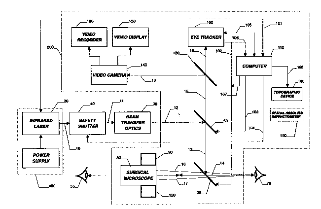
FIG. 1 is a block diagram illustrating the functional
relationship of optical, mechanical, and electrical components of
apparatus incorporating features of the present invention;
FIG. 2 is an expanded schematic diagram of the optical
components of FIG. l;
FIGS. 3(a) and 3(b) illustrate scanning patterns for
the laser beam passing over the cornea;
FIGS. 4(a) and 4(b) illustrate intensity profiles as a
function of the diameter of the focused laser beam, measured at
the cornea;
FIGS. 5(a) and 5(b) illustrate mechanisms for
transferring the laser beam from the laser system to the surgical
apparatus;
-13-
21 85779
FIGS. 6(a) and 6(b) illustrate images of the eye in an
aligned and unaligned position, respectively, with respect to the
detector array of an integral eye tracker; and
FIG. 7 is a schematic diagram of one embodiment of the
electronic circuitry and servo control functions associated with
the eye tracker indicated in FIGS. 1 and 2.
As shown in FIGS. 1 and 2, a surgical apparatus 200
includes an infrared laser source 20 and an optical assembly,
including, in sequence, beam transfer optics 30, discussed below
in conjunction with FIG. 5, a safety shutter 40, and partially-
transmitting mirrors 50 and 60, which cooperate to focus an
output beam 10 upon the cornea of a patient's eye 70, for
correcting curvature of the cornea or for affecting therapeutic
interventions. The laser source 20 is preferably a mid-infrared
laser generating short laser pulses, to yield a tissue removal
mechanism based on photospallation, discussed below. The laser
beam 10 is preferably scanned over a specific central region of
the surface of the cornea in a predefined manner, as discussed
below in conjunction with FIGS. 3(a) and 3(b), so as to
selectively remove tissue at various points within the cornea and
thereby cause the curvature of the cornea to change in a
-14-
21 85779
predictable and controlled fashion.
According to one feature of the invention, the laser
source 20 is preferably a solid state laser, which emits pulsed
radiation in the mid-infrared spectral region with an energy
density capable of causing ablative decomposition of corneal
tissue. As used herein, the term "solid state laser" includes a
diode laser. Preferably, the laser emits radiation at a corneal
absorption peak, i.e., at a wavelength of approximately 3
microns, such as 2.7 to 3.1 microns, corresponding to the maximal
absorption coefficient of water, the main constituent of the
corneal tissue. It has been found that absorption of laser
energy by the corneal tissue of the eye 70 at such a wavelength
results in complete absorption within 1 to 2 microns of tissue
depth.- As discussed further below, it has been found that the
combination of shallow absorption depths and short radiation
pulses reduces the undesirable thermal damage of surrounding
tissue to insignificant levels.
PHOTOSPALLATION
As previously indicated, according to a feature of the
present invention, the surgical technique disclosed herein,
whereby corneal tissue is irradiated with short pulses of a
scanned mid-infrared laser beam, is based on a concept referred
to as photospallation. Generally, photospallation is a
~1 8577q
photomechanical ablation mechanism which results from the
absorption of incident radiation by the corneal tissue. When the
corneal tissue absorbs the infrared radiation, a bipolar
oscillating shock wave is created, which alternately compresses
and stretches the corneal tissue, ejecting the tissue fragments
torn apart during the stretching phase. For a more detailed
discussion of photospallation, see Jacques, S.L., "Laser-Tissue
Interactions: Photochemical, Photothermal, and Photomechanical,"
~asers in General Surgery, 72(3), 531-558 (1992), incorporated by
reference herein. Since photospallation is a mechanical ablation
process, there is very little heat generated in the adjacent
tissue left behind after the ablation.
The laser source 20 may be embodied as a Q-switched
Er:YAG laser, which delivers a beam of mid-infrared radiation at
or near a wavelength of 2.94 microns. Alternatively, the laser
source 20 may be embodied as a Neodemium or Holmium doped laser
which is frequency shifted by an optical parametric oscillator
(OPO) to emit radiation of approximately 3 microns. Of course,
substitution of other alternative laser sources with similar
emission characteristics to that of the Er:YAG laser, as they
become available, are included within the scope of this
invention.
The laser source 20 preferably emits radiation at
-16-
.
21 85779
discrete pulses of less than 50 nanoseconds in duration at a
repetition rate of 5 to 100 hertz. The laser pulses should be
short enough such that lateral thermal damage of tissue adjacent
to the irradiated region is limited to a region smaller than 2
microns wide, consistent with a photospallation process. In
addition, the energy in each pulse of the laser 20 is preferably
on the order of 5 to 30 mJ. Thus, the incident laser beam 14
will ablate tissue locally and thereby remove microscopic
portions of the cornea.
LINE-OF-SIGHT
To correlate the eye's reference frame to that of the
surgical instrument 200, as shown in FIGS. 1 and 2, it is
necessary that the line-of-sight of eye 70 be substantially
coincident with the propagation axis of the incident laser beam
14. As used herein, in accordance with customary definition, the
term "line-of-sight" or ~principal line of vision~ refers to the
chief ray of the bundle of rays passing through the pupil and
reaching the fovea, thus connecting the fovea with the fixation
point through the center of the entrance pupil. It will
therefore be appreciated that the line-of-sight constitutes an
eye metric defined directly by the patient, rather than through
some external measurement of the eye and further, that the line-
of-sight can be defined without ambiguity for a given eye and is
-17-
.
21 8577~
the only axis amenable to objective measurement using cooperative
patient fixation.
Since critical vision is by definition centered on the
line-of-sight of the eye, irrespective of the direction in which
the mechanical axis of symmetry of the eye is pointed, it is
generally acknowledged that for best optical performance, the
point marking the intersection of the line-of-sight with the
cornea establishes the desired center for the optical zone of
refractive procedures seeking to restore visual acuity. It is
noted that the orientation of the line-of-sight of the eye 70, as
shown in FIGS. 1 and 2, may be vertical, horizontal, or
intermediate to those extremes as befitting comfortable
positioning of the patient for surgery without affecting the
validity of the invention.
During preparation for laser surgery on the cornea, the
line-of-sight of the eye 70 must be aligned to coincide with the
laser beam axis by two-axis lateral-translational adjustments, in
a known manner, as directed by the surgeon 55. The surgeon 55
observes the eye 70 through a surgical microscope 80 and judges
the degree of centration of the frontal image of the eye 70 with
respect to a crosshair or other fixed reference mark indicating,
as a result of prior calibration, the location of the axis of
beam 14, in a known manner. The axial location of the eye 70 is
.
218577~
also judged by the surgeon 55 by virtue of the observed degree of
focus of the image of the eye 70 relative to the previously
calibrated and fixed object plane of best focus for microscope
80. Directions from the surgeon 55 allow adjustment of the axial
position of the cornea of eye 70 to coincide with the plane of
best focus.
The required angular orientation of the line-of-sight
of eye 70 is preferably established by directing the patient to
observe and focus attention, i.e., fixate, on two coaxial
illuminated targets (not shown) projected into the eye 70 by a
fixation target device 90, which is preferably integrated into
the microscope 80. The two targets appear to be located at
different axial distances from the eye 70 of the patient and will
have been previously calibrated in a known manner. For a
description of a suitable calibration technique, see PCT
application No. WO 94/07908 to Knopp and Yoder. In this manner,
when the two targets (not shown) appear superimposed, the axis of
the observing eye 70 will be substantially coincident angularly
with the axis of the microscope 80 and also with the axis of
laser beam 14.
In a preferred embodiment, small lateral motions of the
patient~s eye 70, i.e., less than 5 mm in either direction, that
occur after the initial alignment performed in the manner
--19
.
2~85779
described above, and throughout surgical treatment, are rendered
inconsequential by virtue of the function of a two-dimensional
eye tracker 100, discussed further below in conjunction with
FIGS. 6 and 7. The eye tracker 100 senses the motion of the eye
70 and provides signals that are proportional to the errors in
lateral alignment of the eye 70 relative to the axis of the laser
beam 14. The signals generated by eye tracker 100 are converted
into commands for small angular tilts of partially-reflecting
mirror 60 that compensate for errors in the location of the eye
70. The small angular tilts serve to redirect beam 14 so as to
make it coincide with the instantaneous position of the eye 70.
The compensation commands are sent from electronics, discussed
below in conjunction with FIG. 7, within the eye tracker 100 to
mirror 60 by means of one o;r more data connections, collectively
designated 102.
Illumination of the eye 70 to facilitate tracking by
the eye tracker 100 is preferably accomplished by means of a
coaxial illuminator 120, preferably integrated with the
microscope 80, that projects a beam of light 17 at a small angle,
on the order of 8~, with respect to the line-of-sight of the
microscope 80. According to a feature of the invention, the
nature, i.e., the wavelength and temporal modulation frequency,
of the tracking beam 17 generated by illuminator 120 is
-20-
2 1 85779
preferably selected to maximize discrimination by the detectors
and related electronic circuitry within eye tracker 100 of the
tracking beam 17, from ambient room illumination and radiation
from laser 20. In this manner, the ambient illumination and
laser beam 14 will not possess the same temporal modulation nor
spectral characteristics as the tracking beam 17, and will thus
be virtually invisible to the tracking detectors.
In addition, as shown in FIG. 1, the surgical system
200 preferably includes a safety shutter 40 which closes
automatically if the laser beam 14 fails to follow a prescribed
path, if pulse energy-monitoring means provided within laser 20
indicates a malfunction of said laser or if the eye tracker 100
cannot follow the eye motion.
As shown in FIG. 1 and discussed further below, the
surgical apparatus 200 preferably includes a video camera 140
that displays a real-time image of the patient's eye on a monitor
150 during pre-operation alignment and during surgical treatment
and records the video image on a video recorder 160 for
postoperative ex~m;n~tion and documentation of the surgical
procedure.
As shown in FIG. 1, the computer 110 includes multiple
storage and control capabilities. Specifically, the computer 110
communicates and thereby controls the laser source 20 by means of
.
21 85779
a connection 101. In addition, the computer 110 drives the
scanning mirror 50 by means of a connection 103, in accordance
with stored scanning patterns and commands input to the computer
110 by the surgeon 55 or an assistant. A connection 104 between
the computer 110 and the safety shutter 40 affects maximum safety
of the patient, the surgeon, and attending personnel. The
computer 110 monitors the operation and status of the eye tracker
system 100 by means of a connection 105. Alternately, as shown
in FIG 1, computer 110 can be connected to the eye tracker 100 by
means of connection 106 and a separate connection 107 can be
provided from computer 110 to mirror 60 so that the computer 110
could directly control the position of the mirror 60. A further
alternate configuration would allow the computer 110 to combine
the scanning and eye tracking functions together onto a single
mirror, such as the mirror 60, thereby removing the need for
connection 103.
As discussed further below, the surgical apparatus 200
preferably includes a corneal topography device 180 or a
spatially resolved refractometer 190, as shown in FIG. 1. A
corneal topography device 180 may be used for evaluating the
shpae of the corneal tissue to assist in pre-op and post-op
measurements of the eyes' shape or curvature. An alternate
embodiment would include a spatially resolved refractometer (SRR)
.. . . . . .
21 85779
190 to evaluate the refraction of the corneal tissue.
OPTICAL MIRRORS
It may be noted from ex~m-n~tion of FIGS. 1 and 2 that
the partially-reflecting natures of mirrors 50 and 60 play
important roles in the proper function of the invention. In the
case of mirror 50, laser radiation in beam 12 is reflected while
radiation from eye tracker 100 is transmitted. This can be
accomplished, for example, through use of what is commonly called
a '~hot mirror~ coating on the surface of mirror 50. This coating
is dichroic, in other words, the coating has different reflection
and transmission characteristics foJ~ light of differing
wavelengths. The radiation from laser 20 has a wavelength of
approximately 2.9 microns and the mirror 50 should have a high
reflectance at that wavelength. The radiation to eye tracker 100
preferably has a wavelength between 0.8 and 1.0 microns for which
the coating of mirror 50 should have a high transmittance.
Similarly, the dichroic coating on mirror 60 is
preferably selected to have high reflectance at the wavelength of
laser 20 and approximately equal transmittance and reflectance at
the visible wavelengths used by the surgeon's eye in observing
the alignment of the eye with respect to the surgical apparatus
and progress of the surgery, at the wavelength of the fixation
target 90, and at the wavelength of the coaxial illuminator 120.
21 85779
This is possible since the visible range, the fixation target 90,
and the illuminator 120 are adjacent in wavelength and far from
the wavelength of laser 20. At both mirrors 50 and 60 the
transmitted beams suffer small lateral displacements due to
oblique incidence and the finite thickness of the mirror
substrates, but these fixed displacements are easily compensated
for in the design of the apparatus, as would be apparent to a
person of ordinary skill in the art.
In addition, mirror 130, shown between beams 15 and 16
of FIGS. 1 and 2, is also preferably partially transmitting,
although not dichroic. The coating on mirror 130 nominally has
approximately equal reflectance and transmission characteristics
at the wavelengths of the eye tracker light source 120 and
throughout a significant portion of the visible spectral region.
In this manner, a portion of the energy of beam 15 can be
redirected as beam 18 into video camera 140, discussed above. It
is understood that a beamsplitting prism, typically in the form
of a cemented two-element cube with a partially-reflecting
coating on an internal surface can be employed to provide the
function of mirror 130.
SCANNING PATTERNS
As previously indicated, the surgical apparatus 200 of
FIGS. 1 and 2 preferably provides a computer-controlled scanning
-24-
.
2 1 85779
motion of the focused laser beam 14 for sequentially irradiating
contiguous small areas of the central portion of the cornea of
eye 70 with pulses of mid-infrared laser radiation in predefined
patterns, such as those illustrated in FIGS. 3(a) and 3(b). In
each case, the region to be treated has a diameter of up to 9 mm.
The size of the focused spot of laser radiation is preferably on
the order of a 0.5 to 2.0 mm circumscribed diameter.
As shown in FIG. 3(a), a rectilinear or raster-scan 310
of the scanning spot of laser beam 14 covers a square area
centered on the desired treatment region 315. The laser beam 14
is modulated "off" when the computer 110 predicts that the energy
would impinge upon corneal tissue outside the desired treatment
region 315. As shown in FIG. 3(b), the laser beam 14 scans in a
concentric-circle pattern ~22 that is centered on the desired
treatment region 325. While the path of the laser beam 14 may be
continuous from start to finish, as indicated in the illustrative
modes of Figs. 3(a) and 3(b), an alternative operational mode
divides the pattern a list of location coordinates and covers the
entire area in a discontinuous fashion in order to minimize
residual thermal effects of the area adjacent to the scan path by
cumulative irradiation in rapidly sequenced locations of the
beam. In this embodiment, the scanner would have random access
capability to each location.
-25-
~185779
In the illustrative modes shown in FIGS. 3(a) and 3(b),
or in other continuous or discontinuous scan patterns which would
be apparent to persons of ordinary skill in the art, based on the
disclosure herein, adjacent scan paths nominally overlap in a
controlled manner. In this manner, the entire treatment region
315, 325 is uniformly irradiated with m; n; m~l discernible lines
of overexposed or underexposed tissue lying between the scans.
It is noted that the discontinuous property of the sequence
distributes the pulses over the entire area in each time interval
which is short compared to the entire sequence, thereby better
distributing any residual heat to the entire surface and
minimizing the buildup of heat and any temperature rise in any
localized area. Once the pattern is defined by the computer 110,
the implementation of the delivery can be discontinuously
distributed across the corneal surface for maximum surface
smoothness and minimum thermal effect.
Scanning of the laser beam over the cornea surface is
accomplished by a controlled tilting of the partially-reflecting
mirror 50 about two axes so the reflected beam is deviated in an
appropriate manner. This scanning motion is imparted to
electrically-driven tilting mechanisms attached to mirror 50
under control of computer 110 upon initiation of the surgical
treatment.
2 1 85779
The velocity of the scan motion is varied at different
points within the treatment area 315, 325 in accordance with an
algorithm prescribed by the surgeon 55 to cause more or less
ablation to take place locally, thereby causing the desired
changes in refractive power of the cornea's anterior surface to
correct the patient's vision defects. Correction of astigmatic,
or cylindrical, errors can be accomplished by driving the scan
mirror at different speeds as a function of rotational location
about the propagation axis in the pattern. This allows the laser
beam 14 to selectively ablate more tissue near one meridian of
the corneal surface than near the orthogonal meridian. The
nonsymmetric scan motion can be superimposed upon the normal
symmetric pattern to simultaneously correct spherical and
cylindrical refractive errors.
As shown in FIG. 4ta), the intensity profile of the
focused laser beam 14 at the corneal surface ideally is contoured
as a rotationally-symmetric trapezoid, in order to facilitate
uniform irradiation of the treatment region 315, 325. The
essentially gaussian profile shown in FIG. 4(b) approximates the
idealized intensity profile illustrated in FIG. 4(a). It is
noted that for smaller beam diameters, i.e., up to 2 mm,
impinging on the corneal surface, the tissue removal profile for
excimer ablation approximates a gaussian shape, independent from
2 1 85779
the beam intensity profile. For intermediate diameters, however,
i.e., from 2 to 4 mm, the ablation profile approximates the beam
intensity profile of the excimer laser beam. For larger
diameters, i.e., from 4 to 7 mm or more, the ablation profile is
deeper at the edge than the center compared to the beam intensity
profile.
Photospallation is similar to the excimer ablation
mechanism described above in that the beam intensity profile is
generally not critical to the design or ablation pattern when
using a spot size of 2 mm or smaller. Unlike photovaporization,
where the tissue ablation mechanism is photothermal, the tissue
ablation mechanism for photospallation is photomechanical.
Therefore, the ablation pattern depends on the beam diameter,
rather than a specific intensity profile. Thus, as a further
advantage, since the present invention depends on pulse diameter
and is not particularly sensitive to minor variations in the beam
intensity profile, laser design issues may be relaxed.
BEAM TRANSFER OPTICS
As previously indicated, laser beam 10 is transferred
to the main portion of the surgical apparatus 200 by means of
beam transfer optics 30, shown in greater detail in FIGS. 5(a)
and 5(b). It is noted that for the often crowded environment of
an operating room, a flexible arrangement, whereby the beam
-28-
21 85779
delivery is effectively decoupled from the laser system, is
preferred. As shown in FIG. 5(a), the beam transfer optics
preferably includes a focusing lens 160 to condense the laser
beam 10 into the entrance aperture of a decoupled guided means
162, such as a flexible fiber-optic cable. The fiber-optic cable
162 should preferably be capable of transmitting the intense
infrared laser radiation over some distance, i.e., across an
operating room, without damage to the fiber-optic cable itself,
or significant loss of laser energy.
The fiber-optic cable 162 can be embodied as a single-
or multiple-fiber bundle, and comprised of a material that safely
transmits the specific wavelength of the laser 20, such as glass,
sapphire, or another crystal. It is noted that in the infrared
wavelength range, the additlonal losses associated with the added
components required by the decoupled beam transfer optics 30 will
generally be quite small. Alternatively, the laser beam can be
coupled to the scanner system by means of a flexible hollow
waveguide (not shown).
Preferably, the fiber-optic cable 162 connects the
laser 20 to the main portion of the surgical apparatus 200 in a
manner that permits convenient location of the laser 20 in the
vicinity of the surgical apparatus 200, but not necessarily in a
specific location. As shown in FIG. 5(a), the laser radiation
-29-
- . . . . .
21 8577~
exiting the output aperture 163 of the fiber cable 162 is
captured by a relay lens 164 that forms an image of the output
aperture 163. As shown in FIG. 1, this image is then propagated
along paths 11, 12, 13 and 14 by means of partially-reflecting
mirrors 50 and 60, to position the image ~t the anterior surface
of the cornea of eye 70. The image plane of relay lens 164 is
positioned during assembly of the apparatus so as to lie at the
plane of best focus of microscope 80. The fiber-optic cable 162
may be embodied as the SapphIRe product, commercially available
from Saphikon, Inc., or in accordance with the teachings of U.S.
Patent No. 5,349,590.
An alternate embodiment of the beam transfer optics 30
is shown in FIG. S(b). The alternate arrangement of FIG. S(b)
replaces the fiber-optic cable 162 of FIG. S(a) with a flexible
articulated arm 166. The flexibility of the articulated arm 166,
by rotation about axes B-C, C-D, D-E, E-F, and/or F-G, allows
convenient location of the laser source 20 with respect to the
main portion of the surgical apparatus 200, again without
requiring the laser source 20 to occupy a specific location.
Condensing and relaying of the laser radiation at input and
output apertures of the articulated arm are accomplished by means
of lenses 168 and 170 in a manner substantially as described for
the corresponding optical components in FIG. S(a). The
-30-
.
2 1 8~779
articulated arm 166 may be embodied as the Light Guiding Arm,
commercially available from Dantec Measurements Technology, or in
accordance with the teachings of U.S. Patent No. 4,896,015.
Another alternate embodiment for the beam delivery
system would place the laser on the arm of the surgical
microscope in a fixed location with respect to the main portion
of the surgical apparatus 200. Such an arrangement would require
certain rigid relay means to transport the radiation, which may
require greater care in optical alignment, while imposing
additional packaging constraints. For these and other reasons,
the decoupled means of FIG. 5(a) and Fig. 5(b) are preferred.
EYE TRACKER
The importance of proper centration of the treatment is
generally recognized as an important factor for all PRK
procedures. Misalignments of the eye 70 during the procedure are
known to result in irregular astigmatism, glare phenomena, and
decreased visual acuity and contrast sensitivity. Thus, as
previously indicated, the surgical apparatus 200 preferably
includes an eye tracker 100 which senses the motion of the eye 70
and provides signals that are proportional to the errors in
lateral alignment of the eye 70 relative to the axis of the laser
beam 14. An illustrative prior art eye tracking technique is
disclosed in PCT Application No. WO 94/02007 to Knopp, et al,
-31-
.
2~ 85779
incorporated by reference herein.
The eye tracker 100 senses lateral movement of the
patient~s eye 70 by forming an optical image of a significant
feature of the eye on an array 300 of detectors preferably
arranged in the manner depicted in FIG. 6. The eye feature imaged
by the eye tracker 100 is the approximately circular intersection
305 of the transparent cornea with the translucent and white-
colored sclera constituting a structural member of the eyeball
70.
The intersection 305 is commonly known as the limbus of
the eye 70. The limbus is approximately 12 mm in diameter in the
human eye and is easily seen by virtue of its circular geometric
contour and the inherent coloration of underlying ocular tissue
seen through the transparent cornea as compared with the white
sclera. In frontal view, transition at the limbus from the
colored or tinted circular area and the white sclera offers
photometric contrast in an axi-symmetric feature of the eye 70
that lends itself to tracking by the means described here. In a
preferred embodiment, the contrast can be further enhanced by
using red or near infrared filters in front of the detectors to
make blue and green pupils appear as dark as brown pupils to the
detector array 300.
When the limbus feature of the eye 70 is perfectly
-32-
.
2 1~ ~577~
centered with respect to the axis of laser beam 14, the image of
the limbus formed by lens 320 is centered on the detector array
300, as shown in FIG. 6(a). Under this centered condition, the
four detectors comprising the array 300 each receive essentially
equal amounts of energy from the image of the limbus 305 and,
with the assistance of associated electronic means (not shown),
create a null signal that is transmitted to tracking mirror 60
via connection 102 which serves to hold the mirror 60 stationary
in its current position.
When the eye 70 is not perfectly centered with respect
to the axis of the laser beam 14, however, the image of the
limbus 305 formed at the detector array 300 is more or less
decentered, as indicated schematically in FIG. 6(b). Under such a
decentered condition, unequal amounts of light energy are
deposited on the four detector elements comprising the array 300
and error signals proportional to the lateral displacement are
created by the aforementioned associated electronics. These
error signals are transmitted to the drive mechanism of mirror 60
causing the mirror 60 to deflect as required to return the image
to its centered position.
Accordingly, the function of the eye tracker 100 is to
maintain a centered condition between the axis of beam 14 and the
cornea of eye 70. In this manner, the laser radiation delivered
-33-
2 1 85779
through beam 14 is applied to the cornea as if the eye had not
moved from its nominal centered position. In addition, in order
to allow for real-time tracking, the above-described tracking
algorithm is preferably performed at least once for each
interpulse duration. Thus, in the illustrative embodiment, where
each pulse has a duration of less than 50 nanoseconds, at a
repetition of 100 Hertz, there will be 10 milliseconds between
pulses and the eye tracking response time is preferably less than
10 milliseconds.
It is understood that the scanning algorithm which is
applied to mirror 50, in the manner described above, and the eye
tracking function which is applied to mirror 60, could be
combined and applied onto a single mirror, such as the mirror 60.
In this embodiment, the mirror 50 would be a fixed mirror/beam
splitter. This configuration could reduce hardware cost but
would complicate the logical operation of the system and could
increase the angular range requirements of the single mirror 60.
Using two separate mirrors reduces the range requirements for
each mirror and simplifies the design, manufacture, and testing
of the separate scanning and eye tracking functions.
As previously indicated, frontal illumination of the
eye 70, which is essential to proper functioning of the eye
tracker 100, is provided by the coaxial illuminator 120 which may
-34-
.. . . . . .
~ 1 ~5779
be integrated with the microscope 80. The illuminator 120
projects a tracking beam 17 onto the eye 70. Light reflected and
scattered differentially by the cornea and underlying tissue and
the adjacent sclera at the limbus 305 constitutes the object
imaged by lens 320 at an appropriate magnification onto the
detector array 300.
In a preferred embodiment of the invention, the
wavelength of the illuminator beam 17 is chosen in the near
infrared range of wavelength at approximately 0.8 to 1.0 microns.
The sensitivity of the human eye is very low at those wavelengths
so the portion of the beam 17 reflected by the cornea surface
back into the microscope 80 will be so small as to not affect
observation of the patient's eye by the surgeon 55 through the
microscope 80. In addition! because of its low visibility to the
eye 70, the near infrared frontal illumination also will not
interfere with fixation of the eye by the patient upon the
visible light sources, or targets, located within fixation target
device 90.
Further, the intensity of the light source within
illuminator 120 can be modulated at some convenient temporal
frequency so as to further facilitate discrimination from
unmodulated room ambient illumination or laser beam 14 by
appropriate synchronous filtering within the electronics
.
21 85779
associated with the detectors of array 300. The detectors of the
array 300 are not sensitive to the infrared radiation from laser
source 20, so will not respond to laser beam 14 during operation
of the eye tracker 100.
By virtue of the near angular coincidence of tracking
beam 17 and laser beam 14, the specular reflection of tracking
beam 17 from the cornea occurs near the center of the cornea and
well inside the limbus 305. This reflection will therefore not
interfere with the eye motion sensing function of the eye tracker
system since it will not be imaged by lens 320 onto the detectors
comprising array 300. It has been found that the use of a
temporally modulated infrared light source, and the favorable
choice of angular incidence of the beam 17 of illumination from
said source onto the cornea of eye 70 constitute distinct
improvements in the state of the art as represented by PCT
Application No. WO 94/02007.
FIG. 7 shows, in schematic form, one embodiment of a
servo system 500 used to drive the tracking mirror 60, along with
the associated input signals from tracking detectors 300
contained within eye tracker 100 and related controls. In a
preferred embodiment of the invention, the four detectors,
collectively labeled 300 in FIG. 2, each comprise a single
element PIN silicon photodetector, although dual-element
-36-
.
2 ~ 85779
detectors may alternately be selected, based upon specific
functional requirements of the instrument.
Voltage signals 301 received from the detectors are
subsequently fed into amplifier set 330, with the amplified
signals 331 channeled directly into a demodulator 340. This
demodulator is temporally synchronized, as indicated by control
122, with the tracking light source 120 used to illuminate the
eye to ensure that only light of the appropriate frequency is
selected for the tracking signals. As previously indicated, this
synchronization constitutes a means for temporal differentiation
of reflected light used for tracking, thus further enhancing
signal levels over ambient light background. The gated signals
341 emerging from the demodulator are then fed into the logic
circuit 510. ;
The logic circuit 510 comprises a key element of the
servo subsystem, and serves as the central switchboard of the
closed tracking feedback loop. The logic circuit converts the
amplified and demodulated signals from the detectors of the array
300, corresponding to target position, into commands for
controlling the tracking element, in this case, the tracking
mirror 60. It is to be understood that diametrically opposing
pairs of detectors produce varying electrical outputs as the
image of the limbus 305 moves with respect to the X and Y axes,
zt85779
as indicated in FIG. 6.
The arithmetic difference between signals from each
pair of opposing detectors is substantially proportional to the
displacement of the image from the centered or null position in
the corresponding axis. The signal differences produced within
logic circuit 510 and further processed by the circuit 510
constitute mirror displacement commands indicated by controls
511. These displacement commands are relayed to the servo
drivers 520 which, in turn, activate actuators 550 which are
mechanically linked to mirror 60, thus causing the mirror 60 to
pivot about its axes. In this manner, the angular orientation of
the mirror 60 may be modified as required to pursue the target
motion in two dimensions.
Transducers 540 a-re also mechanically connected to
mirror 60 to provide feedback to logic circuit 510 via
connections 541. The transducers 540 generally are comprised of
position-sensing elements which, in a preferred embodiment, are
simple, readily-available components. The transducers S40 allow
stabilization of the motion of the tracking element, in this case
mirror 60, referenced to a pre-selected default position. In
addition, the transducers 540 sense when the tracking mirror 60
is at the end of its range and will no longer track the motion.
This enables the computer 110 to stop the laser source 20, or to
-38-
.
21 85779
close shutter 40, when the tracker is no longer able to follow
the eye motion.
In the preferred embodiment, the reference position of
the mirror 60 corresponds to alignment of the patient's line-of-
sight with the optical axis of the instrument, as previously
discussed. This reference position can be selected by the
computer 110, when the surgeon 55 indicates that the patient is
aligned. Note that the collection of signals shown in FIG. 7,
designated 301, 521, and 541 from the eye tracker 300 to the
tracking mirror 60 were denoted collectively as connection 102 in
FIG. 1. It is noted that for visual clarity, FIG. 7 illustrates
only two of each of the four servo drivers 520, transducers 540
and actuators 550 that would be included in the illustrative
servo system.
Like most servo systems, the system shown in FIG. 7 is
an off-null measurement system based on returning the errors
signals to zero. There may be alternative implementations of a
servo control system other than the one depicted in FIG. 7 which
would still allow the accurate measurement and/or control of eye
displacements at sufficiently high rates. Such alternative servo
systems are therefore included within the scope of the present
invention.
-39-
21 8-5779
TOPOGRAPHIC MEASUREMENTS
As previously indicated, a corneal topography device
180 may be used to assist in pre-op and post-op measurements of
the eyes' shape or curvature. Any commercially available
topographic instrument may be used for this purpose as long as it
is modified to include reference targets for fixation as utilized
by the present invention. An alternate embodiment would include
in this location a Spatially Resolved Refractometer (SRR) 190 to
measure true refraction across the cornea.
The ability to establish a common reference frame
between different ophthalmic instruments is of further importance
in consideration of the desirability of integrating the method of
corneal surgery that is the subject of the invention with
independent refractive and/sr topographic measurements of the
cornea. It is generally recognized that accurate measurement and
determination of the refractive status of the eye is desirable
for a successful outcome of any refractive surgical procedure.
Corneal topographic devices, such as those manufactured
by EyeSys and Computed Anatomy, have had some utility in
providing evaluation of pre- and post-operative shape of the
cornea. Other instruments that have recently become available,
such as the OrbScan product by Orbtek, Inc., may provide
information about the local shape of the cornea which can be
-40-
21 85779
highly useful for optimizing the correction of certain types of
refractive errors, such as astigmatism. For any of these
instruments to be effective, however, it must be compatible with
repeated measurements being referenced to the same location in
the eye. This aspect can be provided by an eye tracking or
fixation technique, in the manner described above, that is unique
to a patient and not to an instrument. Inclusion of such an
alignment feature may also allow intraoperative measurement of
corneal topography which could be used as an active feedback
during the procedure for the purpose of enhancing the precision
of surgery and eliminating undesirable variables affecting
predictability. Prior art as described by U.S. Patent No.
5,350,374 to Smith shows the possibility of integrating an active
feedback control loop based on a particular type of topographic
instrument with a corneal surgery procedure.
In various embodiments, the present invention also
seeks to include topographic feedback that is compatible with any
number of available corneal measurement devices thus
incorporating many of the advantageous features of the prior art
devices, but enlarging their scope to include PRK surgery with a
mid-infrared laser using a scanning beam delivery system.
An alternative to the shape mapping of these topography
devices is the refraction mapping device and method called
-41-
2 1 8~5779
Spatially Resolved Refractometer (SRR). For a detailed
discussion of SRR, see Webb, R.H., Murray Penny, C., Thompson,
K.P., "Measurement of Ocular Local Wavefront Distortion with a
Spatially Resolved Refractometer," Applied Optics, 31, 19, 3678-
3686 (1992). The SRR device measures the refraction at each
point on the cornea over the pupil by having a patient align two
fixation sources through a small pinhole. This pinhole is
translated across the cornea to map each point of the cornea with
a separate refraction measurement. Since the purpose of PRK is
to correct the refractive error of a patient, the SRR map is the
ideal input for correction by the PRK system, providing an
improvement over the refraction measured in a refracting lane, as
well as the power map from a topography system. This
preoperative input data may be used to help define the ablation
profile and pattern. Alternatively, SRR may to used to map the
eye during a procedure.
It is to be understood that the embodiments and
variations shown and described herein are illustrative of the
principles of this invention only and that various modifications
may be implemented by those skilled in the art without departing
from the scope and-spirit of the invention.
-42-
- . . . . . . . .