Some of the information on this Web page has been provided by external sources. The Government of Canada is not responsible for the accuracy, reliability or currency of the information supplied by external sources. Users wishing to rely upon this information should consult directly with the source of the information. Content provided by external sources is not subject to official languages, privacy and accessibility requirements.
Any discrepancies in the text and image of the Claims and Abstract are due to differing posting times. Text of the Claims and Abstract are posted:
| (12) Patent: | (11) CA 2191427 |
|---|---|
| (54) English Title: | INTRAMEDULLARY NAIL |
| (54) French Title: | CLOU INTRAMEDULLAIRE |
| Status: | Term Expired - Post Grant Beyond Limit |
| (51) International Patent Classification (IPC): |
|
|---|---|
| (72) Inventors : |
|
| (73) Owners : |
|
| (71) Applicants : |
|
| (74) Agent: | SMART & BIGGAR LP |
| (74) Associate agent: | |
| (45) Issued: | 2007-05-01 |
| (86) PCT Filing Date: | 1995-06-09 |
| (87) Open to Public Inspection: | 1995-12-21 |
| Examination requested: | 2002-05-23 |
| Availability of licence: | N/A |
| Dedicated to the Public: | N/A |
| (25) Language of filing: | English |
| Patent Cooperation Treaty (PCT): | Yes |
|---|---|
| (86) PCT Filing Number: | PCT/GB1995/001355 |
| (87) International Publication Number: | WO 1995034248 |
| (85) National Entry: | 1996-11-27 |
| (30) Application Priority Data: | ||||||
|---|---|---|---|---|---|---|
|
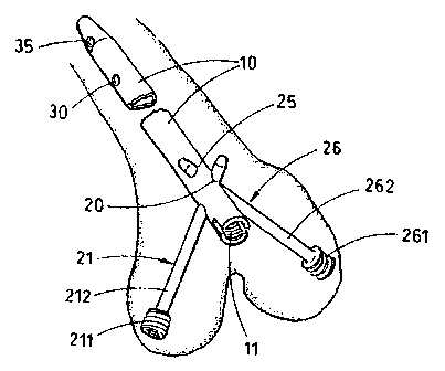
This invention relates to a surgical intramedullary mail (10), for stabilisation of condylar and supracondylar fractures of the femur. It
incorporates a Cruciate arrangement of two obliquely crossing locking bolts (21 and 26) such that each condyle of the femur is gripped by
an individual bolt. By this means both femoral condyles are stabilised with respect to the shaft of the femur. The bolts are oriented so that
each passes through the main extra-articular mass of each condyle, Further predrilled holes are provided (30, 35) for insertion of proximal
locking bolts to stabilise the nail with respect to the shaft of the femur. The nail is intended for retrograde insertion from distal to proximal
into the intramedullary canal of the femur. Insertion of the locking bolts may be facilitated by the use of a temporary jig which is attached
securely to the distal end of the nail (11). Nails of similar design can be used for stabilisation of equivalent fractures of the humerus.
L'invention concerne un clou intramédullaire chirurgical (10) destiné à stabiliser des fractures condyliennes ou supracondyliennes du fémur. Il comprend un dispositif cruciforme composé de deux boulons (21, 26) de blocage à croisement oblique, de façon que chaque condyle du fémur soit maintenu par l'un d'entre eux. Les deux condyles du fémur sont ainsi stabilisés par rapport au corps du fémur. Ces boulons sont orientés de façon à traverser la masse extra-articulaire principale de chaque condyle. D'autres perforations pré-percées (30, 35) sont prévues pour l'insertion de boulons de blocage proximaux destinés à stabiliser le clou par rapport au corps du fémur. Ce clou est conçu pour une insertion rétrograde, de l'extrémité distale à l'extrémité proximale, dans le canal intramédullaire du fémur. L'insertion des boulons de verrouillage peut être facilitée par l'utilisation d'un gabarit de couplage temporaire solidement fixé à l'extrémité distale du clou (11). Des clous de conception similaire permettent de stabiliser des fractures équivalentes de l'humérus.
Note: Claims are shown in the official language in which they were submitted.
Note: Descriptions are shown in the official language in which they were submitted.
2024-08-01:As part of the Next Generation Patents (NGP) transition, the Canadian Patents Database (CPD) now contains a more detailed Event History, which replicates the Event Log of our new back-office solution.
Please note that "Inactive:" events refers to events no longer in use in our new back-office solution.
For a clearer understanding of the status of the application/patent presented on this page, the site Disclaimer , as well as the definitions for Patent , Event History , Maintenance Fee and Payment History should be consulted.
| Description | Date |
|---|---|
| Inactive: Expired (new Act pat) | 2015-06-09 |
| Letter Sent | 2013-06-26 |
| Inactive: Single transfer | 2013-05-17 |
| Grant by Issuance | 2007-05-01 |
| Inactive: Cover page published | 2007-04-30 |
| Inactive: Final fee received | 2007-02-21 |
| Pre-grant | 2007-02-21 |
| Notice of Allowance is Issued | 2006-08-24 |
| Letter Sent | 2006-08-24 |
| Notice of Allowance is Issued | 2006-08-24 |
| Inactive: Approved for allowance (AFA) | 2006-06-09 |
| Amendment Received - Voluntary Amendment | 2006-04-03 |
| Inactive: S.30(2) Rules - Examiner requisition | 2005-11-08 |
| Amendment Received - Voluntary Amendment | 2005-08-12 |
| Letter Sent | 2005-07-18 |
| Reinstatement Requirements Deemed Compliant for All Abandonment Reasons | 2005-06-28 |
| Deemed Abandoned - Failure to Respond to Maintenance Fee Notice | 2005-06-09 |
| Inactive: S.30(2) Rules - Examiner requisition | 2005-02-17 |
| Inactive: Application prosecuted on TS as of Log entry date | 2002-05-30 |
| Letter Sent | 2002-05-30 |
| Inactive: Status info is complete as of Log entry date | 2002-05-30 |
| Request for Examination Requirements Determined Compliant | 2002-05-23 |
| All Requirements for Examination Determined Compliant | 2002-05-23 |
| Inactive: Delete abandonment | 2000-07-14 |
| Inactive: Office letter | 2000-07-14 |
| Deemed Abandoned - Failure to Respond to Maintenance Fee Notice | 2000-06-09 |
| Small Entity Declaration Determined Compliant | 1996-11-27 |
| Application Published (Open to Public Inspection) | 1995-12-21 |
| Abandonment Date | Reason | Reinstatement Date |
|---|---|---|
| 2005-06-09 | ||
| 2000-06-09 |
The last payment was received on 2006-05-17
Note : If the full payment has not been received on or before the date indicated, a further fee may be required which may be one of the following
Please refer to the CIPO Patent Fees web page to see all current fee amounts.
Note: Records showing the ownership history in alphabetical order.
| Current Owners on Record |
|---|
| BIOMET TRAUMA, LLC |
| Past Owners on Record |
|---|
| MICHAEL GORDON MATTHEWS |

