Note: Descriptions are shown in the official language in which they were submitted.
21 gl 822
_~170mos.ein 27/11/96
STEREOTACTIC RADIOSURGERY
FIELD OF THE INVENTION
The present invention relates to radiosurgery generally, and particularly to
stereotactic radiosurgery.
BACKGROUND OF THE INVENTION
Stereotactic radiosurgery provides a dose of radiation in a target volume in a
patient. The target is irradiated at a multiplicity of orientations with finely collimated beams.
The use of stereotactic radiosurgery to render tissue necrotic is well established
and various systems are currently used for stereotactic radiosurgery. The prior art recognizes the
need to confine radiation as much as possible to the target volume being treated. Generation of a
desired dose pattem at the target volume is the objective of a treatment plan which takes into
account limitations of the particular radiosurgical system used. Typical types include a Gamma
Unit which utilizes a multiplicity of Cobalt-60 sources arranged on a spherical surface, a linear
accelerator (LINAC) which utilizes a photon beam source mounted on a rotating gantry, and a
stationary generator for a beam of charged particles. These radiosurgical systems, as well as
associated methods, characteristics and performances are described in various publications, e.g.,
in Stereotactic Radiosurgery, Alexander E. et al., McGraw-Hill, 1993; and in Neurosurgery
Clinics of North America, vol. 3, no. 1, Lunsford L. D. (editor), W.B. Saunders Co., Jan. 1992.
Treatment planning capabilities include a selection of a dose level to the target, a
choice of collimators for beam shaping and a detellllindlion of beam orientations at which radia-
tion is applied to the target volume. In order to reduce the dose deposited in healthy tissue
outside the target volume, it is generally desirable to spread beam orientations over a wide range.
The prior art describes beam orientations of the Gamma Unit as being fixed
relative to the stationary unit. The orientation of the beam with respect to the target may be
determined only by selecting the initial elevation angle of the patient's head relative to the unit.
Radiation intensities and exposure times of all unplugged bearns are identical for all orientations.
Dose pattems may be shaped only by the elimination of selected beams through plugging the
corresponding collimators prior to treatment.
21gl822
-
A typical implementation of LINAC sc~nnin~ involves rotating the LINAC gantry
about its horizontal axis which orthogonally intersects the beam and the target volume at an
isocenter. Such rotation causes a beam of radiation to trace an arc on a sphere surrounding the
target. A multiplicity of non co-planar arcs is produced by consecutive gantry rotations, each one
associated with an increment of the azimuthal orientation of the patient. The number of arcs is
typically between 4 and 11. Th beam intensity for each arc stays constant throughout the contin-
uous arc irradiation. The heavy rotating gantry is associated with added expense and reduced
accuracy.
Another prior art implementation of LrNAC beam scanning is by rotating the
patient about a vertical axis which intersects the target and irradiating with a beam which is angled
with respect to the axis. Each such rotation is geometrically equivalent to the beam forming a
conical surface of radiation. Incrementing the beam slant angle between consecutive rotations
produces a multiplicity of coaxial conical radiation surfaces having the target in the focus. Here
too, the beam intensity for each conical scan stays constant throughout the irradiation.
Yet another prior art implementation of LINAC beam scanning is by simultaneous
rotation of the LINAC gantry and of the patient turntable. The orthogonal axes of rotation
intersect the radiation beam at an isocenter coinciding with the target. The rotational speed about
each axis is constant. The beam intensity, however, remains constant throughoutthe continuous
irradiation. The heavy rotating gantry is associated with added expense and reduced accuracy.
Charged particles stereotactic radiosurgery uses a di~erelll approach. Since thehorizontal radiation beam is stationary, beam orientations are obtained by incrementing the
~7im~1th~1 orientation of the patient as well as the roll angle of the patient about a longitudinal
horizontal axis. The axes of rotation intersect the radiation beam orthogonally at an isocenter
coinciding with the target. Irradiation is discrete (i.e. non-continuous) from a small number of
orientations. Time con~llmin~ positioning ofthe patient is required for each such orientation, thus
preventing the use of a large number of orientations.
- 2191822
SUMMARY OF THE ~NVENTION
The present invention seeks to provide improved apparatus and techniques for
radiosurgery which represent a radical departure from the prior art.
The present invention provides a treatment modality utilizing any conventional or
other radiation source and introduces treatment planning options and parameters which are not
reflected in the prior art.
In the present invention, in contrast with the prior art, a large number of
orientations may be used and the dose delivered from each orientation is controllable. Unlike the
prior art, a continuous orientations scan and dose control may be implemented utilizing a radiation
source which is stationary during irradiation.
The present invention provides apparatus for selectively irra~i~ting a target volume
of tissue within a patient. A target is identified in a patient and a desired radiation dose pattern for
therapeutic irradiation of the target volume is selected.
A collimator is selected in accordance with the treatment plan. A radiation source,
such as a LINAC, heavy particle generator or cobalt-60 source, emits a radiation beam through
the selected collimator. The collim~ted radiation source is positioned at predetermined
orientations relative to the target volume so that the desired radiation dose to the target volume is
achieved in increments from di~elenl beam paths.
The positioning of the collim~ted radiation beam relative to the target volume is
achieved by means of a positioner attached to an immobilizing device, such as a stereotactic
frame, which is in turn rigidly attached to the patient. The positioner moves and rotates the patient
:im~lth~11y and elevationally relative to the radiation beam during irradiation.The positioner comprised in the present invention permits the positioning of thetarget tissue volume in different orientations relative to the radiation beam by rotating the patient
a7imllth~11y and elevationally relative to the radiation beam during irradiation. This permits the
implementation of more versatile and varied irradiation plans than are available in the prior art.
As distinguished from the prior art, the present invention includes a motion
controller which permits control over the dwell time of the irradiation from each orientation
relative to the target volume. This provides the flexibility of delivering different doses of radiation
from difrel en~ orientations.
2191822
-
During radiation treatment, there may be varying beam attenuations caused by
different tissue volumes in the beam path intervening between the radiation source and the target
volume. The ability to vary the doses of radiation from different orientations compensates for the
varying beam attenuations.
The ability to vary the time dwell time of the radiation beam at different
orientations relative to the tar~et volume facilitates the calculation and implementation of more
versatile and varied radiation plans than are available in the prior art.
There is thus provided in accordance with a preferred embodiment of the present
invention, a positioning system for use in radiosurgery including a frame rigidly attachable to a
part of a patient, a positioner operative to move the frame together with the part of the patient
rigidly attached thereto, in at least one of six degrees of freedom, sensing apparatus operative to
measure position and orientation of the frame with reference to a center of rotation, a motion
controller operative to control the position and the orientation of the frame relative to the center
of rotation in a time domain according to a predetermined formula.
In accordance with a prefelled embodiment of the present invention, the positioner
is operative to rotate the frame and part of the body attached thereto around two axes of rotation,
and wherein the first axis of rotation is generally perpendicular to the second axis of rotation.
There is also provided in accordance with a plef~lled embodiment of the present
invention, an irr~rli~ting system for use in radiosurgical treatment including a source of radiation
arranged to produce a radiation beam operative to irradiate a target volume in a patient and a
positioning system as described hereinabove.
In accordance with a preferred embodiment of the present invention, the irra~i~ting
system also includes a beam controller for controlling the intensity of the radiation beam
according to the position and the orientation of the frame.
The source of radiation is preferably a linear accelerator (L~NAC).
There is also provided in accordance with a preferred embodiment of the present
invention, a method for radiosurgical treatment of a patient including the steps of:
selecting a set of orientations of radiation beams operative to irradiate a target
volume in the patient;
2191822
calculating a corresponding set of dose quotas delivered by each radiation beam at
each orientation, wherein an accumulation of the dose quotas provides a desired dose pattern in
and around the target volume;
determining for each dose quota a corresponding radiation intensity and exposuretime which together produce the dose quota; and
irra~i~ting the, target volume from each orientation according to each
corresponding radiation intensity and exposure time.
In accordance with a prefelled embodiment of the present invention, the method
further includes providing an irra~i~tin~ system, as described above, wherein the source of
radiation is substantially stationary during irradiation, positioning the frame and the part of the
patient rigidly attached thereto such that the target volume of the patient substantially coincides
with the center of rotation of the positioning system, and using the positioning system to move the
target volume according to the set of orientations of radiation beams while irr~ ting the target
volume according to the corresponding radiation intensity and exposure time.
2191822
BRIEF DESCRIPTION OF THE DRAWINGS
The present invention will be understood and appreciated more fully from the
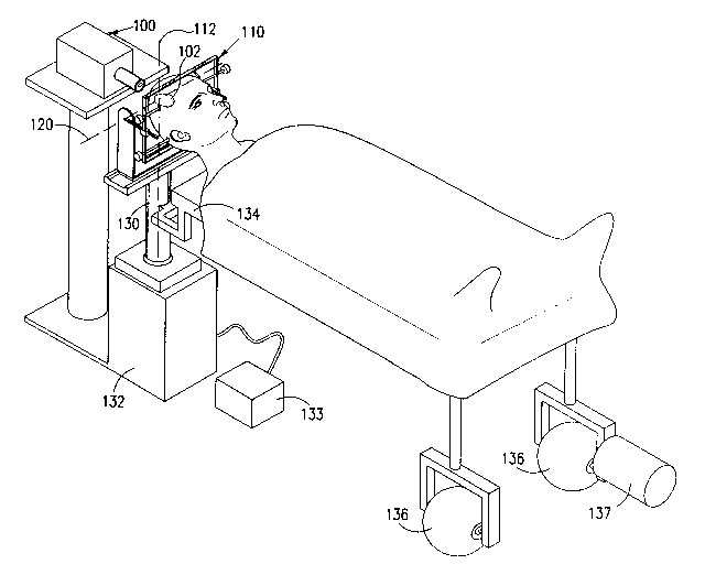
following detailed description, taken in conjunction with the drawings in which:Fig. I is a simplified pictorial illustration of treatment of a patient using apparatus
constructed and operative in accordance with a preferred embodiment of the present invention;
Fig. 2A is a simplified pictorial illustration of part of the apparatus of Fig. I;
Fig. 2B is an illustration of use of the apparatus of Fig. 2A in treatment of a
patient;
Fig. 3 is a simplified pictorial illustration of rotation of a patient about an elevation
axis using apparatus illustrated in Fig. 1;
Fig. 4 is a simplified pictorial illustration of rotation of a patient about an ~7im~1th~1
axis using apparatus illustrated in Fig. l;
Fig. 5 is a simplified pictorial illustration of a pattern of irradiation impinging on a
patient's skull, using the apparatus of Fig. l; and
Figs. 6A and 6B are simplified pictorial illustrations of treatment of a patient using
apparatus constructed and operative in accordance with another preferred embodiment of the
present invention.
2191822
DETAILED DESCRIPTION OF PREFERRED EMBODIMENTS
Reference is now made to Fig. 1, which is a simplified pictorial illustration oftreatment of a patient using apparatus constructed and operative in accordance with a preferred
embodiment of the present invention. Preferably an interchangeable collimator radiation
generator 100, is employed to direct a beam of radiation onto a target location 102 in a patient's
head.
In the present embodiment, the patient's head is securely and precisely held in
position preferably by means of a stereotactic frame assembly 110, such that the target location
102 lies along a vertical axis 112, about which the entire patient is rotatable. The stereotactic
frame assembly 110 defines an elevation axis 120, which intersects the vertical axis 112 at the
target location 102.
The stereotactic frame assembly 110 is mounted on a post 130 which is aligned
along and rotatable about axis 112. Post 130 is tr~n.cl~t~ble in a plane perpendicular to axis 112,
and is also selectably raisable and lowerable along axis 112, by means of an X-Y-Z translator 132.
The motion of translator 132 is preferably controlled in a closed control loop by a motion
controller 133 and appropriate sensors (not shown). Fixedly mounted onto post 130 is a patient
support platform 134 which is provided with wheeled supports 136 at an end thereof opposite to
post 130. An ~7imllth motor 137 may be mounted on one of the supports 136 for rotating
platform 134 about axis 112.
Referring now additionally to Fig. 2A, it is seen that the stereotactic frame
assembly 110 comprises a generally horizontal base member 150 which is fixedly mounted onto
post 130 and is tr~n~l~t~ble and rotatable together therewith. Horizontal base member 150 is
formed with a track 152 along which a support element 154 is selectably displaceable in one
dimension along the track. The elevation axis 120 is defined with respect to element 154 and
intersects a pair of upst~ndina arms 156 and 158 thereofat locations 160 and 162 respectively.
A pair of pivot arrns 164 and 166 are pivotably mounted onto upstanding arms 156and 158 at locations 160 and 162 respectively. Slidably mounted onto each of pivot arms 164 and
166 are respective mounting sliders 168 and 170. Slidably mounted onto mounting sliders 168 and
170 are respective side portions 172 and 174 of a head mounting frame 176. Head mounting
frame 176 is preferably slidable with respect to sliders 168 and 170 along an axis, indicated by
21~1~22
ar~ws 178, which is perpendicular to the longi~utlin~l axes of pivot arms 164 and 166,
respectively indicated by arrows 180 and 182.
A plurality of mounting screws 184, which may be threadably mounted onto head
mounting frame 176, are preferably employed to rigidly engage the skull of a patient and thus
securely mount it with respect to frame 176, as seen in Fig. 2B. It is appreciated that stereotactic
frame assembly 110 may be used not only for the skull, but for irr~ ting any other portion of the
patient's body.
In operation, frame 176 is securely ~tt~hed to the skull of a patient. The target
location 102, for example, of a tumor, is determined by any suitable method, such as
computerized tomography. Frame 176 is then positioned with respect to interchangeable
collimator radiation generator 100 (Fig. 1) by suitable adj--strnent of any or all of support element
154, and mounting sliders 168 and 170, such that a center of rotation 190, located at the
intersection of axes 112 and 120, is located substantially at the target location 102.
Reference is now made to Fig. 3 which is a simplified pictorial illustration of
rotation of a patient about elevation axis 120 using app~L-ls illustrated in Fig. 1. It is noted that
the location of the center of rotation 190 with respect to a beam 192, generated by
interchangeable collimator radiation generator 100 and directed at center of rotation 190, remains
substantially unchanged during any elevational rotation.
Reference is now made to Fig. 4 which is a simplified pictorial illustration of
rotation of a patient about ~imllth~l axis 112 using app~s illustrated in Fig. 1. It is noted that
the location of the center of rotation 190 with respect to beam 192 remains substantially
unchanged during any ~7imllth~l rotation.
Reference is now made to Fig. 5 which is a simplified pictorial illustration of an
irradiation matrix projected on a patient's skull, using the apparatus of Fig. 1, and by suitable
elevational and ~7imllth~l movement of the patient. Each cell of the matrix is irradiated with a
dose quota delivered by a set of radiation beams, wherein an ~cc.-m--l~tion of the dose quotas
provides a desired dose pattern in and around the target volume. For each dose quota there is a
corresponding pair of a radiation intensity and an exposure time, which together produce the dose
quota.
2191822
Reference is now made to Figs. 6A and 6B which are simplified pictorial
illustrations of treatment of a patient using apparatus constructed and operative in accordance
with another preferred embodiment of the present invention. A linear accelerator 200 is preferably
housed in a gantry 202 and is operative to irradiate a patient via a collimator 204.
The stereotactic frame assembly 110, described hereinabove with reference to Figs.
I - 4, may be used with linear~accelerator 200 to irradiate a patient. A patient may be rotated
elevationally about an axis 206 and azimuthally about an axis 208, in a manner similar to that
described hereinabove with reference to Figs. 3 and 4. In Fig. 6A, azimuthal movement of the
patient may be restricted generally in an angular region designated by reference numeral 210. By
rotating gantry 202 about an axis 211 to the position shown in Fig. 6B, the patient may be rotated
an additional angular region 212.
It is appreciated that various features of the invention which are, for clarity,described in the contexts of separate embodiments may also be provided in combination in a
-single embodiment. Conversely, various features of the invention which are, for brevity, described
in the context of a single embodiment may also be provided separately or in any suitable
subcombination.
It will be appreciated by persons skilled in the art that the present invention is not
limited to what has been particularly shown and described hereinabove. Rather, the scope of the
present invention is defined only by the claims that follow:
