Note: Descriptions are shown in the official language in which they were submitted.
~ W096/039~2 ' ~1 95949 Pcr~sgsll017g
FEMALE INCONTINENCE DEVIOE INCLUDING
ELECTRONIC SENSORS
R ~ ''U O I~I IJ
This i6 a continuation-in-part application of U.S.
application Serial No. 07/888,597 filed on May 27, 1992
now US Patent 5,352,1a2 issued Oct. 4, 1994. The present
invention relates to a device and a method to control
urination in an incontinent female and simultaneously
monitor bladder pressure and urine chemistry.
A wide spread medical problem suffered by at least
11 million American adults, particularly women is
incontinence. Many more suffer from the problem but,
because of embarrassment or because the problem is only
intermittent,, don't disclose their lack of bladder
control. There are numerous causes including pregnancy,
stress, as well as the normal aging process.
There presently are no adequate non-surgical
techniques for treating this problem. Catheters with an
attached bags are uncomfortable and are known to lead to
urinary infection. Pads may be effective for small
urinary leakage, such as occurs under stress, but are not
suitable for large volumes of urine evacuated from a full
bladder. Additionally, the use of pads requires the user
to carry a large supply of replacement pads. Urethral
plugs are unacceptable replacements because the user
frequently will soil her hands trying to remove the
device, reinsertion of the nonsterile device may lead to
a bladder infection and the frequent insertion of the
~ plug, possibly a do~en times a day, can damage the
urethra and may cause bladder spasms. Plugs may also be
W0961~3~2 ~ q ~ ~4 9 PCT~59~/1017
dangerous because they totally obstruct the urethra and
may result in excessive retention of urine.
A further problem which exists with certain
bedridden female patients is that they may not have the
ability to reach a bathroom to void and may need nursing
assistance or they may not have the ability to sense when
their bladder i8 full so that they can urinate without
soiling themselves or the bed linen.
Thus there i6 a need for a convenient, relatively
clean, and frequently repeatable procedure which can be
practiced by the woman and nursing personnel and devices
which can be used in the procedure. The devices must
also be safe to use, discrete, and reliable so that the
woman can participate in a normal life style without fear
of embarrassing herself by accidentally voiding the
contents of her bladder or constantly running to the
bathroom to change pads. Still further, there i8 a need
for medical personnel to conduct routine urinalysis on a
clean urine sample so they can be aware of the onset of
urinary infection or other medical events which can be
identified by periodic urinaly8is.
S~MNARY
The present invention is directed to a device and
method that sUpplies these needs and eliminates the
deficiencies of prior devices and systems.
The device of the invention comprises a valved
drainage catheter for temporary placement in the female
urethra. A~d;tl~nAlly, the system also includes a sizing
device for selecting the proper length catheter, a
~ placement device to assure that the catheter is properly
po8itioned and instruments to aid in opening the valve in
~ W096/03942 ;~ 2 i q5949 PCT~S9j/10179
the catheter.
he catheter comprises a hollow tube with an
extendable mushroom head on the internal end, a mushroom
shaped cap on the external end and a manually openable
valve between the internal end and the external end or
within the mushroom shaped cap on the external end to
prevent urine from exiting the catheter prematurely.
Also, the catheter includes one or more sensors to
measure bladder pressure and chemical and physical
properties of the urine, means to transmit information
generated by the sensors to a remote location and, if
nece3sary, energy sources to power the sensOrs and
transmission device. The sizing device is of similar
shape as the drainage catheter except the catheter has a
longer length, the outer surface has measurement indicia
spaced along its length and the mushroom cap is replaced
by a removable disc shaped similar to the mushroom cap.
After insertion of the sizing device in the urethra, the
disc is slid along the external portion of the catheter
ur.til it rest snugly against the perineal area. The
indicia exposed below the disc indicates the correct
catheter length for a proper fit.
o place the catheter, the system inclu'~ ~ stylet
for insertion into the c.~ neter. ~-&~nage is
accom~ shed by using specially designed valve openers.
DRANINGS
These and other features, aspects and
advantages of the present invention will become better
understood with reference to the following description,
appended claims, and accompanying drawings, where:
WO 9611)3g~2 ~ f ~ ~ 9 ~ ~ 4 9 ~usg~ 7~ ~
Figure l is a side view showins the drainage
catheter placed in the urethra of a female, the female
~ody ~eing shown in cross section.
Figure 2 is a cutaway side view of the drainage
catheter taken along line 2-2 of Figure l.
Figure 3 is a top view of a first embodiment of the
lOinternal end of the drainage catheter of Figure 2.
Figure 4 is a top view of a second em'oodlment of the
internal end of the drainage catheter of Figure 2.
15Figure S is a cutaway side view of the drainage
catheter taken along line 2-2 of Figure 1, the catheter
being extended for placement.
Figure 6 is partial cutaway view of a sizing device.
Figure 7 is an enlarged cutaway side view of the
valve section of the drainage catheter of FigUre 2 with
a drainage straw inserted.
25Figure 8~ is an enlarged cutaway side view of the
valve section of the drainage catheter of Figure 2
showing an alternative construction with a plug inserted.
Figure 9~;is an enlarged cutaway side view of the
30valve section of the drainage catheter cf Figure 2
showing an al~ernative construction with a magnetic ball
valve inserted, the valve being in its closed positio~.
Figure 10 is an enlarged cutaway side view of the
35alternative valve 5ection of Figure 9 in its open
WO9~/0394~ 'r,3~ 2l9 59 4q PCT~lS9~/l0l7g
position.
igure 11 is an enlarged cutaway view of the valve
section of the drainage catheter of Figure 2 showing a
second alternative ball valve structure in its closed
structure.
Figure 12 is an enlarged cutaway side view of the
second alternative valve section of Figure ll in its open
position.
Figure 13 is a side view of a second version of a
drainage catheter for controlling female incontinence.
Figure 14 is a cutaway side view of the second
version of the drainage catheter taken along line 14 -14
of Figure 13.
Figure 15 is an enlarged cutaway side view of the
sensor ring portion of the second version of the drainage
catheter taken along line 14 -14 of Figure 13.
Figure 16 is an enlarged cutaway side view of the
pressure transducer mounted in the sensor ring portion of
the second version of the drainage catheter taken along
line 16 -16 of Figure 15.
Figure 17 i6 a cutaway side view of the second
version of the drainage catheter taken along line 14-14
of Figure 13 showing a further valve design.
DESCRIPTION
Figures 1 through 16 show drainage catheters
embodying features of the invention.~5
W096l03942 '' ~ t 9~94~ PCT~ 0179
The drain~ge catheter lO includes a tubular center
section 12 with a sealing portion on the internal end 14
and a cap 16 or: the external end. ln the P~o~;m~nt
sho~n in Figure 2, the sealing portion is a mushroom
shaped crown 18 which can be extended for placement of
the catheter. The crown has several drainage holes 20
located in its upper surface. Enclosed in the lumen 22
of the catheter is a one WAy valve 24 which can be opened
by the woman using the catheter lO. In the center of t~.e
cap is an drainage outlet 32.
Alternate designs for the top surface of the crown
18 are shown in Figures 3 and 4. Figure 3 shows six
large drainage holes 20 separated by spokes 26. Figure
4 shows a similar cro~n 18 design having smaller holes 20
in the crown surface 28. Shown in phantom in Figure 4
are struts which depend from the lower or inner surface
of the crown 18 to ~s~ure t.hat drainage through the holes
20 and into the lumen 22 i.s not ~locked.
The valva 24 shown in Figures 2, 5 and 7 is of A
duck bill design which prevents fluid from descending
down the tube unless the valve is purposefully opened.
Figures 8 - 12 ~how two alternztive valve structures and
Figure 14 incorporates a slit valve structure which will
be discussed ~elow.
To insert tha cathater 10 into the female urethra a
stylet 30 is inserted through the drainage outlet 32 and
valve 24 until it comes in contact with the reinforced
center 34 of the crown 18. The ~tylet 3C is then
advanced further extending the crown 18 until the
diameter of the outer surface of the crown i~ about the
~ same as the diameter of the catheter 10. The extended
crown 18 is then inserted through the extarnal opening of
~ wog6/03~42 ' $~ 2~95949 PCrlUS9~/10179
the urethra anà advanced until it enters the bladder. If
the catheter is properly sized, the inner surface 36 of
the cap 16 should be resting snugly against the skin
surrounding the external opening of the urethra.
Insertion of the catheter 10 may be assisted by applying
a small amount of a sterile lubricant to the crown 18.
The stylet 30 is then removed while the cap 16 is held in
place. Removal of the stylet 30 allows the crown 18 to
return to its normal shape with the crown inner surface
38 resting against the bladder surface as shown in Figure
1.
In order to minimize or eliminate leakage around the
catheter 10 the length L from the crown inner surface 38
to the cap inner surface 36 should be accurately
determined. To do so the sizing device 50, shown in
Figure 5, is used. The sizer 50 is of substantially the
same shape and has the same outer dimensions as the
catheter 10 with the exception that the sizer is longer
in length than the catheter 10. However, in place of the
cap 16 the sizer 50 has a removable disk 52 which can
slide along the outer surface of the sizer tube 54. At
least a portion of the tube outer surface has indicia 56
thereon for use in selecting the proper catheter length
L. Using the stylet 30 the sizer 50 is inserted into the
urethra and the crown is allowed to prolapse against the
bladder neck. The disc 52 is then slid along the sizer
outer surface until it rests snugly against the tissue
surrounding the urethra and the measurement marked on the
outer surface of the tube 54 is read. The measurement
indicates the catheter size to use for proper fit.
Figure 7 shows the valve section of the catheter of
Figure 2 greatly enlarged to show the functioning of the
valve during a drainage procedure. To drain the bladder
W096~39~2 ~ 9 5 9 ~ 9 PCT~S9.~110179
a sterile spike 60 is inserted through the drainage
outlet 32 in the external end of the implanted catheter
10 . The spike 60 has a center portion 64 sized to fit
snugly in the drainage outlet 32 and to open the valve
24. The spike 60 has a tapered head 62 on the top end of
the tubular center portion 64 and an enlarged diameter
handle 66 on the lower portion. Ports 72 are located at
the juncture of the head 62 with the central portion 64.
~ central lumen 68 starts at the ports 72 and runs the
length of the spike 60 terminating in an opening 70 at
the base of the handle 66. While the diameter of the
central portion 64 is sized to fit snugl~ ~through the
drainage port 32, the handle diameter i8 chosen 80 that
it will not easily enter the drsinage port 32, thus
preventing the head 62 of the spike from being inserted
to far into the catheter 10 and damaging the crown 18 or
the bladder. Additionally, the combined length of the
head 62 and the central portion 64 is chosen 80 that when
the top end ?4 of the handle 66 rests against the
drainage opening 32, the head 62 pierces the drainage
outlet 32 and the valve 24, exposing the ports 70 to a
standing column of urine in the catheter 10 above the
valve 24. This cooperation of parts allows the user to
drainage the bladder without soiling her hands from
leaking urine, Once drainage is complete the spike is
withdrawn and the valve clo9es and seals. The spike can
then De disposed of or resterilized.
Figure ~ shows and alternate valve rs~h~n; cm
comprising a plug 80 sized to ~it in the drainage opening
32. The plug can be used in place of the valve 24 (as
shown in Figure 8) or in combination with the valve 24 as
additional protection against leakage (not shown~. The
plug is shown with a draw string 82 and a pull tab 84.
~ wo g6/03942 ~ 2 1 9 5 9 4 9 PCT~S9~1~179
Figures 9 and lO show the catheter 10 with a ball
valve mechanism 90 inserted in the external end of a
drainage catheter 10. The catheter 10 is shown to have
a uniform inner diameter along it entire length. After
insertion of the catheter 10 using the stylet 30 and
removal of the stylet the ball valve 90 is placed and
secured in the catheter lO. The ball valve 90 comprises
a ball 92 which is attracted by a magnetic disc 94 placed
over the drainage outlet 96. Also enclosed in the valve
90 is a spring 98 which lifts the ball 92 off the seat
lO0 when the magnetic disc 94 is removed. To raise the
ball 92 off the seat 100 the tab 102 is grasped and
pulled downward. The removal of the magnetic force
allows the spring to lift the ball 92 unsealing the
drainage outlet 96 so that the urine can flow through the
valve openings 104 ar.d out the catheter 10.
Figures ll and 12 show the catheter 10 with a ball
valve mechanism 110 inserted in the external end of a
drainage catheter 10. Like the ~ ' ~;mFnt shown in
Figures 9 and 10, the catheter is also shown to have a
uniform inner diameter along its entire length. The
valve mechanism 110 is inserted into the catheter 10
after its placement in the urethra. The ball valve 110
comprises a ball 112 which is held against the drainage
opening 114 by the spring 116. To raise the ball 112 off
the seat 113 a spike 120 is inserted through the drainage
opening 114 until the shoulder 122 contacts the bottom
124 of the valve, compressing the spring 116 allowing
urine to flow through the valve 110, ports 126 and
drainage channel 128 through the center of the spike 120.
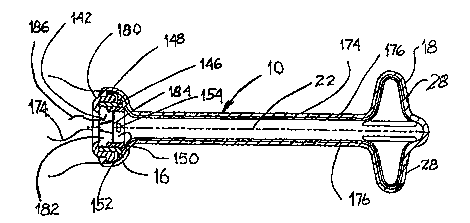
Figures 13 through 16 show the catheter 10 with a
slit valve r--h~ni~m 140 inserted in cap 16 at the
external end of a drainage catheter 10. Like the
WOs6~J3g42 ~ 0~ ~..f.~ 5949 PCT~Ss5ll0179
embodiments shown in Figures 1 khrough 12, the catheter
10 is also shown to have a uniform inner diameter alons
its entire length. The slit valve 140 includes a center
portion 142 curved inward towards the catheter lumen 22
with a slit 144 located in the center thereof. The slit
144 is a one way ~alve which acts in ~he same manner as
the duck bill valve discussed above in regard to ~igures
1 - 7. To ~apcn the slit 144 a spike 120 is inserted
through the slit 144 unti]. the shoulder 122 contacts the
center portion 142 o~ the valve allowing urine to flow
through the ports 72, drainage channel 128 and through
the center of the spike 60.
To aid in holdiny the slit valve 140 in the cap 16,
i5 a locking ring 146 is positioned against the internal
surface of the slit valve 140, locking the edge 148 of
the slit valve 140 against the internal surface of the
cap 16. Positioned lnternally of the locking ring 146 is
a sensor ring 150 enlarged and shown in a cutaway view in
Figure 15. As the bladder fills with urine the area
within the catheter 10, including the lumen 22 and volume
152 enclosed by the cap 16, also fills with urine and the
pres8ure within the bladder is uniformly distributed
throughout the volume o~ urine. Mounted in the sensor
ring 150 is a pressure sensor 154 such as a pressure
transducer or piezoelectric chip 156. An example of a
pressure sensor using a piezoelectric chip is shown in
Figure 16 wherein in the chip 156 is mounted between two
edge supports 1513 over an enclosed air space 160.
Pressure against the ~ace 162 of the chip causes the chip
156 to bend into the air space and, in turn, create an
electrical signal which can be detected, measured, and
correlated with the pressure applied to the iace 162.
The signal transmitted along electrical lead 164 can then
be ied directly to a moritoring device or to a signal
~ W096~3942 ~ 2 t 9 5 9 4 9 PCT~S9S/10179
11
transmitter 166, such as an FM transmitter, for
monitoring at a remote location. The transmitter can be
powered by a power source 168 also mounted in the sensor
ring 150 or external of the catheter. In a still further
embodiment, the sensor ring may include positive and
negative electrodes 170, 172 composed of dissimilar
metals. The normal urine is usually acidic with a pH of
5.5 - 6.5 and thus can act as a conductive medium which
can serve as the electrolyte creating a battery in
conjunction with the positive and negative electrodes
170, 172. ~bnormal urine may have a pH from 5 to 8.5.
The assembly creates an electrical current which can be
stored in the power source 168 or directly power the
transmitter 166.
Imbedded in the wall 174 and extending along the
length of the catheter 10 are signal transmission means
176, such as electrical wires or optical fibers, which
are connected on a first end to sensors 178 and on a
second to a signal receiver (not shown). The sensors are
located in the crown 18 with their sensing surfaces being
exposed to the urine in the bladder. The sensors 178 can
have various different capabilities which one skilled in
the art of urinalysis would recognize and find valuable
in monitoring a patient. For example, the sensors 178
can electrically or optically detect pH, temperature,
pressure and various different chemical constituents of
urine such as sodium, potassium, glucose, drug markers
and metabolites, specific gravity, proteins, leukocyte
esterase, nitrites, urobilinogen, whole or crenated blood
cells, ketones, bilirubin, turbidity and color. Figures
13 and 14 show a crown 18 with four struts 28 and four
sensors. However, this is not intended to place a limit
on the number of sensors as more than one signal
transmission means can be placed along a strut 18, more
~0~6l03942 ~ 95~ PCT~Sg~110179
than four struts 18 can be used, as shown in Figures 3
and 4, or a single sensor can detect various different
materials. As an example, an optical fiber capable of
transmitting I~ or W light can be used. Different
frequency light transmitted along the signal transmission
means can directly, or as a result of signal responsive
fluorescence,~quantitatively or qualitatively determine
chemical constituents in the urine in the bladder.
While Figure 1~ shows the fiignal transmission means
176 exiting through the cap 16, it is also contemplated
that they can be connected directly to the sensor ring
150 and the signal transmitted as described aoove. ~s a
further alternative, the sensors 178 can be mounted
directly on the sensor rir.g 150. This presents a
particular advantage for optical sensing as transmitter
and receiver fiber ends can be mounted opposite each
other to detect and analyze light transmitted through the
urine.
~ particular advantage o~ the embodiment
incorporating sensors and a signal collection and~or
transmission means is that nursing personnel can readily
monitor a bedridden patient or a patient unable to
manipulate the drainage device. Pressure signals from
the pressure sensor 154 will indicate when the patient
has to void so~that assistance can be provided before the
be~l;n~nc are soiled. Abnormal temperature readings can
be indications of and early warnings of bladder or
systemic infection so that treatment can be instituted at
an early stage. The online, continuous chemical analysis
of the urine can be a valuable addition to control of
drug treatment or a diagr.ostic tone in the indication of
the need for additional or changed treatment.
wog6/03942 ~ ; 2 1 95949 PCTlUSg~ 7g
13
A still further version of the urinary drainage
catheter, shown in Figure 17, includes an electrically
activated valve 180. The valve, which comprises a valve
body 182 and a movable plug 184, can be opened by an
electrical signal transmitted along an input lead 186 or
by way of a radïo signal (i.e., a short range FM signal)
transmitted to a receiver ~not shown~ in the valve 180 or
in the sensor ring 150, the receiver being operatively
connected to the movable plug 184.
The catheter 18 can be fabricated from a broad range
of materials presently used for forming urinary catheter
including, but not limited to natural and synthetic
rubbers, silicone rubbers, thermoplastic elastomers,
latex, polyvinyl chloride, polyethylene, and PTFE with or
without coatings such as silicone materials, Teflon,
hydrophilic compounds and other materials which improve
the compatibility with mucosal tissue. Additionally,
antibacterials, anti-inflammatory drugs, antibiotics or
other drugs can be coated on the catheter surface or
absorbed into the coatings on the catheter surface. In
the embodiment of Figures g and 10, the ball 92 is a
magnetic material, preferentially a plastic material
having magnetic materials or magnetizable materials
dispersed therein or ceramometallic materials. The spike
66 or 120 may be formed from a broad range of materials.
Stiffness during use is the primary design criteria.
Secondly, since the spike is intended to be disposable,
the material should be inexpensive. While materials like
polyethylene or polypropylene are suitable, a
particularly preferred material is a material slowly
dissolvable in water or biodegradable so that the spike
can be disposed of into the toilet without clogging the
plumbing system. The sensors, signal transmission means,
power source and other cnmpnn~nts of the embodiment of
wo~a3912 ~ 21~5949 PCT~595/ln179
Figure 13 - 16 can be selected from a broad range of
components commonly used for these purposes.
The dimensior.s of the catheter are dependent on the
dimensions of the anatomy of the patient into which the
catheter is being placed. The outer diameter of the
tubular section 12 of the catheter is about 8 mm and the
effective length between the cap 1~ and the crown 1~ is
between about 2.5 and 4.5 cm. However, as indicated, the
dimensions can be selected to create a non-leak seal with
the patients urinary tract. The diameter of the cap and
the crown is from about 12 to 17 mm.
The present invention has been described in
considerable detail with re~erence to certair. preferred
versions and uses thereof, other versions and uses are
possible. Other valve designs, dimensionfi, mate}ials or
crown desisns may be used without ~h~ngl r~ the inventive
concept. Additionally the sensor CPr~h; l; ty described
for one particular embodimer.t of the valved catheter can
be incorporated in the other versions shown herein or in
prior art indwelling urinary catheters. Therefore, the
scope of the appended claims should not be limited to the
description of the preferred versions contained herein.