Note: Descriptions are shown in the official language in which they were submitted.
2195955.
WO 96/05309 I PCTIUS95/10479
MODULATORS OF BODY WEIGHT, CORRESPONDING NUCLEIC
ACIDS AND PROTEINS, AND DIAGNOSTIC AND THERAPEUTIC USES
THEREOF
TECHNICAL FIELD OF THE INVENTION
The present invention relates genei.illy to the control of body weight of
mammals
including animals and humans, and more particularly to materials identified
herein as
modulators of weight, and to the diagnostic and therapeutic uses to which such
modulators may be put.
BACKGROUND OF THE INVENTION
Obesity, defined as an excess of body fat relative to lean body mass, is
associated
with important psychological and medical morbidities, the latter including
hypertension, elevated blood lipids, and Type II or non-insulin-dependent
diabetes
melitis (NIDDM). There are 6-10 million individuals with NIDDM in the U.S.,
including 18% of the population of 65 years of age [Harris et al., Int. J.
Obes.,
11:275-283 (1987)]. Approximately 45 % of males and 70% of females with NIDDM
are obese, and their diabetes is substantially improved or eliminated by
weight
reduction [Harris, Diabetes Care, 14(3):639-648 (1991)]. As described below,
both
obesity and NIDDM are strongly heritable, though the predisposing genes have
not
been identified. The molecular genetic basis of these metabolically related
disorders
is an important, poorly understood problem.
The assimilation, storage, and utilization of nutrient energy constitute a
complex
homeostatic system central to survival of metazoa. Among land-dwelling
mammals,
storage in adipose tissue of large quantities of metabolic fuel as
triglycerides is crucial
for surviving periods of food deprivation. The need to maintain a fixed level
of
energy stores without continual alterations in the size and shape of the
organism
requires the achievement of a balance between energy intake and expenditure.
However, the molecular mechanisms that regulate energy balance remain to be
elucidated. The isolation of molecules that transduce nutritional information
and
WO 96105309 2195955
PCT/US95/10479
2
control energy balance will be critical to an understanding of the regulation
of body
weight in health and disease.
An individual's level of adiposity is, to a large extent, genetically
determined.
Examination of the concordance rates of body weight and adiposity amongst mono-
and dizygous twins or adoptees and their biological parents have suggested
that the
heritability of obesity (0.4-0.8) exceeds that of many other traits commonly
thought
to have a substantial genetic component, such as schizophrenia, alcoholism,
and
atherosclerosis [Stunkard et al., N. Engl. J. Med., 322:1483-1487 (1990)].
Familial
similarities in rates of energy expenditure have also been reported [Bogardus
et al.,
Diabetes, 35:1-5 (1986)]. Genetic analysis in geographically delimited
populations
has suggested that a relatively small number of genes may account for the 30-
50%
of variance in body composition [Moll et al., Am. J. Hum. Genet., 49:1243-1255
(1991)]. However, none of the genes responsible for obesity in the general
population have been genetically mapped to a definite chromosomal location.
Rodent models of obesity include seven apparently single-gene mutations. The
most
intensively studied mouse obesity mutations are the ob (obese) and db
(diabetes) genes. When present on the same genetic strain background, ob and
db
result in indistinguishable metabolic and behavioral phenotypes, suggesting
that these
genes may function in the same physiologic pathway [Coleman et al.,
Diabetologia,
14:141-148 (1978)]. Mice homozygous for either mutation are hyperphagic and
hypometabolic, leading to an obese phenotype that is notable at one month of
age.
The weight of these animals tends to stabilize at 60-70 g (compared with 30-35
g in
control mice). ob and db animals manifest a myriad of other hormonal and
metabolic
changes that have made it difficult to identify the primary defect
attributable to the
mutation [Bray et al., Am. J. Clin. Nutr., 50:891-902 (1989)].
Each of the rodent obesity models is accompanied by alterations in
carbohydrate
metabolism resembling those in Type II diabetes in man. In some cases, the
severity
of the diabetes depends in part on the background mouse strain [Leiter,
?195955
= WO 96105309 PCT/U595/10479
3
Endocrinology, 124:912-922 (1989)]. For both ob and db, congenic C57BL/Ks mice
develop a severe diabetes with ultimate i cell necrosis and islet atrophy,
resulting in
a relative insulinopenia. Conversely, congenic C57BL/6J ob and db mice develop
a
transient insulin-resistant diabetes that is eventually compensated by 0 cell
hypertrophy resembling human Type II diabetes.
The phenotype of ob and db mice resembles human obesity in ways other than the
development of diabetes - the mutant mice eat more and expend less energy than
do
lean controls (as do obese humans). This phenotype is also quite similar to
that seen
in animals with lesions of the ventromedial hypothalamus, which suggests that
both
mutations may interfere with the ability to properly integrate or respond to
nutritional
information within the central nervous system. Support for this hypothesis
comes
from the results of parabiosis experiments [Coleman, Diabetologia, 9:294-298
(1973)]
that suggest ob mice are deficient in a circulating satiety factor and that db
mice are
resistant to the effects of the ob factor (possibly due to an ob receptor
defect). These
experiments have led to the conclusion that obesity in these mutant mice may
result
from different defects in an afferent loop and/or integrative center of the
postulated
feedback mechanism that controls body composition.
Using molecular and classical genetic markers, the ob and db genes have been
mapped to proximal chromosome 6 and midchromosome 4, respectively [Bahary et
al., Proc. Nat. Acad. Sci. USA, 87:8642-8646 (1990); Friedman et al.,
Genomics,
11:1054-1062 (1991)]. In both cases, the mutations map to regions of the mouse
genome that are syntenic with human, suggesting that, if there are human
homologs
of ob and db, they are likely to map, respectively, to human chromosomes 7q
and lp.
Defects in the db gene may result in obesity in other mammalian species: in
genetic
crosses between Zucker fa/fa rats and Brown Norway +/+ rats, the fa mutation
(rat
chromosome 5) is flanked by the same loci that flank db in mouse [Truett et
al.,
Proc. Natl. Acad. Sci. USA, 88:7806-7809 (1991)].
WO 96105309 2; 9 5 9 5 5 PCr/US95/10479 =
4 7 7
Because of the myriad factors that seem to impact body weight, it has not been
possible to predict which factors and, more particularly, which homeostatic
mechanisms, are primarily determinative of body weight. Thus, the principal
problem underlying the present invention is to provide modulators of body
weight
which allow the control of adiposity and fat content of mammals.
SUMMARY OF THE INVENTION
According to the present invention the problem of control of adiposity and fat
content
of animals, particularly mammals, has been solved through the provision of
obesity
(OB) polypeptides and nucleic acid molecules coding for these polypeptides as
disclosed herein. The present invention provides, for the first time, isolated
polypeptides useful for modulation, i.e., control and regulation, of body
weight and
adiposity as well as nucleic acid sequences encoding such polypeptides which
not only
allow for recombinant production of the OB polypeptides but are themselves
useful
in modulation of body weight.
Obesity (OB) polypeptides of the present invention have about 145 to about 167
amino acids, are capable of modulating body weight in an animal, particularly
a
mammal, and include allelic variants or analogs, including fragments, thereof
having
the same biological activity. The polypeptides can be prepared by recombinant
or
chemical synthetic methods. Presently preferred OB polypeptides include those
having the amino acid sequence of SEQ ID NOS: 2, 4, 5 or 6, or allelic
variants or
analogs, including fragments, thereof.
Immunogenic fragments of OB polypeptides of the invention include: Val-Pro-Ile-
Gln-Lys-Val-Gln-Asp-Asp-Thr-Lys-Thr-Leu-Ile-Lys-Thr (SEQ ID NO: 18); Leu-His-
Pro-Ile-Leu-Ser-Leu-Ser-Lys-Met-Asp-Gln-Thr-Leu-Ala (SEQ ID NO: 19); Ser-Lys-
Ser-Cys-Ser-Leu-Pro-Gln-Thr-Ser-Gly-Leu-Gln-Lys-Pro-Glu-Ser-Leu-Asp (SEQ ID
NO: 20); and Ser-Arg-Leu-Gln-Gly-Ser-Leu-Gln-Asp-Ile-Leu-Gln-Gln-Leu-Asp-Val-
Ser-Pro-Glu-Cys (SEQ ID NO: 21).
21, . WO 96/05309 PCT/US95/10479
r r 1 S I 11-f r_f , y t-~ .
Human OB polypeptide analogs include those having the human amino acid
sequences
of SEQ ID NOS: 4 and 6, wherein one or more of amino acids 53, 56, 71, 85, 89,
92, 95, 98, 110, 118, 121, 122, 126, 127, 128, 129, 132, 139, 157, 159, 163,
and
166 (according to the numbering of SEQ ID NO: 4) is substituted with another
amino
5 acid such as the divergent amino acid of the mouse OB polypeptide as set out
in SEQ
ID NO: 2, or an alanine. Such analogs also include those wherein: (a) the
serine
residue at position 53 is substituted with glycine, alanine, valine, cysteine,
methionine, or threonine; (b) the serine residue at position 98 is substituted
with
glycine, alanine, valine, cysteine, methionine, or threonine; and (c) the
arginine
residue at position number 92 is substituted with asparagine, lysine,
histidine,
glutamine, glutamic acid, aspartic acid, serine, threonine, methionine, or
cysteine.
An OB polypeptide analog according to the invention preferably has 83 percent
or
greater amino acid sequence homology to the human OB polypeptide amino acid
sequence set out in SEQ ID NO: 2, 4, 5 or 6.
Additional human OB polypeptide analogs according to the invention have the
amino
acid sequence of SEQ ID NOS: 4 and 6 and have: (a) one or more aspartic acid
residues substituted with glutamic acid; (b) one or more isoleucine residues
substituted
with leucine; (c) one or more glycine or valine residues substituted with
alanine; (d)
one or more arginine residues substituted with histidine; (e) one or more
tyrosine or
phenylalanine residues substituted with tryptophan; (f) one or more of
residues 121
through 128 (according to the numbering of SEQ ID NO:4) substituted with
glycine
or alanine; and (g) one or more residues at positions 54 through 60 or 118
through
166 (according to the number of SEQ ID NO: 4) substituted with lysine,
glutamic
acid, cysteine, or proline.
Presently preferred human OB polypeptide truncated analogs according to the
invention include those wherein (according to the numbering of SEQ ID NO: 4):
(a)
one or more residues at positions 121 to 128 are deleted; (b) residues 1-116
are
deleted; (c) residues 1-21 and 54 to 167 are deleted; (d) residues 1-60 and
117 to 167
are deleted; (e) residues 1-60 are deleted; (t) resides 1-53 are deleted; and,
(g) an
CA 02195955 2011-04-20
6
analog of subpart (a) wherein residues 1-21 are deleted. OB polypeptides and
ob
polypeptide analogs of the invention which lack the 21 amino acid "signal"
sequence
(e.g., amino acids I through 21 of SEQ ID NO: 4)can have an N-terminal amino
acid
or amino acid sequence such as (1) methionine, (2) a glycine-serine-histidine-
methionine sequence (SEQ ID NO: 38), (3) a methionine-glycine-serine-serine-
histidine-histidine-histidinehistidine-histidine-histidine-serine-serine-
glycine-leucine-
valine-proline-arginine-glycine-serine-histidine-methionine sequence (SEQ ID
NO:
98), (4) a leucine-glutamic acid-lysine-arginine-glutamic acid-alanine-
glutamic acid-
alanine sequence (SEQ ID NO: 26), (5) a glutamic acid-alanine-glutamic acid-
alaaine
sequence (SEQ ID NO: 27), (6) a leucine-glutamic acid-lysine-arginine sequence
(SEQ ID NO: 28); (7) a methionine-glycine-serine-serine-histidine-histidine-
histidine-
histidine-histidine-histidine-serine-serine-glycine-leucine-valine praline-
arginine-
glycine-serine-proline sequence (SEQ ID NO: 99), and (8) a glycine-serine-
proline
sequence.
In one aspect, there is provided an OB polypeptide selected from the group
consisting of
(a) a polypeptide comprising the amino acid sequence of SEQ ID NOS: 2, 4,
or 6; and
(b) a biologically active polypeptide that is a polypepetide analog of SEQ ID
NOS: 2, 4, 5 or 6, wherein said polypeptide analog reduces the body weight
of an animal, and wherein said polypeptide analog is selected from the group
consisting of-
(i) a human OB polypeptide analog of SEQ ID NO: 4, wherein one or more
amino acids selected from the group consisting of amino acids 53, 56, 71,
85, 89, 92, 95, 98, 110, 118, 121, 122, 126, 127, 128, 129, 132, 139, 157,
159, 163, and 166 is substituted with an amino acid other than that
occurring at the same position in SEQ ID NO: 4; and
(ii) a human OB polypeptide analog of SEQ ID NO:6, wherein one or
more of amino acids selected from the group consisting of amino acids 52, 55,
70,
4, 88, 91, 94, 97, 109, 117, 120, 121, 125, 126, 127, 128, 131, 138, 156, 158,
162,
and 165 is substituted with an amino acid other than that occurring at the
same
position in SEQ ID NO: 6.
1 I I f I - 1
CA 02195955 2011-04-20
6a
Derivatives of an OB polypeptide according to the invention have one or more
chemical moieties attached thereto including water-soluble polymers such as
polyethylene glycol. Polyethylene glycol derivatized derivatives can be mono-,
di-,
tri- or tetrapegylated e.g., N-terminal monopegylated. Preferred N-terminal
monopeglyated derivatives of ob polypeptides of the invention include OB
polypeptides comprising the amino acid residues 22 through 167 of SEQ ID NO:4
or
residues 22 through 166 of SEQ ID NO: 6, optionally having a (pegylated)
methionine at position 21.
Isolated nucleic acid molecule provided by the present invention encode an OB
polypeptide, allelic variant, or analog, including fragments, as described
above.
Specifically provided are DNA molecules for use in securing expression of an
OB
polypeptide having the biological activity of modulating body weight in a
mammal,
and selected from the group consisting of: (a) the DNA molecules set out in
SBQ ID
NOS: 1 and 3 or fragments thereof; (b) DNA molecules which hybridize to the
DNA
molecules defined in (a) or hybridizable fragments thereof; and (c) DNA
molecules
-i (y ~~ f; ¾ s f 6
= WO 96/05309 21925.955 PCT/US95/10479
7
that code on expression for the amino acid sequence encoded by any of the
foregoing
DNA molecules. Illustrative of such molecules is the human genomic DNA
molecule
of SEQ ID NOS: 22 and 24.
Preferred DNA molecules according to the invention encode a polypeptide having
an
amino acid sequence as set out in: (a) SEQ ID NO: 2; (b) amino acids 22
through
167 of SEQ ID NO: 2; (c) SEQ ID NO: 4; (d) amino acids 22 through 167 of SEQ
ID NO: 4; (e) SEQ ID NO: 5; (f) amino acids 22 through 166 of SEQ ID NO: 5;
and
(g) SEQ ID NO: 6; and (h) amino acid 22 through 166 of SEQ ID NO: 6, as well
as
polypeptides which have an N-terminal amino acid or amino acid sequence as
previously noted. Illustratively, a preferred DNA molecule has the sequence
set out
as the protein coding sequence of SEQ ID NO: 3 and particularly has the
sequence
set out as the sequence encoding amino acids 22 through 167.
Detectably labeled nucleic acid molecules hybridizable to a DNA molecule of
the
invention are also provided and include nucleic acid molecules hybridizable to
a non-
coding region of an OB nucleic acid, which non-coding region is selected from
the
group consisting of an intron, a 5' non-coding region, and a 3' non-coding
region.
The present invention also provides oligonucleotide primers for amplifying
human
genomic DNA encoding an ob polypeptide such as oligonucleotides set out in SEQ
ID NOS: 29 through 32.
Vectors provided by the invention comprise a DNA molecule according to the
invention as described above and preferably have the form of an expression
vector
which comprises the DNA molecule to operatively associated with an expression
control sequence. Unicellular host cells of the invention are transformed or
transfected with a DNA molecules of the invention or with a vector as
described
above. Preferred host cells include bacteria, yeast, mammalian cells, plant
cells,
insect cells, and human cells in tissue culture. Illustratively, such host
cells are
selected from the group consisting of E. coil, Pseudomonas, Bacillus,
Streptomyces,
yeast, CHO, R1.1, B-W, L-M, COS 1. COS 7, BSCI, BSC40, BMT10, and Sf9
21759/ 55
WO 96/05309
FGT/US95/10479 =
8
cells. Presently preferred yeast hosts include Saccharomyces, Pichia, Candida,
Hansenula and Torulopsis. Also provided are mammalian cells containing an ob
polypeptide encoding DNA sequence and modified in vitro to permit higher
expression of ob polypeptide by means of a homologous recombinational event
consisting of inserting an expression regulatory sequence in functional
proximity to
the ob polypeptide encoding sequence. The expression regulatory sequence can
be
an ob polypeptide expression or not and can replace a mutant ob polypeptide
regulatory sequence in the cell.
The present invention provides methods for preparing an ob polypeptide
comprising:
(a) culturing a cell as described above under conditions that provide for
expression
of the ob polypeptide; and (b) recovering the expressed ob polypeptide. This
procedure can also be accompanied by the steps of. (c) chromatographing the
polypeptide on a Ni-chelation column; and (d) purifying the polypeptide by gel
filtration. In a preferred embodiment, after step (c) and before step (d), the
method
includes chromatographing the ob polypeptide on a strong cation exchanger
column.
The present invention also provides labeled and unlabeled monoclonal and
polyclonal
antibodies specific for ob polypeptides of the invention and immortal cell
lines that
produce a monoclonal antibody of the invention. Antibody preparation according
to
the invention involves: (a) conjugating an ob polypeptide to a carrier
protein; (b)
immunizing a host animal with the OB polypeptide fragment-carrier protein
conjugate
of step (a) admixed with an adjuvant; and (c) obtaining antibody from the
immunized
host animal.
The invention provides methods for measuring the presence of an OB polypeptide
in
a sample, comprising: (a) contacting a sample suspected of containing an OB
polypeptide with an antibody (preferably bound to a solid support) that
specifically
binds to the OB polypeptide under conditions which allow for the formation of
reaction complexes comprising the antibody and the OB polypeptide; and (b)
detecting
'In P'IP
= WO 96105309 2195955 PCT/US95/10479
9
the formation of reaction complexes comprising the antibody and ob polypeptide
in
the sample, wherein detection of the formation of reaction complexes indicates
the
presence of OB polypeptide in the sample. Correspondingly provided are in
vitro
methods for evaluating the level of OB polypeptide in a biological sample
comprising:
(a) detecting the formation of reaction complexes in a biological sample
according to
the method noted above; and (b) evaluating the amount of reaction complexes
formed,
which amount of reaction complexes corresponds to the level of OB polypeptide
in
the biological sample. When detecting or diagnosing the presence of a disease
associated with elevated or decreased levels of OB polypeptide according to
the
invention, an evaluation as above is made and the level detected is compared
to a
level of OB polypeptide present in normal subjects or in the subject at an
earlier time.
An increase in the level of OB polypeptide as compared to normal or prior
levels
indicates a disease associated with elevated levels of OB polypeptide and a
decreased
level of OB polypeptide as compared to normal levels indicates a disease
associated
with decreased levels of OB polypeptide. Correspondingly provided are in vitro
methods for monitoring therapeutic treatment of a disease associated with
elevated or
decreased levels of OB polypeptide in a mammalian subject comprising
evaluating,
as describe above, the levels of OB polypeptide in a series of biological
samples
obtained at different time points from a mammalian subject undergoing such
therapeutic treatment.
Pharmaceutical compositions according to the invention comprise an OB
polypeptide
as described above together with a pharmaceutically acceptable carrier and are
useful
in therapeutic methods for reducing the body weight of an animal. Additional
pharmaceutical compositions of the invention for use in therapeutic methods
for
increasing the body weight of an animal comprise an antagonist of an OB
polypeptide,
preferably selected from the group consisting of an antibody that binds to and
neutralizes the activity of the OB polypeptide, a fragment of the ob
polypeptide that
binds to but does not activate the OB polypeptide receptor, and a small
molecule
antagonist of the OB polypeptide. The present invention also provides
corresponding
body appearance improving cosmetic compositions for reducing or increasing the
WO 96/05309 2195955 PCTIUS95/10479
body weight of an individual, which compositions are useful in cosmetic
processes
for improving the body appearance of an individual. Such cosmetic compositions
are
administered to the individual in a dose amount sufficient to modulate the
individual's
body weight to a desired level.
5 Also addressed by the present invention is the use of nucleic acid moles of
the
invention, as well as antisense nucleic acid molecules hybridizable to a
nucleic acid
encoding an OB polypeptide according to the invention, for manufacture of a
medicament for (e.g., gene therapy) modification body weight of an animal.
Also
provided is the use of an OB polypeptide or antagonist according to the
invention for
10 the manufacture of a medicament for modification of the body weight of an
animal.
Medicaments so developed can be employed for modification of the body weight
of
a mammal in treating a disorder selected from the group consisting of
diabetes, high
blood pressure and high cholesterol and as part of combinative therapy with a
medicament for treating such disorders. Such medicaments can be employed in
therapeutic methods involving intravenous, intraarterial, intraperitoneal,
intramuscular, subcutaneous, nasal, oral or pulmonary delivery systems.
Data presented herein show that the establish that the OB polypeptides of the
invention in their form are secreted primarily from mammalian adipocytes and
that
the polypeptides function as hormones.
The Examples herein demonstrate that the OB polypeptide, alternatively termed
herein
"leptin," circulates in mouse, rat, and human plasma. Leptin is absent in
plasma
from ob/ob mice, and is present at ten-fold higher concentrations in plasma
from
db/db mice, and twenty-fold higher concentrations in fa/fa rats. Most
significantly,
daily injections of recombinant leptin dramatically reduces the body mass of
ob/ob
mice, significantly affects the body weight of wild-type mice, and has no
effect on
db/db mice.
WO 96/05309 2 PCf/US95110479
2195955
11
In a further aspect, the ob polypeptide from one species is biologically
active in
another species. In particular, the human OB polypeptide is active in mice.
In a first instance, the modulators of the present invention comprise nucleic
acid
molecules, including recombinant DNA molecules (e.g., cDNA or a vector
containing
the cDNA or isolated genomic DNA) or cloned genes (i. e. , isolated genomic
DNA),
or degenerate variants thereof, which encode polypeptides themselves serving
as
modulators of weight control as hereinafter defined, or conserved variants or
fragments thereof, particularly such fragments lacking the signal peptide
(alternatively
referred to herein as mature OB polypeptide), which polypeptides possess amino
acid
sequences such as set forth in FIGURE IA through E (SEQ ID NO:2), FIGURE 3
(SEQ ID NO:4), FIGURE 5 (SEQ ID NO:5) and FIGURE 6 (SEQ ID NO:6). In
specific embodiments, amino acid sequences for two variants of murine and
human
ob polypeptides are provided. Both polypeptides are found in a form with
glutamine
49 deleted, which may result from an mRNA splicing anomaly. The OB
polypeptides
from various species may be highly homologous; as shown in Figure 4, murine
and
human OB polypeptides are greater than 80% homologous.
The nucleic acid molecules, recombinant DNA molecules, or cloned genes, may
have
the nucleotide sequences or may be complementary to DNA coding sequences shown
in FIGURE IA through E (SEQ ID NO:1) and FIGURE 2A and B (SEQ ID NO:3).
In particular, such DNA molecules can be cDNA or genomic DNA isolated from the
chromosome. Nucleic acid molecules of the invention may also correspond to 5'
and
3' flanking sequences of the DNA and intronic DNA sequences. Accordingly, the
present invention also relates to the identification of a nucleic acid having
a nucleotide
sequence selected from the sequences of Figure IA through E (SEQ ID NO: 1) and
Figure 2A and B (SEQ ID NO:3) herein, and degenerate variants, allelic
variations,
= and like cognate molecules.
WO 96/05309 R
2195955 PCI1US95/10479
12
A nucleic acid molecule of the invention can be DNA or RNA, including
synthetic
variants thereof having phosphate or phosphate analog, e.g., thiophosphate,
bonds.
Both single-stranded and double-stranded sequences are contemplated herein.
The present invention further provides nucleic acid molecules for use as
molecular
probes, or as primers for polymerase chain reaction (PCR) amplification, i.e.,
synthetic or natural oligonucleotides having a sequence corresponding to a
portion of
the sequences shown in Figure 1A through E (SEQ ID NO:1), Figure 2A and B (SEQ
ID NO:3) and Figure 20A through C (SEQ ID NOs:22 and 24); or the 5' and 3'
flanking sequences of the coding sequences; or intronic sequences of the
genomic
DNA. In particular, the invention contemplates a nucleic acid molecule having
at
least about 10 nucleotides, wherein a sequence of the nucleic acid molecule
corresponds to a nucleotide sequence of the same number of nucleotides in the
nucleotide sequences of Figure 1A through E (SEQ ID NO: 1), Figure 2A and B
(SEQ
ID NO:3) and Figure 20A through C (SEQ ID NO:22), or a sequence complementary
thereto. More preferably, the nucleic acid sequence of the molecule has at
least 15
nucleotides. Most preferably, the nucleic acid sequence has at least 20
nucleotides.
In an embodiment of the invention in which the oligonucleotide is a probe, the
oligonucleotide is detectably labeled, e.g., with a radionuclide (such as
32P), or an
enzyme.
In further aspects, the present invention provides a cloning vector, which
comprises
the nucleic acids of the invention that encode the ob polypeptide; and a
bacterial,
insect, or a mammalian expression vector, which comprises the nucleic acid
molecules of the invention encoding the ob polypeptide, operatively associated
with
an expression control sequence. Accordingly, the invention further relates to
a host
cell, such as a bacterial cell, yeast cell, insect cell, or a mammalian cell,
transfected
or transformed with an appropriate expression vector, and correspondingly, to
the use
of the above mentioned constructs in the preparation of the modulators of the
invention.
WO 96/05309 2.1 9 5 9 5 5 PCT/US95110479
13
In yet a further aspect, the present invention relates to antibodies that bind
to the ob
polypeptide. Such antibodies may be generated against the full-length
polypeptide,
or antigenic fragments thereof. In one aspect, such antibodies inhibit the
functional
(i.e., body weight and fat composition modulating) activity of the ob
polypeptide.
In another aspect, antibodies can be used to determine the level of
circulating ob
polypeptide in plasma or serum. In yet a further aspect, region-specific
antibodies,
particularly monoclonal antibodies, can be used as probes of OB polypeptide
structure.
All of the foregoing materials are to be considered herein as modulators of
body
weight and fat composition, and as such, may be used in a variety of contexts.
Specifically, the invention contemplates both diagnostic and therapeutic
applications,
as well as certain agricultural applications, all contingent upon the use of
the
modulators defined herein, including both nucleic acid molecules and peptides.
Moreover, the modulation of body weight carries specific therapeutic
implications and
benefits, in that conditions where either obesity or, conversely, cachexia
represent
undesired bodily conditions, can be remedied: by the administration of one or
more
of the modulators of the present invention.
Thus, a method for modulating body weight of a mammal is proposed that
comprises
controlling the expression of the protein encoded by a nucleic acid having a
nucleotide
sequence selected from the sequence of Figure 1A through E (SEQ ID NO:1), the
sequence of Figure 2A and B (SEQ ID NO:3) and degenerate and allelic variants
thereof. Such control may be effected by the introduction of the nucleotides
in
question by gene therapy into fat cells of the patient or host to control or
reduce
obesity. Conversely, the preparation and administration of antagonists to the
nucleotides, such as anti-sense molecules, would be indicated and pursued in
the
instance where conditions involving excessive weight loss, such as anorexia
nervosa,
cancer, or AIDS are present and under treatment. Such constructs would be
introduced in a similar fashion to the nucleotides, directly into fat cells to
effect such
changes.
WO 96/05309 2 } 9 5 9 5 5 PCT/US95/10479 .
14
Correspondingly, the proteins defined by Figures IA through E, 3, 5, and 6
(SEQ ID
NO:1, SEQ ID NO:4, SEQ ID NO:5, and SEQ ID NO:6), conserved variants, active
fragments thereof, and cognate small molecules could be formulated for direct
administration for therapeutic purposes, to effect reduction or control of
excessive
body fat or weight gain. Correspondingly, antibodies and other antagonists to
the
stated protein materials, such as fragments thereof, could be prepared and
similarly
administered to achieve the converse effect. Accordingly, the invention is
advantageously directed to a pharmaceutical composition comprising an OB
polypeptide of the invention, or alternatively an antagonist thereof, in an
admixture
with a pharmaceutically acceptable carrier or excipient.
In addition, the OB polypeptide of the invention may be administered for its
cosmetic
effects, e.g., to improve body appearance by reducing fat deposits. The OB
polypeptide can be used independently or in conjunction with other cosmetic
strategies, e.g., surgery, for its cosmetic effects.
The diagnostic uses of the present nucleotides and corresponding peptides
extend to
the use of the nucleic acids to identify further mutations of allelic
variations thereof,
so as to develop a repertoire of active nucleotide materials useful in both
diagnostic
and therapeutic applications. In particular, both homozygous and heterozygous
mutations of the nucleotides in question could be identified that would be
postulated
to more precisely quantitate the condition of patients, to determine the at-
risk
potential of individuals with regard to obesity. Specifically, heterozygous
mutations
are presently viewed as associated with mild to moderate obesity, while
homozygous
mutations would be associated with a more pronounced and severe obese
condition.
Corresponding DNA testing could then be conducted utilizing the aforementioned
ascertained materials as benchmarks, to facilitate an accurate long term
prognosis for
particular tendencies, so as to be able to prescribe changes in either dietary
or other
personal habits, or direct therapeutic intervention, to avert such conditions.
WO 96/05309 2195955 PCT/US95/10479
The diagnostic utility of the present invention extends to methods for
measuring the
presence and extent of the modulators of the invention in cellular samples or
biological extracts (or samples) taken from test subjects, so that both the
nucleic acids
(genomic DNA or mRNA) and/or the levels of protein in such test samples could
be
5 ascertained. Given that the increased activity of the nucleotide and
presence of the
resulting protein reflect the capability of the subject to inhibit obesity,
the physician
reviewing such results in an obese subject would determine that a factor other
than
dysfunction with respect to the presence and activity of the nucleotides of
the present
invention is a cause of the obese condition. Conversely, depressed levels of
the
10 nucleotide and/or the expressed protein would suggest that such levels must
be
increased to treat such obese condition, and an appropriate therapeutic
regimen could
then be implemented.
Further, the nucleotides discovered and presented in Figures IA through E and
2A
and B represent cDNA which, as stated briefly above, is useful in the
measurement
15 of corresponding RNA. Likewise, recombinant protein material corresponding
to the
polypeptides of Figures lA through E and 3 may be prepared and appropriately
labeled, for use, for example, in radioimmunoassays, for example, for the
purpose
of measuring fat and/or plasma levels of the OB protein, or for detecting the
presence
and level of a receptor for OB on tissues, such as the hypothalamus.
Yet further, the present invention contemplates not only the identification of
the
nucleotides and corresponding proteins presented herein, but the elucidation
of the
receptor to such materials. In such context, the polypeptides of Figures IA
through
E, 3, 5, and/or 6 could be prepared and utilized to screen an appropriate
expression
library to isolate active receptors. The receptor could thereafter be cloned,
and the
receptor alone or in conjunction with the ligand could thereafter be utilized
to screen
for small molecules that may possess like activity to the modulators herein.
Yet further, the present invention relates to pharmaceutical compositions that
include
certain of the modulators hereof, preferably the polypeptides whose sequences
are
2195955
WO 96/05309
PCI'/QS95/10479
16
presented in SEQ ID NO:2, SEQ ID NO:4, SEQ ID NO:5 and SEQ ID NO:6, their
antibodies, corresponding small molecule agonists or antagonists thereof, or
active
fragments prepared in formulations for a variety of modes of administration,
where
such therapy is appropriate. Such formulations would include pharmaceutically
acceptable carriers, or other adjuvants as needed, and would be prepared in
effective
dosage ranges to be determined by the clinician or the physician in each
instance.
Accordingly, it is a principal object of the present invention to provide
modulators
of body weight as defined herein in purified form, that exhibit certain
characteristics
and activities associated with control and variation of adiposity and fat
content of
mammals.
It is a further object of the present invention to provide methods for the
detection and
measurement of the modulators of weight control as set forth herein, as a
means of
the effective diagnosis and monitoring of pathological conditions wherein the
variation
in level of such modulators is or may be a characterizing feature.
It is a still further object of the present invention to provide a method and
associated
assay system for the screening of substances, such as drugs, agents and the
like, that
are potentially effective to either mimic or inhibit the activity of the
modulators of the
invention in mammals.
It is a still further object of the present invention to provide a method for
the
treatment of mammals to control body weight and fat content in mammals, and/or
to
treat certain of the pathological conditions of which abnormal depression or
elevation
of body weight is a characterizing feature.
It is a still further object of the present invention to prepare genetic
constructs for use
in genetic therapeutic protocols and/or pharmaceutical compositions for
comparable
therapeutic methods, which comprise or are based upon one or more of the
CA 02195955 2007-10-02
17
modulators, binding partners, or agents that may control their production, or
that may
mimic or antagonize their activities.
Other objects and advantages will become apparent to those skilled in the art
from a
review of the ensuing description which proceeds with reference to the
following
illustrative drawings.
BRIEF DESCRIPTION OF THE DRAWINGS
FIGURE 1 (A through B) depicts the nucleic acid sequence (SEQ ID NO: !) and
deduced amino acid sequence (SEQ ID NO:2) derived for the murine OB cDNA. A
39 base pair 5' leader was followed by a predicted 167 amino acid open reading
frame and an approximately 3.7 kb 3' untranslated sequence. (In previously
filed
Patent No. 5,935,810 filed November 30, 1994 and Patent No.
6,429,290j, filed May 10, 1995, an additional 58-base 5' non-coding sequence
was
determined subsequently, to be a cloning artifact. This artifact has no
bearing on the
coding region, the 39 base 5' non-coding region presently depicted in FIGURE
1, or
3' non-coding region of the gene.) A total of about 2500 base pairs of the 3'
untranslated sequence is shown. Analysis of the predicted protein sequence by
observation and using the SigSeq computer program indicates the presence of a
signal
sequence (underlined). Microheterogeneity of the cDNA was noted in that
approximately 70% of the cDNAs had a glutamine codon at codon 49 and 30% did
not (see FIGURES 5 and 6, infra). This amino acid is underlined, as is the
arginine
codon that is mutated in C57BU6J ob/ob mice (1J mice).
FIGURE 2 (A and B) depicts the nucleic acid sequence (SEQ ID NO:3) derived for
the human OB cDNA. The nucleotides are numbered from I to 701 with a start
site
at nucleotide 46 and a termination at nucleotide 550.
FIGURE 3 depicts the full deduced amino acid sequence (SEQ ID NO:4) derived
for
the human OB gene corresponding to the nucleic acid sequence of FIGURE 2A and
2 195955
WO 96/05309 PGT/U595/10479
18
B. The amino acids are numbered from 1 to 167. A signal sequence cleavage site
is located after amino acid 21 (Ala) so that the mature protein extends from
amino
acid 22 (Val) to amino acid 167 (Cys).
FIGURE 4 depicts the comparison between the murine (SEQ ID NO:2) and human
(SEQ ID NO:4) deduced amino acid sequences. The sequence of the human OB
deduced amino acid sequence was highly homologous to that of mouse.
Conservative
changes are noted by a dash, and non-conservative changes by an asterisk. The
variable glutamine codon is underlined, as is the position of the nonsense
mutation
in C57BL/6J ob/ob (1J) mice. Overall, there is 83% identity at the amino acid
level,
although only eight substitutions were found between the valine at codon 22
(immediately downstream of the signal sequence overage) and the cysteine at
position
117.
FIGURE 5 depicts the full length amino acid sequence (SEQ ID NO:5) derived for
the murine OB gene as shown in FIGURE 3, but lacking glutamine at position 49.
The amino acids are numbered from 1 to 166. A signal sequence cleavage site is
located after amino acid 21 (Ala) (and thus, before the glutamine 49 deletion)
so that
the mature protein extends from amino acid 22 (Val) to amino acid 166 (Cys).
FIGURE 6 depicts the full deduced amino acid sequence (SEQ ID NO:6) derived
for
the human OB gene as shown in FIGURE 4, but lacking glutamine at position 49.
The amino acids are numbered from 1 to 166. A signal sequence cleavage site is
located after amino acid 21 (Ala) (and thus, before the glutamine 49 deletion)
so that
the mature protein extends from amino acid 22 (Val) to amino acid 166 (Cys).
FIGURE 7. (A) Physical map of the location of ob in the murine chromosome, and
the YAC and P1 cloning maps. "M and N" corresponds to Mull and Notl
restriction
sites. The numbers correspond to individual animals that were recombinant in
the
region of ob of the 1606 meioses that were scored. Met, Pax 4, D6Rck39,
D6Rckl3,
and Cpa refer to locations in the region of ob that bind to the DNA probes.
YACs
= WO 96105309 9 5 9
" ` PCTIUS95/10479
19
were isolated using D6Rckl3 and Pax-4 as probes, and the ends were recovered
using
vectorette PCR and/or plasmid end rescue and used in turn to isolate new YACs.
(B)
The resulting YAC contig. One of the YACs in this contig, Y902A0925, was
chimeric. Each of the probes used to genotype the recombinant animals is
indicated
in parentheses. (6) Corresponds to YAC 107; (5) corresponds to M16(+) (or
M16(pLUS)); (4) corresponds to adu(+); (3) corresponds to aad(pICL); (2)
corresponds to 53(pICL); and (1) corresponds to 53(+). (C) The PI contig of
bacteriophage Pl clones isolated with selected YAC end probes. The ob gene was
isolated in a P1 clone isolated using the distal end of YAC YB6S2F12 (end (4))
(alternatively termed herein adu(+)).
FIGURE 8 presents a photograph of an ethidium bromide stain of 192 independent
isolates of the fourth exon trapping experiment that were PCR amplified and
characterized.
FIGURE 9 is a photograph of an ethidium bromide stain of PCR-amplified clones
suspected of carrying ob. Each of the 7 clones that did .n41 carry the
artifact was
reamplified using PCR and electrophoresed on a I % agarose gel in TBE and
stained
with ethidium bromide. The size markers (far left unnumbered lane) are the
commercially available "1 kB ladder". Lane I -- clone 1D12, containing an "HIV
sequence." Lane 2 -- clone IFI, a novel clone outside of the ob region. Lane 3
--
clone 1H3. Lane 4 -- clone 2B2, which is the identical to IFI. Lane 5 -- clone
2G7,
which contains an ob exon. Lane 6 -- clone 2GI1, which is identical to 1FI.
Lane
7 -- clone 2H1, which does not contain an insert.
FIGURE 10 presents the sequence of the 2G7 clone (SEQ ID NO:7), which includes
an exon coding for a part of the OB gene. The primer sequences used to amplify
this
exon are boxed in the figure (SEQ ID NOS:8 and 9).
FIGURE 11 (A) Reverse transcription-PCR analysis of mRNA from different
tissues of the same mouse with the 2G7 primers and actin primers. The RT-PCR
-, `
WO 96/05309 2195955 ~ "t r " 4 t PCT/U595/10479
reactions were performed using 100 ng of total RNA reverse transcribed with
oligo
dT as a primer for first strand cDNA synthesis. PCR amplification was
performed
for 35 cycles with 94 denaturation for 1'; 55 hybridization for 1'; and 72 C
extensions for 2' with a 1' second autoextension per cycle. RT-PCR products
were
5 resolved in a 2 % low melting point agarose gel run in lx TBE buffer. (B)
Northern
blot of mRNA from different organs of the mouse using PCR labeled 2G7 as a
probe.
Ten jig of total RNA from each of the tissues was electrophoresed on an
agarose gel
with formaldehyde. The probe was hybridized at 65 C in Rapid Hybe (Amersham).
Autoradiographic signals were apparent after I hour of exposure; the
experiment
10 shown was the result of a 24 hour exposure.
FIGURE 12 (A) An ethidium bromide stain from an RT-PCR reaction on fat cell
(white adipose tissue) RNA from each of the mouse strains listed. Total RNA
(100
ng) for each sample was reverse transcribed using oligo dT and reverse
transcriptase,
and the resulting single-stranded cDNA was PCR amplified with the 2G7 primers
15 (lower bands) or actin primers (upper bands). Both the 2G7 and actin
primers were
included in the same PCR reaction. The products were run on a I % agarose TBE
gel. (B) Northern analysis corresponding to (A). Ten jig of fat cell (white
adipose
tissue) RNA from each of the strains indicated were run out and probed with
the PCR
labeled 2G7 probe as in Figure I IB, above. An approximately 20-fold increase
in
20 the level of 2G7 mRNA was apparent in white fat RNA from the C57BIJ6J ob/ob
(1J) strain relative to lean littermates. In both the RT-PCR and Northern
experiments
there was no detectable signal in 2G7 RNA from the SM/Ckc-+D"ob27/ob2/ (2J)
mice
even after a 2 week exposure. A 24 hour autoradiographic exposure is shown.
The
same filter was hybridized to an actin probe (bottom portion of the panel).
FIGURE 13 is a Northern analysis of additional 2J animals and control animals
that
confirms the absence of the ob mRNA from 2J animals. The Northern analysis was
performed as in Figures 11 and 12. In this case, the control RNA was ap2, a
fat
specific transcript. There is no significance to the varying density of the
ap2 bands.
= WO 96/05309 C ]- 2 1 9 5 9 5 5 PCTIUS95/10479
21
FIGURE 14 compares the DNA sequence of the C57BL/6J (normal) and the
C57BL/63 ob/ob (13) mice in the region of the point mutation that leads to
introduction of a premature stop codon (nonsense mutation) in the mutant
strain
cDNA. The oblob mice had a C- T mutation that changed an arginine residue at
position 105. This base change is shown as the output from the automated DNA
sequencer. RT-PCR was performed using white fat RNA from both strains (+/+
and ob/ob) using primers from the 5' and 3' untranslated regions. The PCR
reaction
products were gel purified and directly sequenced manually and using an
Applied
Biosystems, Inc. 373A automated sequencer with primers along both strands of
the
coding sequence.
FIGURE 15 (A) Southern blot of genomic DNA from each of the mouse strains
listed. Approximately 5 ug of DNA (derived from genomic DNA prepared from
liver, kidney or spleen) was restriction digested with the restriction enzyme
indicated.
The DNA was then electrophoresed in a I % agarose TBE gel and probed with PCR
labeled 2G7. Restriction digestion with BglII revealed an increase in the size
of an
approximately 9 kB (the largest) BgM fragment in SM/Ckc-+ a obv/ob" (23) DNA.
RFLPs were not detectable with any other restriction enzymes. Preliminary
restriction mapping of genomic DNA indicated that the polymorphic Bg1II site
is
about 7 kB upstream of the transcription start site. None of the other enzymes
tested
extend past the mRNA start site. (B) Segregation of a BgllI polymorphism in
the
SM/Ckc-+ `obv/abut strain. Six obese and five lean progeny from the same
generation of the coisogenic SM/Ckc-+Datobv/obv (23) colony were genotyped by
scoring the BgM polymorphism as shown in (A). All of the phenotypically obese
animals were homozygous for the larger allele of the polymorphic Bgll
fragment.
The DNA in the "control" lane was prepared from an unrelated SM/Ckc-+ s+/+
mouse, bred separately from the SM/Ckc-+ acobv/obv colony.
FIGURE 16 is a Southern blot of EcoRI digested genomic DNA from the species
listed, using an OB cDNA as a probe (i.e., a zoo blot). Hybridization signals
were
detectable in every vertebrate sample, even after a moderate stringency
hybridization.
2195955
WO 96/05309 =
r x k PCTIUS95/10479
22
The cat DNA in this experiment was slightly degraded. The restricted DNA was
nm
on a I % agarose TBE gel, and transferred to an imobilon membrane for probing.
The filter was hybridized at 65 C and washed in 2X SSC/0.2% SDS at 65 C twice
for twenty minutes and exposed for 3 days using Kodak (Rochester, N.Y.) X-OMAT
film.
FIGURE 17 presents the expression cloning region of vector pET-15b (Novagen)
(SEQ ID NOS.: 11 and 12).
FIGURE 18 presents analysis of the eluate from a His-binding resin (Ni) column
for
a recombinant mature murine ob fusion to a His-tag (A) and mature human OB
fusion
to a His-tag (B). Bacteria were transformed with vectors pETM9 and pETH14,
respectively. Upon induction with 1 mM 1PTG at optimal conditions, the
transformed
bacteria were able to produce 100-300 gg/ml of OB fusion protein, primarily in
the
inclusion bodies. The inclusion bodies were solubilized with 6M guanidine-HC1
or
urea, and fusion protein (present in the lysis supernatant) was loaded on the
His-
binding resin (Ni) column in 10 ml of 1x binding buffer with urea. The column
was
eluted stepwise with 5 ml aliquots of 20 M, 60 AM, and 300 AM imidazole, and
finally with strip buffer. The aliquots were analyzed for the presence of OB
polypeptide fusion on a 15% acrylamide gel. Each lane contains the equivalent
of
100 pl of bacterial extract.
FIGURE 19 (A) In vitro translation of OB RNA. A human OB cDNA was
subcloned into the pGEM vector. The plasmid was linearized and plus strand RNA
was synthesized using Sp6 polymerase. The in vitro synthesized RNA was
translated
in the presence or absence of canine pancreatic microsomal membranes. An
approximately 18 kD primary translation product was seen after in vitro
translation.
The addition of microsomal membranes to the reaction led to the appearance of
a
second translation product about 2 kD smaller than the primary translation
product.
The size of the translation product of interleukin-la RNA, which lacks an
encoded
signal sequence, was unchanged by the addition of microsomal membranes. These
WO 96/05309 2 19 5 9 5 5 PCT/US95/10479
23 J
data indicated the presence of a functional signal sequence. (B) In vitro
translation
in the presence or absence of proteinase K. Protease treatment resulted in
complete
proteolysis of the 18 kD primary translation product, while the 16 kD
processed form
was unaffected. Permeabilization of the microsome with 0.1 % TRITON-X100
rendered the processed form protease sensitive. These results indicate that
the
product had translated into the lumen of the microsome.
FIGURE 20 (A through E) The sequence of the human OB gene (SEQ ID NOs:22
and 24). (F) A schematic diagram of the murine OB gene. (G) A schematic
diagram
of the human OB gene. In both (F) and (G), the start and stop codons are
underlined.
There is no evidence of a first intron homologous to the mouse fast intron in
the
human gene, but its existence cannot be excluded.
FIGURE 21 presents a schematic drawing of one of the cloning strategies
employed
to achieve recombinant expression of OB in Pichia yeast. (A) Expression vector
of
OB with an a-mating factor signal sequence. (B) Schematic drawing of the
structure
of the recombinant fusion protein, including the amino acid sequence (SEQ ID
NO:26) showing the Xhol site and putative KEX-2 and STE-13 cleavage sites, and
the N-terminal surplus amino acids present after KEX-2 cleavage (SEQ ID
NO:27).
(C) An alternative strategy for producing mature OB involves preparing a
construct
with an amino acid sequence corresponding to a XhoI cleavage site and a KEX-2
cleavage site immediately upstream of the mature ob polypeptide sequence (SEQ
ID
NO:28).
FIGURE 22 Alternative expression strategy in Pichia. (A) Expression vector of
an
OB fusion with a His-tag adopted from the pET expression system under control
of
the a-mating factor signal sequence (SEQ ID NO:33). (B) Schematic drawing of
the
structure of the recombinant OB fusion protein containing a His-tag, which
includes
the a-mating factor signal sequence, putative KEX-2 and STE-13 cleavage sites,
the
His-tag, and a thrombin cleavage site, which would yield OB with three surplus
N-
terminal amino acid residues.
CA 02195955 2007-10-02
24
FIGURE 23 (A) PAGE analysis of expression of murine OB (both the
microheterogenous forms, i.e., containing and missing Gin 49) in transformed
pichia
yeast. The expected band of approximately 16 kD is visible in the transformed
yeast
culture fluid (second and third lanes), but not in culture fluid from non-
transformed
yeast (first lane). (B) PAGE analysis of partially purified recombinant OB
polypeptide on carboxymethyl cellulose, a weak cation exchanger. A band of
about
16 kD is very visible in fractions 3 and 4 from the column, which was eluted
with
250 mM NaCl. Lane I -- loaded sample; lane 2 -- flow through; lanes 3-5 --
fractions eluted with 250 mM NaCl.
FIGURE 24 shows that the OB protein circulates in mouse plasma. (A)
Immunoprecipitations from mouse blood. 0.5 ml of mouse plasma was pre-cleared
with unconjugated sepharose and incubated overnight with immunopurified anti-
OB
antibodies conjugated to sepharose 4B beads. The immunoprecipitate was.
separated
on a 15 % SDS-PAGE gel, transferred and Western blotted with an anti-OB
antibody.
The protein migrated with a molecular weight of approximately 16 kD, to the
same
position as the mature mouse ob protein expressed in yeast. The protein was
absent
in plasma from C57BIJ6J ob/ob mice and increased ten-fold in plasma from
C57BLB/Ks db/db mice relative to wild type mice. db mice have been suggested
to
overproduce the OB protein, secondary to resistance to its effects. (B)
Increased
levels of OB in fatty rats. The fatty rat is obese as a result of a recessive
mutation
on rat chromosome 5. Genetic data has suggested a defect in the same gene
mutated
in db mice. Plasma from fatty rats and lean littermates was immunoprecipitated
and
run on Western blots. A twenty-fold increase in the circulating level of OB is
seen
in the mutant animals, (C). Quantitation of the OB protein in mouse plasma.
Increasing amounts of the recombinant mouse protein were added to 100 X of
plasma
from ob mice and immunoprecipitated. The signal intensity on Western blots was
compared to that from 1001, of plasma from wild-type mice. A linear increase
in
signal intensity was seen with increasing amounts of recombinant protein
demonstrating that the immunoprecipitations were performed under conditions of
antibody excess. Similar signals were seen in the wild-type plasma sample and
the
* Trademark
'1 rv rM ' I'n
= WO 96/05309 2 1 9 5 9 5 5 PCT/US95/10479
25 F 7
sample with 2 ng of recombinant protein indicating the circulating level in
mouse
plasma is approximately 20 ng/ml. (D) OB protein in adipose tissue extracts.
Cytoplasmic extracts of mouse adipose tissue were prepared from db and wild-
type
mice. Western blots showed increased levels of the 16 kD protein in extracts
prepared from db mice.
FIGURE 25 shows that the OB protein circulates at variable levels in human
plasma.
(A) Western blots of human plasma. Plasma samples were obtained from six lean
volunteers. Immunoprecipitation and Western blotting revealed the presence of
an
immunoreactive 16 kD protein, identical in size to a recombinant 146 amino
acid
human protein expressed in yeast. Variable levels of the protein were seen in
each
of the six samples. (B) An ELISA (Enzyme Linked Immunoassay) for human ob.
Microtiter plates were coated with immunopurified anti-human OB antibodies.
Known amounts of recombinant protein were added to the plates and detected
using
immunopurified biotinylated anti-ob antibodies. Absorbance at 414 nm was
plotted
against known concentrations of OB to yield a standard curve. The resulting
standard
curve showed that the assay was capable of detecting 1 ng/ml or more of the
human
OB protein. (C) Quantitation of the OB protein in human plasma. An ELISA
immunoassay was performed using 100 X of plasma from the six lean volunteers
and
the standards used in panel B. Levels of the OB protein ranging from 2 ng/ml
in
BPI to 15 ng/ml in HP6 were seen. These data correlated with the Western blot
data
in panel A.
FIGURE 26 shows that the OB protein forms inter- or intramolecular disulphide
bonds. (A) Western blots under reducing and non-reducing conditions. The
Western
blots of mouse and human plasma were repeated with and without the addition of
reducing agents to the sample buffer. When B-mercaptoethanol is omitted from
the
sample buffer, immunoprecipitates from iffi plasma migrate with an apparent
molecular mass of 16 kD and 32 kD. Addition of B-mercaptoethanol to the buffer
leads to the disappearance of the 32 kD moiety (see Figure 24). This result is
recapitulated when the mouse protein is expressed in the yeast, Pichia
pastoris. In
2195955
WO 96105309 . ;, PCT1U595110479
< ti i h r
26
this case, the mouse OB protein migrates to the position of a dimer. Under
reducing
conditions the purified recombinant mouse protein migrates with an apparent
molecular weight of 16 kD, indicating that the 32 kD molecular form is the
result of
one or two intermolecular disuphide bonds. The human protein expressed in vivo
and
in Pichia pastoris migrates with a molecular mass of 16 kD under both reducing
and
non-reducing conditions (data not shown). (B) The human protein expressed in
yeast
contains an intramolecular disulphide bond. Secreted proteins generally assume
their
correct conformation when expressed in the Pichia pastoris expression system.
The
146 amino acid mature human protein was expressed in Pichia pastoris and
purified
from the yeast media by a two-step purification protocol involving IMAC and
gel
filtration. The purified recombinant protein was subjected to mass
spectrometry
before and after cyanogen bromide cleavage. Cyanogen bromide cleaves at the
carboxy terminus of methionine residues. The molecular mass of the recombinant
yeast protein was 16,024 3 Da (calculated molecular mass = 16,024 Da).
Cyanogen bromide cleaves after the three methionines in the protein sequence
at
amino acids 75, 89, and 157. The cyanogen bromide fragment with measured mass
8435.6 Da corresponds to amino acids 90-157 and 158-167 joined by a disulphide
linkage between cys-117 and cys-167 (calculated molecular mass = 8434.5 Da).
N.D. = note detected.
FIGURE 27 depicts the preparation of the bioactive recombinant protein. The
nucleotide sequence corresponding to the 145 amino acid mature mouse OB
protein
was cloned into the pET 15b expression vector. This pET vector inserts a
polyhistidine tract (His-tag) upstream of the cloned sequence which allows
efficient
purification using Immobilized Metal Affinity Chromatography (IMAC). The
recombinant bacterial protein initially partitioned in the insoluble membrane
fraction
after bacterial lysis. The membrane fraction was solubilized using guanidium
hydrochloride and loaded onto an IMAC column. The protein was eluted stepwise
with increasing concentrations of imidazole as shown. The eluted protein was
refolded and treated with thrombin to remove the His-tag, as described below.
The
final yield of soluble protein was 45 ng/ml of bacterial culture.
PCTIUS95/10479
= WO 96/05309 2195955
27
FIGURE 28 shows the biologic effects of the OB protein. Time course of food
intake (panels A-C) and body weight (panels D-F). Groups of ten animals
received
either daily intraperitoneal injections of the OB protein at a dose of 5
mg/kg/day
(solid squares), daily injections of PBS (solid circles) or no treatment
(solid triangles).
The treatment groups included C57B1/6J ob/ob mice (panels A and D), C57B1/Ks
db/db mice (panels B and E) and CBA/J+/+ mice (panels C and F). The food
intake of the mice was measured daily and the body weight was recorded at
three to
four day intervals as indicated. (The scale of the body weight in grams is
different
for the wild-type mice vs. the ob and db mice.) The food intake of the ob mice
receiving protein was reduced after the first injection and stabilized after
the fourth
day at a level approximately 40% of that seen in the sham injected group (p<
.001).
The body weight of these animals decreased an average of 1.3 grams/day and
stabilized after three weeks to a level approximately 60 % of the starting
weight (p <
.0001). No effect of the protein was demonstrable in db mice. Small but
significant
effects on body weight were observed in CBA/J mice at two early time points (p
<
.02). The standard error of each measure is depicted by a bar and the
statistical
significance of these results is shown in Table 1.
FIGURE 29 shows the results of pair feeding of ob mice. (A) A group of four
C57B1/6J ob/ob mice were fed an amount of food equal to that consumed by the
group of ob mice receiving recombinant protein. The weight loss for both
groups
was calculated after five, eight, and twelve days. The food-restricted mice
lost
(hatched bar) less weight than the ob mice receiving protein (solid bar) (p<
.02).
This result indicates that the weight-reducing effect of the OB protein is the
result of
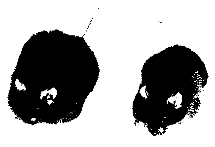
effects on both food intake and energy expenditure. (B) Photograph of a
treated ob
mouse. Shown are two C57Bl/6J ob/ob mice. The mouse on the left received PBS
and weighed 65 grams, which was the starting weight. The mouse on the right
received daily injections of the recombinant OB protein. The starting weight
of this
animal was also 65 grams, and the weight after three weeks of protein
treatment was
38 grams. (C) Livers from treated and untreated ob mice. Shown are livers from
treated and untreated C57B1/6J ob/ob mice. The liver from the mouse receiving
PBS
WO 96/05309 2195955 PCr/US95/10479 =
28
had the gross appearance of a fatty liver and weighed 5.04 grams. The liver
from
the mouse receiving the recombinant ob protein had a normal appearance and
weighed 2.23 grams.
Figure 30 shows the in situ hybridization of ob to adipose tissue. Sense and
antisense
ob RNA was labeled in vitro using Sp6 and T7 polymerase and digoxigenin. The
labeled RNAs were hybridized to paraffin embedded sections of adipose tissue
from
epididymal fat pads of eight week old C57Bi/Ks mice (labeled wild-type) and
C57BI/Ks db/db mice (labeled db). In the figure, the lipid droplets appear as
unstained vacuoles within cells. The cytoplasm is a thin rim at the periphery
of the
cells and is indistinguishable from the cell membrane. Hybridization to all
the
adipocytes in the field was detected in the wild-type sections only using the
antisense
probe and greatly increased levels were seen in the tissue sections from the
db/db
animals.
FIGURE 31 shows that OB RNA is expressed in adipocytes in vivo and in vitro.
Total RNA (10 micrograms) from several different sources was electophoresed on
blotted and hybridized to an ob probe. Firstly, differences in cell buoyancy
after
collagenase digestion was used to purify adipocytes. OB RNA was present only
in
the adipocyte fraction. Lane S indicates the stromovascular fraction and A
indicates
the adipocyte fraction. In addition, OB RNA was not expressed in the
undifferentiated 3T3-442 preadipocyte cells lane U. Differentiated adipocytes
from
these cell lines expressed clearly detectable levels of OB mRNA (lane D).
FIGURE 32 shows that OB RNA is expressed in all adipose tissue depots. All of
the
adipose tissue depots tested expressed ob RNA. The inguinal fat pad expressed
somewhat lower RNA levels, although there was variability in the level of
signals in
different experiments. (Figure 31A) Lanes (1) epididymal (2) inguinal (3)
abdominal
(4) parametrial fat pads. Brown fat also expressed a low level of OB RNA.
(Figure
31B) The level of OB expression in brown fat was unchanged in animals housed
at
= WO 96/05309 219:5955 PCT/US95/10479
29
4 C for one week while the abundance of the brown fat specific UCP RNA, known
to be cold inducible, increased five-fold.
FIGURE 33 depicts the expression of OB RNA in db/db and gold thioglucose
treated
mice. Total RNA from the parametrial fat pads of gold thioglucose (GTG) and
db/db
treated mice was electrophoresed and Northern blotted. GTG administered as a
single
dose is known to cause obesity by inducing specific hypothalamic lesions. (A)
One
month old CBA female mice were treated with GTG (.2 mg/g), with a resulting
increase of >20 g in treated animals relative to control animals (<5 g). (B)
Hybridization of an OB probe to RNA from db/db and GTG treated mice revealed a
twenty-fold increase in the abundance of ob RNA relative to control RNA (actin
or
GAPDH). FIGURE 34 represents a Northern blot analysis of human RNA. Northern
blots
containing 10 mg of total RNA from human adipose tissue (FAT, panel A) and 2
mg
of polyA+ RNA from other human tissues (panel B) were hybridized to human ob
or human li-actin probes as indicated. An intense signal at approximately 4.5
kb was
seen with the adipose tissue total RNA. Hybridization to the polyA + RNA
revealed
detectable signals in heart (HE) and placenta (PL), whereas OB RNA was not
detected in brain (BR), lung (LU), liver (LI), skeletal muscle (SM), kidney
(KI), and
pancreas (PA). In each case, the length of the autoradiographic exposure is
indicated.
Of note, the genesis of the lower molecular bands seen in placental RNA (e.g.,
alternate splicing, RNA degradation) is not known.
FIGURE 35 represents YAC contig containing the human OB gene and 8
microsatellite markers. The YAC-based STS-content map of the region of
chromosome 7 containing the human OB gene is depicted, as deduced by
SEGMAP/Version 3.29 [Green et at., PCR Methods Applic., 1:77-90 (1991)]. The
19 uniquely-ordered STSs (see Table 3) are listed along the top. The 8
microsatellite-
specific STSs are indicated with stars (see Table 4). Also indicated are the
STSs
corresponding to the Pax4 and OB genes as well as the predicted positions of
the
WO 96/05309 2195955 PCr/us95/10479
centromere (CEN) and 7q telomere (TEL) relative to the contig. Each of the 43
YAC clones is depicted by a horizontal bar, with its name given to the left
and
estimated YAC size (in kb, measured by pulsed-field gel electrophoresis)
provided
in parenthesis. The presence of an STS in a YAC is indicated by a darkened
circle
5 at the appropriate position. When an STS corresponds to the insert end of a
YAC,
a square is placed around the corresponding circle, both along the top (near
the STS
name) and at the end of the YAC from which it was derived. For the 5 YACs at
the
bottom (below the horizontal dashed line), 1 or more STS(s) expected to be
present
(based on the established STS order) was not detected (as assessed by testing
the
10 individual YACs with the corresponding STS-specific PCR assay(s) at least
twice),
and these are depicted as open circles at the appropriate positions. Most of
the YACs
were isolated from a human-hamster hybrid cell-derived library [Green et at.,
Genomics, 25:170-183 (1995)1, with their original names as indicated. The
remaining
YACs were isolated from total human genomic libraries, and their original
library
15 locations are provided in Table 3. Boxes are placed around the names of the
3 YACs
(yWSS691, yWSS999, and yWSS2935) that were found by FISH analysis to map to
7g31.3. The contigis displayed in its `uncomputed' form, where YAC sizes are
not
used to estimate clone overlaps or STS spacing, and all of the STSs are
therefore
spaced in an equidistant fashion. In the `computed' form, where YAC sizes are
used
20 to estimate the relative distance separating each pair of adjacent STSs as
well as the
extent of clone overlaps, the total YAC contig appears to span just over 2 Mb.
DETAILED DESCRIPTION
The present invention relates to the elucidation and discovery of a protein,
termed
herein ob polypeptide or leptin, nucleic acids encoding the protein, including
25 degenerate variations thereof, e.g., that incorporate optimal codons for
expression in
a particular expression system, which protein demonstrates the ability to
participate
in the control of mammalian body weight. The nucleic acids in object represent
the
coding sequences corresponding to the murine and human OB polypeptide, which
is
postulated to play a critical role in the regulation of body weight and
adiposity. Data
= WO 96/05309 2 19 5 9 5 5 PCT/U895/10479
31
presented herein indicate that the polypeptide product of a nuceic acid of the
invention
is secreted by the cells that express it, and that the polypeptide functions
as a
hormone. Additional experimental data demonstrate that the OB polypeptide is
very
effective in treating obesity in mice carrying a mutation of the ob gene. In
addition,
high bolus doses or moderate continuous doses of OB polypeptide effect weight
reduction in normal (wild-type) mice.
In addition, the Examples herein demonstrate that the OB polypeptide,
alternatively
termed herein "leptin," circulates in mouse, rat, and human plasma. Leptin is
absent
in plasma from ob/ob mice, and is present at ten-fold higher concentrations in
plasma
from db/db mice, and twenty-fold higher concentrations in fa/fa rats. Most
significantly, daily injections of recombinant leptin dramatically reduce the
body mass
of oblob mice, significantly affects the body weight of wild-type mice, and
has no
effect on db/db mice.
In a further aspect, the OB polypeptide from one species is biologically
active in
another species. In particular, the human OB polypeptide is active in mice.
In its primary aspect, the present invention is directed to the identification
of
materials that function as modulators of mammalian body weight. In particular,
the
invention concerns the isolation, purification and sequencing of certain
nucleic acids
that correspond to the OB gene or its coding region in both mice and humans,
as well
as the corresponding polypeptides expressed by these nucleic acids. The
invention
thus comprises the discovery of nucleic acids having the nucleotide sequences
set
forth in FIGURE IA through E (SEQ ID NO:1) and FIGURE 2A and B (SEQ ID
NO:3), and to degenerate variants, alleles and fragments thereof, all
possessing the
activity of modulating body weight and adiposity. The correspondence of the
present
nucleic acids to the OB gene portends their significant impact on conditions
such as
obesity as well as other maladies and dysfunctions where abnormalities in body
weight are a contributory factor. The invention extends to the proteins
expressed by
the nucleic acids of the invention, and particularly to those proteins set
forth in
W096105309 21M55
PCTMS95110479
32
FIGURE IA through E (SEQ ID NO:2), FIGURE 3 (SEQ ID NO:4), FIGURE 5
(SEQ ID NO:5), and FIGURE 6 (SEQ ID NO:6), as well as to conserved variants,
active fragments, and cognate small molecules.
As discussed earlier, the weight control modulator peptides or their binding
partners
or other ligands or agents exhibiting either mimicry or antagonism to them or
control
over their production, may be prepared in pharmaceutical compositions, with a
suitable carrier and at a strength effective for administration by various
means to a
patient experiencing abnormal fluctuations in body weight or adiposity, either
alone
or as part of an adverse medical condition such as cancer or AIDS, for the
treatment
thereof. A variety of administrative techniques may be utilized, among them
oral
administration, nasal and other forms of transmucosal administration,
parenteral
techniques such as subcutaneous, intravenous and intraperitoneal injections,
catheterizations and the like. Average quantities of the recognition factors
or their
subunits may vary and in particular should be based upon the recommendations
and
prescription of a qualified physician or veterinarian.
In accordance with the above, an assay system for screening potential drugs
effective
to mimic or antagonize the activity of the weight modulator may be prepared.
The
weight modulator may be introduced into a test system, and the prospective
drug may
also be introduced into the resulting cell culture, and the culture thereafter
examined
to observe any changes in the activity of the cells, due either to the
addition of the
prospective drug alone, or due to the effect of added quantities of the known
weight
modulator.
As stated earlier, the molecular cloning of the OB gene described herein has
led to
the identification of a class of materials that function on the molecular
level to
modulate mammalian body weight. The discovery of the modulators of the
invention
has important implications for the diagnosis and treatment of nutritional
disorders
including, but not limited to, obesity, weight loss associated with cancer and
the
treatment of diseases associated with obesity such as hypertension, heart
disease, and
= WO 96/05309 2195955
PCTIUS95/10479
33
Type II diabetes. In addition, there are potential agricultural uses for the
gene
product in cases where one might wish to modulate the body weight of domestic
animals. Finally, to the extent that one or more of the modulators of the
invention
are secreted molecules, they can be used biochemically to isolate their
receptor using
the technology of expression cloning. The discussion that follows with
specific
reference to the OB gene bears general applicability to the class of
modulators that
comprise a part of the present invention, and is therefore to be accorded such
latitude
and scope of interpretation.
As noted above, the functional activity of the OB polypeptide can be evaluated
transgenically. In this respect, a transgenic mouse model can be used. The ob
gene
can be used in complementation studies employing transgenic mice. Transgenic
vectors, including viral vectors, or cosmid clones (or phage clones)
corresponding to
the wild type locus of candidate gene, can be constructed using the isolated
ob gene.
Cosmids may be introduced into transgenic mice using published procedures
[Jaenisch, Science, 240:1468-1474 (1988)]. The constructs are introduced into
fertilized eggs derived from an intercross between Fl progeny of a C57BL/6J
ob/ob
X DBA intercross. These crosses require the use of C57BL/6J oblob ovarian
transplants to generate the Fl animals. DBA/2J mice are used as the
counterstrain
because they have a nonagouti coat color which is important when using the
ovarian
transplants. Genotype at the ob loci in cosmid transgenic animals can be
determined
by typing animals with tightly linked RFLPs or microsatellites which flank the
mutation and which are polymorphic between the progenitor strains.
Complementation will be demonstrated when a particular construct renders a
genetically obese F2 animal (as scored by RFLP analysis) lean and nondiabetic.
Under these circumstances, final proof of complementation will require that
the ob/ob
or dbldb animal carrying the transgene be mated to the oblob or db/db ovarian
transplants. In this cross, all N2 animals which do not carry the transgene
will be
obese and insulin resistant/diabetic, while those that do carry the transgene
will be
lean and have normal glucose and insulin concentrations in plasma. In a
genetic
sense, the transgene acts as a suppressor mutation.
It n
W096/05309 2195955 - - ' s PCTYUS95/10479
34
Alternatively, OB genes can be tested by examining their phenotypic effects
when
expressed in antisense orientation in wild-type animals. In this approach,
expression
of the wild-type allele is suppressed, which leads to a mutant phenotype.
RNARNA
duplex formation (antisense-sense) prevents normal handling of mRNA, resulting
in
partial or complete elimination of wild-type gene effect. This technique has
been
used to inhibit TK synthesis in tissue culture and to produce phenotypes of
the
Kruppel mutation in Drosophila, and the Shiverer mutation in mice Izant et
al., Cell,
36:1007-1015 (1984); Green et al., Annu. Rev. Biochem., 55:569-597 (1986);
Katsuki
et al., Science, 241:593-595 (1988). An important advantage of this approach
is that
only a small portion of the gene need be expressed for effective inhibition of
expression of the entire cognate mRNA. The antisense transgene will be placed
under control of its own promoter or another promoter expressed in the correct
cell
type, and placed upstream of the SV40 polyA site. This transgene will be used
to
make transgenic mice. Trsgenic mice will also be mated ovarian transplants to
test
whether ob heterozygotes are more sensitive to the effects of the antisense
construct.
In the long term, the elucidation of the biochemical function of the OB gene
product
(the OB polypeptide or protein) is useful for identifying small molecule
agonists and
antagonists that affect its activity.
Various terms used throughout this specification shall have the definitions
set out
herein, for example, below.
The term "body weight modulator", "modulator", "modulators", and any variants
not
specifically listed, may be used herein interchangeably, and as used
throughout the
present application and claims refers in one instance to both nucleotides and
to
proteinaceous material, the latter including both single or multiple proteins.
More
specifically, the aforementioned terms extend to the nucleotides and to the
DNA
having the sequences described herein and presented in Figure IA through E
(SEQ
ID NO:1), and Figure 2A and B (SEQ ID NO:3). Likewise, the proteins having the
2195955
WO 96/05309 PCTIUS95/10479
amino acid sequence data described herein and presented in Figure IA through E
(SEQ ID NO:2), and Figure 3 (SEQ ID NO:4) are likewise contemplated, as are
the
profile of activities set forth with respect to all materials both herein and
in the
claims. Accordingly, nucleotides displaying substantially equivalent or
altered
5 activity are likewise contemplated, including substantially homologous
analogs and
allelic variations. Likewise, proteins displaying substantially equivalent or
altered
activity, including proteins modified deliberately, as for example, by site-
directed
mutagenesis, or accidentally through mutations in hosts that produce the
modulators
are likewise contemplated.
10 A composition comprising "A" (where "A" is a single protein, DNA molecule,
vector, recombinant host cell, etc.) is substantially free of "B" (where "B"
comprises
one or more contaminating proteins, DNA molecules, vectors, etc., but
excluding
racemic forms of A) when at least about 75% by weight of the proteins, DNA,
vectors (depending on the category of species to which A and B belong) in the
15 composition is "A". Preferably, "A" comprises at least about 90% by weight
of the
A+B species in the composition, most preferably at least about 99 % by weight.
It
is also preferred that a composition, which is substantially free of
contamination,
contain only a single molecular weight species having the activity or
characteristic of
the species of interest.
20 The OB Polypentides
The terms "protein," which refers to the naturally occurring polypeptide, and
"polypeptide" are used herein interchangeably with respect to the ob gene
product and
variants thereof. The term "mature protein" or "mature polypeptide"
particularly
refers to the OB gene product with the signal sequence (or a fusion protein
partner)
25 removed.
As noted above, in specific embodiments ob polypeptides of the invention
include
those having the amino acid sequences set forth herein e.g., SEQ ID NOS: 2, 4,
5,
6, etc., including the ob polypeptide modified with conservative amino acid
WO 96/05309 3 2195955 PCr/US95/10479
36
substitutions, as well as biologically active fragments, analogs, and
derivatives
thereof. The term "biologically active," is used herein to refer to a specific
effect of
the polypeptide, including but not limited to specific binding, e.g., to a
receptor,
antibody, or other recognition molecule; activation of signal transduction
pathways
on a molecular level; and/or induction (or inhibition by antagonists) of
physiological
effects mediated by the native ob polypeptide in vivo. OB polypeptides,
including
fragments, analogs, and derivatives, can be prepared synthetically, e.g.,
using the
well known techniques of solid phase or solution phase peptide synthesis.
Preferably,
solid phase synthetic techniques are employed. Alternatively, OB polypeptides
of the
invention can be prepared using well known genetic engineering techniques, as
described infra. In yet another embodiment, the OB polypeptide can be
purified,
e.g., by immunoaffinity purification, from a biological fluid, such as but not
limited
to plasma, serum, or urine, preferably human plasma, serum, or urine, and more
preferably from a subject who overexpresses the polypeptide, such as an obese
person
suffering from a mutation in the OB receptor or from obesity related to a
mutation
corresponding to "fatty."
Fragments of the OB Polypeptide
In a particular embodiment, the present invention contemplates that naturally
occurring fragments of the OB polypeptide may be important. The peptide
sequence
includes a number of sites that are frequently the target for proteolytic
cleavage, e.g.,
arginine residues. It is possible that the full length polypeptide may be
cleaved at one
or more such sites to form biologically active fragments. Such biologically
active
fragments may either agonize or antagonize the functional activity of the OB
polypeptide to reduce body weight.
Analogs of the OB Polypeptide
The present invention specifically contemplates preparation of analogs of the
OB
peptide, which are characterized by being capable of a biological activity of
OB
polypeptide, e.g., of binding to a specific binding partner of ob peptide,
such as the
OB receptor. In one embodiment, the analog agonizes OB activity, i.e., it
functions
..;,.;, 2195955
= WO 96105309 PCT/US95/10479
37
similarly to the ob peptide. Preferably, an OB agonist is more effective than
the
native protein. For example, an OB agonist analog may bind to the OB receptor
with
higher affinity, or demonstrate a longer half-life in vivo, or both.
Nevertheless, OB
peptide agonist analogs that are less effective than the native protein are
also
contemplated. In another embodiment, the analog antagonizes OB activity. For
example, an OB analog that binds to the OB receptor but does not induce signal
transduction can competitively inhibit binding of native OB to the receptor,
thus
decreasing OB activity in vivo. Such an OB antagonist analog may also
demonstrate
different properties from ob peptide, e.g., longer (or shorter) half-life in
vivo, greater
(or lesser) binding affinity for the OB receptor, or both.
In one embodiment, an analog of OB peptide is the OB peptide modified by
substitution of amino acids at positions on the polypeptide that are not
essential for
structure or function. For example, since it is known that human OB peptide is
biologically active in mouse, substitution of divergent amino acid residues in
the
human sequence as compared to the murine amino acid sequence will likely yield
useful analogs of OB peptide. For example, the serine residue at position 53
or
position 98, or both (in the unprocessed peptide sequence depicted in Figure
4) from
human may be substituted, e.g., with glycine, alanine, valine, cysteine,
methionine,
or threonine. Similarly, the arginine residue at position number 92 (Figure 4)
may
be substituted, e.g., with asparagine, lysine, histidine, glutamine, glutamic
acid,
aspartic acid, serine, threonine, methionine, or cysteine. Referring still to
Figure 4,
other amino acids in the human OB peptide that appear to be capable of
substitution
are histidine at position 118, tryptophan at position 121, alanine at position
122,
glutamic acid at position 126, threonine at position 127, leucine at position
128,
glycine at position 132, glycine at position 139, tryptophan at position 159,
and
glycine at position 166. In another embodiment, it may be possible to
substitute one
or more of residues 121 to 128 (as depicted in Figure 4), e.g., with glycines
or
alanines, or substituting some of the residues with the exceptions of serine
as position
123, or leucine at position M.
WO 96/05309
2195955 PCT/US95/10479
38
In another embodiment, an analog of the OB polypeptide, preferably the human
OB
polypeptide, is a truncated form of the polypeptide. For example, it has
already been
demonstrated that the glutamine at residue 49 is not essential, and can be
deleted from
the peptide. Similarly, it may be possible to delete some or all of the
divergent
amino acid residues at positions 121-128. In addition, the invention
contemplates
providing an OB analog having the minimum amino acid sequence necessary for a
biological activity. This can be readily determined, e.g., by testing the
activity of
fragments of OB for the ability to bind to OB-specific antibodies, inhibit the
activity
of the native OB polypeptide, or agonize the activity of the native OB
peptide. In one
embodiment, the invention provides a truncated OB polypeptide consisting of
the loop
structure formed by the disulfide bond that forms between cysteine residues
117 and
167 (as depicted in Figure 4). In another embodiment, the truncated analog
corresponds to the amino acids from residue 22 (which follows the putative
signal
peptide cleavage site) to 53 (the amino acid residue immediately preceding a
flexible
loop region detected with limited proteolysis followed by mass spectrometric
analysis
of the OB polypeptide; see Cohen et al., Protein Science, 4:1088 (1995). In
another
embodiment, the truncated analog corresponds to amino acids from residue 61
(the
residue immediately following the flexible loop region as detected with the
limited
proteolysis/mass spec. analysis of the OB polypeptide) to amino acid residue
116 (the
residue immediately preceding the first cysteine residue). In yet another
embodiment,
the truncated analog corresponds to amino acids from residue 61 to amino acid
residue 167.
Furthermore, one or more of the residues of the putative flexible loop at
residues
number 54 to 60 are substituted. For example, one or more of the residues may
be
substituted with lysine, glutamic acid, or cysteine (preferably lysine) for
cross linking,
e.g., to a polymer, since flexible loop structures are preferred sites for
derivatization
of a protein. Alternatively, the residues at the flexible loop positions may
be
substituted with amino acid residues that are more resistant to proteolysis
but that
retain a flexible structure, e.g., one or more prolines. In yet another
embodiment,
WO 96/05309 2 1/ 5/ 5 5 PCr/US95/10479
39
substitutions with amino acid residues that can be further derivatized to make
them
more resistant to degradation, e.g., proteolysis, is contemplated.
It will be appreciated by one of ordinary skill in the art that the foregoing
fragment
sizes are approximate, and that from one to about five amino acids can be
included
or deleted from each or both ends, or from the interior of the polypeptide or
fragments thereof, of the recited truncated analogs, with the exception that
in the
disulfide bonded loop analogs, the cysteine residues must be maintained.
It has been found that murine OB peptide contains 50% a-helical content, and
that
the human OB polypeptide contains about 60% a-helical content, as detected by
circular dichroism of the recombinant peptides under nearly physiological
conditions.
Accordingly, in another embodiment, amino acid residues can be substituted
with
residues to form analogs of OB polypeptide that demonstrate enhanced
propensity for
forming, or which form more stable, a-helix structures. For example, a-helix
structure would be preferred if Glu, Ala, Len, His, Tip are introduced as
substitutes
for amino acid residues found in the native OB polypeptide. Preferably,
conservative
amino acid substitutions are employed, e.g., substituting aspartic acid at
residue(s)
29, 30, 44, 61, 76, 100, and/or 106 (as depicted in Figure 4) with glutamic
acid(s)
(Glu); substituting isoleucine(s) with leucine; substituting glycine or
valine, or any
divergent amino acid, with alanine (e.g., serine at position 53 of the human
OB
polypeptide with alanine), substituting arginine or lysine with histidine, and
substituting tyrosine and/or phenylalanine with tryptophan. Increasing the
degree, or
more importantly, the stability of a-helix structure may yield an OB analog
with
greater activity, increased binding affinity, or longer half-life. In a
specific
embodiment, the helix forming potential of the portion of the OB peptide
corresponding to amino acid residues 22 through 53 is increased. In another
embodiment, the helix-forming potential or stability of the amino acid
residues 61-116
is increased. In yet another embodiment, the helix forming potential of the
disulfide
loop structure corresponding to amino acids 117 to 167 is increased. Also
contemplated are OB analogs containing enhanced a-helical potential or
stability in
WO 96105309 2 1 9 5 9 5 5 PCT/US95/10479 =
more than one of the foregoing domains. In a further embodiment, truncated OB
polypeptide analogs are generated that incorporate structure-forming, e.g.,
helix-
forming, amino acid residues to compensate for the greater propensity of
polypeptide
fragments to lack stable structure.
5 Analogs, such as fragments, may be produced, for example, by pepsin
digestion of
weight modulator peptide material. Other analogs, such as muteins, can be
produced
by standard site-directed mutagenesis of weight modulator peptide coding
sequences.
Analogs exhibiting "weight modulator activity" such as small molecules,
whether
functioning as promoters or inhibitors, may be identified by known in vivo
and/or in
10 vitro assays. -
Small Molecule Analogs and Pepridomimetics of OB Polypeptide
The structure of the ob polypeptide, preferably human OB polypeptide, can be
analyzed by various methods known in the art. The protein sequence can be
characterized by a hydrophilicity analysis [e.g., Hopp et al., Proc. Natl.
Acad. Sci.
15 USA, 78:3824 (1981)]. A hydrophilicity profile can be used to identify the
hydrophobic and hydrophilic regions of the OB polypeptide, which may indicate
regions buried in the interior of the folded polypeptide, and regions
accessible on the
exterior of the polypeptide. In addition, secondary structural analysis [e.g.,
Chou et
al, Biochem., 13:222 (1974)] can also be done, to identify regions of OB
polypeptide
20 that assume specific secondary structures. Manipulation of the predicted or
determined structure, including secondary structure prediction, can be
accomplished
using computer software programs available in the art.
By providing an abundant source of recombinant OB polypeptide, the present
invention enables quantitative structural determination of the polypeptide. In
25 particular, enough material is provided for nuclear magnetic resonance
(NMR),
infrared (IR), Raman, and ultraviolet (UV), especially circular dichroism
(CD),
spectroscopic analysis. In particular NMR provides very powerful structural
analysis
of molecules in solution, which more closely approximates their native
environment
WO 96/05309 2 1 / 5 9 5 5 PCT/US95/10479
41 /
[Marion et al., Biochim. Biophys. Res. Comm., 113:967-974 (1983); Bar et al.,
J.
Magn. Reson., 65:355-360 (1985); Kimura et al., Proc. Natl. Acad. Sci. USA,
77:1681-1685 (1980)]. Other methods of structural analysis can also be
employed.
These include but are not limited to X-ray crystallography [Engstom, Biochem.
Exp.
Biol., 11:7-13 (1974)].
In yet a further embodiment, an analog of OB polypeptide can be tested to
determine
whether it cross-reacts with an antibody specific for native OB polypeptide,
or
specific fragments thereof. The degree of cross-reactivity provides
information about
structural homology or similarity of proteins, or about the accessibility of
regions
corresponding to portions of the polypeptide that were used to generate
fragment-
specific antibodies.
Screening for OB Analogs
Various screening techniques are known in the art for screening for analogs of
polypeptides. Various libraries of chemicals are available. Accordingly, the
present
invention contemplates screening such libraries, e.g., libraries of synthetic
compounds
generated over years of research, libraries of natural compounds, and
combinatorial
libraries, as described in greater detail, infra, for analogs of OB
polypeptide. In one
embodiment, the invention contemplates screening such libraries for compounds
that
bind to anti-OB polypeptide antibodies, preferably anti-human ob polypeptide
antibodies. In another aspect, once the OB receptor is identified (see infra),
any
screening technique known in the art can be used to screen for OB receptor
agonists
or antagonists. The present invention contemplates screens for small molecule
ligands
or ligand analogs and mimics, as well as screens for natural ligands that bind
to and
agonize or antagonize activate OB receptor in vivo.
Knowledge of the primary sequence of the receptor, and the similarity of that
sequence with proteins of known function, can provide an initial clue as to
the
agonists or antagonists of the protein. Identification and screening of
antagonists is
further facilitated by determining structural features of the protein, e.g.,
using X-ray
crystallography, neutron diffraction, nuclear magnetic resonance spectrometry,
and
CA 02195955 2007-10-02
42
other techniques for structure determination. These techniques provide for the
rational design or identification of agonists and antagonists.
Another approach uses recombinant bacteriophage to produce large libraries.
Using
the "phage method" [Scott et al., Science, 249:386-390 (1990); Cwirla et al.,
Proc.
Natl. Acad. Sci. USA, 87:6378-6382 (1990); Devlin et al., Science, 249:404-406
(1990)], very large libraries can be constructed (106-1(` chemical entities).
A second
approach uses primarily chemical methods, of which the Geysen method [Geysen
et
al., Molecular Immunology, 23:709-715 (1986); Geysen et al., J. Immunologic
Method, 102:259-274 (1987)] and the recent method of Fodor et al., Science,
251:767-773 (1991) are examples. Furka et al. 14th International Congress of
Biochemistry, Volume 5, Abstract FR:013 (1988); Furka, Int. J. Peptide Protein
Res., 37:487-493 (1991)]; Houghton (U.S. Patent No. 4,631,211, issued December
1986); and Rutter et al. (U.S. Patent No. 5,010,175, issued April 23, 1991)
describe
methods to produce a mixture of peptides that can be tested as agonists or
antagonists.
In another aspect, synthetic libraries [Needels et al., Proc. Natl. Acad. Sci.
USA,
90:10700-10704 (1993); Lam et al., International Patent Publication No. WO
92/00252 ] , and the
like can be used to screen for OB receptor ligands according to the present
invention.
With such libraries, receptor antagonists can be detected using cells that
express the
receptor without actually cloning the OB receptor.
Alternatively, assays for binding of soluble ligand to cells that express
recombinant
forms of the OB receptor ligand binding domain can be performed. The soluble
ligands can- be provided readily as recombinant or synthetic OB polypeptide.
The screening can be performed with recombinant cells that express the OB
receptor,
or alternatively, using purified receptor. protein, e.g., produced
recombinantly, as
described above. For example, the ability of labeled, soluble or solubilized
OB
WO 96/05309
219 5 9 5 5 PC /US95/10479
43
receptor, that includes the ligand-binding portion of the molecule, to bind
ligand can
be used to screen libraries, as described in the foregoing references.
Derivatives of OB Polypeptides
Generally, the present protein (herein the term "protein" is used to include
"polypeptide," unless otherwise indicated) may be derivatized by the
attachment of
one or more chemical moieties to the protein moiety. The chemically modified
derivatives may be further formulated for intraarterial, intraperitoneal,
intramuscular,
subcutaneous, intravenous, oral, nasal, rectal, bucal, sublingual, pulmonary,
topical,
transdermal, or other routes of administration. Chemical modification of
biologically
active proteins has been found to provide additional advantages under certain
circumstances, such as increasing the stability and circulation time of the
therapeutic
protein and decreasing immunogenicity. See U.S. Patent No. 4,179,337, Davis et
at, issued December 18, 1979. For a review, see Abuchowski et al., "Soluble
Polymer-Enzyme Adducts", in Enzymes as Drugs, pp. 367-383, Holcenberg and
Roberts, eds., Wiley-Interscience, New York, NY, (1981). A review article
describing protein modification and fusion proteins is Francis, Focus on
Growth
Factors, 3:4-10 (1992).
Chemical Moieties For Derivatization
The chemical moieties suitable for derivatization may be selected from among
water
soluble polymers. The polymer selected should be water soluble so that the
protein
to which it is attached does not precipitate in an aqueous environment, such
as a
physiological environment. Preferably, for therapeutic use of the end-product
preparation, the polymer will be pharmaceutically acceptable. One skilled in
the art
will be able to select the desired polymer based on such considerations as
whether the
polymer/protein conjugate will be used therapeutically, and if so, the desired
dosage,
circulation time, resistance to proteolysis, and other considerations. For the
present
proteins and peptides, these may be ascertained using the assays provided
herein.
* I ny ` WO 96/05309 2 1 9 5 7 5 5 PCTIUS95110479
44
Polymer Molecules
The water soluble polymer may be selected from the group consisting of, for
example, polyethylene glycol, copolymers of ethylene glycol/propylene glycol,
carboxymethylcellulose, dextran, polyvinyl alcohol, polyvinyl pyrrolidone,
poly-1,
3-dioxolane, poly-I,3,6-trioxane, ethylene/maleic anhydride copolymer,
polyaminoacids (either homopolymers or random copolymers), and dextran or
poly(n-vinyl pyrrolidone)polyethylene glycol, propropylene glycol
homopolymers,
polypropylene oxide/ethylene oxide co-polymers, polyoxyethylated polyols and
polyvinyl alcohol. Polyethylene glycol propionaldenhyde may provide advantages
in
manufacturing due to its stability in water.
The polymer may be of any molecular weight, and may be branched or unbranched.
For polyethylene glycol, the preferred molecular weight is between about 2kDa
and
about 100kDa (the term "about" indicating that in preparations of polyethylene
glycol,
some molecules will weigh more, some less, than the stated molecular weight)
for
ease in handling and manufacturing. Other sizes may be used, depending on the
desired therapeutic profile (e.g., the duration of sustained release desired,
the effects,
if any on biological activity, the ease in handling, the degree or lack of
antigenicity
and other known effects of the polyethylene glycol to a therapeutic protein or
analog).
Polymer/Protein Ratio
The number of polymer molecules so attached may vary, and one skilled in the
art
will be able to ascertain the effect on function. One may mono-derivatize, or
may
provide for a di-, tri-, tetra- or some combination of derivatization, with
the same or
different chemical moieties (e.g., polymers, such as different weights of
polyethylene
glycols). The proportion of polymer molecules to protein (or peptide)
molecules will
vary, as will their concentrations in the reaction mixture. In general, the
optimum
ratio (in terms of efficiency of reaction in that there is no excess unreacted
protein
or polymer) will be determined by factors such as the desired degree of
derivatization
(e.g., mono, di-, tri-, etc.), the molecular weight of the polymer selected,
whether
the polymer is branched or unbranched, and the reaction conditions.
CA 02195955 2007-10-02
Attachment of the Chemical Moiety to the Protein
The polyethylene glycol molecules (or other chemical moieties) should be
attached
to the protein with consideration of effects on functional or antigenic
domains of the
protein. There are a number of attachment methods available to those skilled
in the
5 art, e.g., EP 0 401 384 (coupling PEG to G-CSF).
See also Malik et al., Exp. Hematol., 20:1028-1035 (1992) (reporting
pegylation of
GM-CSF using tresyl chloride). For example, polyethylene glycol may be
covalently
bound through amino acid residues via a reactive group, such as a free amino
or
carboxyl group. Reactive groups are those to which.an activated polyethylene
glycol
10 molecule may be bound. The amino acid residues having a free amino group
may
include lysine residues and the N-terminal amino acid residues, those having a
free
carboxyl group may include aspartic acid residues glutamic acid residues and
the
C-terminal amino acid residue. Sulfhydry groups may also be used as a reactive
group for attaching the polyethylene glycol molecule(s). Preferred for
therapeutic
15 purposes is attachment at an amino group, such as attachment at the N-
terminus or
lysine group. Attachment at residues important for receptor binding should be
avoided if receptor binding is desired.
N-terminally Chemically Modified Proteins.
One may specifically desire N-terminally chemically modified protein. Using
20 polyethylene glycol as an illustration of the present compositions, one may
select
from a variety of polyethylene glycol molecules (by molecular weight,
branching,
etc.), the proportion of polyethylene glycol molecules to protein (or peptide)
molecules in the reaction mix, the type of pegylation reaction to be
performed, and
the method of obtaining the selected N-terminally pegylated protein. The
method of
25 obtaining the N-terminally pegylated preparation (i. e., separating this
moiety from
other monopegylated moieties if necessary) may be by purification of the
N-terminally, pegylated material from a population of pegylated protein
molecules.
Selective N-terminal chemical modification may be accomplished by reductive
alkylation which exploits differential reactivity of different types of
primary amino
2195955
WO 96/05309 PCi'/US95110479
46
groups (lysine versus the N-terminus) available for derivatization in a
particular
protein. Under the appropriate reaction conditions, substantially selective
derivatization of the protein at the N-terminus with a carbonyl group
containing
polymer is achieved. For example, one may selectively N-terminally pegylate
the
protein by performing the reaction at a pH which allows one to take advantage
of the
pK, differences between the a-amino groups of the lysine residues and that of
the a-
amino group of the N-terminal residue of the protein. By such selective
derivatization attachment of a water soluble polymer to a protein is
controlled: the
conjugation with the polymer takes place predominantly at the N-terminus of
the
protein and no significant modification of other reactive groups, such as the
lysine
side chain amino groups, occurs. Using reductive alkylation, the water soluble
polymer may be of the type described above, and should have a single reactive
aldehyde for coupling to the protein. Polyethylene glycol propionaldehyde,
containing
a single reactive aldehyde, may be used.
Nucleic Acids Associated With OB Polypeeptide
As noted above, the present invention is directed to nucleic acids encoding ob
polypeptides, as well as associated genomic non-coding sequences 5', 3', and
intronic
to the OB gene. Thus, in accordance with the present invention there may be
employed conventional molecular biology, microbiology, and recombinant DNA
techniques within the skill of the art. Such techniques are explained fully in
the
literature. See, e.g., Sambrook et al., Molecular Cloning: A Laboratory
Manual,
Second Edition, Cold Spring Harbor Laboratory Press, Cold Spring Harbor, New
York (1989); Glover ed., DNA Cloning: A Practical Approach, Volumes I and II,
NERL Press, Ltd., Oxford, U.K. (1985); Gait ed., Oligonucleotide Synthesis,
Oxford
University Press (1984); Haines et al., eds., Nucleic Acid Hybridization,
Springer-
Verlag (1985); Haines et al., eds. Transcription And Translation, Oxford
University
Press (1984)]; Freshney ed., Animal Cell Culture, Oxford University Press
(1986)];
Immobilized Cells And Enzymes, IRL Press (1986)]; Perbal, A Practical Guide To
Molecular Cloning, Wiley, New York (1984). Of particular relevance to the
present
invention are strategies for isolating, cloning, sequencing, analyzing, and
= WO 96/05309 2 1 9 5 9 5 5 PCTIUS95/10479
47
characterizing a gene or nucleic acid based on the well known polymerase chain
reaction (PCR) techniques.
A "replicon" is any genetic element (e.g., plasmid, chromosome, virus) that
functions
as an autonomous unit of DNA replication in vivo, i.e., capable of replication
under
its own control.
A 'vector" is a replicon, such as a plasmid, phage or cosmid, to which another
DNA
segment may be attached so as to bring about the replication of the attached
segment.
A "cassette" refers to a segment of DNA that can be inserted into a vector at
specific
restriction sites. The segment of DNA encodes a polypeptide of interest, and
the
cassette and restriction sites are designed to ensure insertion of the
cassette in the
proper reading frame for transcription and translation.
"Heterologous" DNA refers to DNA not naturally located in the cell, or in a
chromosomal site of the cell. Preferably, the heterologous DNA includes a gene
foreign to the cell.
A cell has been "transfected" by exogenous or heterologous DNA when such DNA
has been introduced inside the cell. A cell has been "transformed" by
exogenous or
heterologous DNA when the transfected DNA effects a phenotypic change.
Preferably, the transforming DNA should be integrated (covalently linked) into
chromosomal DNA making up the genome of the cell.
A "clone" is a population of cells derived from a single cell or common
ancestor by
mitosis.
A "nucleic acid molecule" refers to the phosphate ester polymeric form of
ribonucleosides (adenosine, guanosine, uridine or cytidine; "RNA molecules")
or
2195955
WO 96/05309 PCTIUS95/10479 =
48
deoxyribonucleosides (deoxyadenosine, deoxyguanosine, deoxythymidine, or
deoxycytidine; "DNA molecules") in either single-stranded form, or a double-
stranded
helix. Double-stranded DNA-DNA, DNA-RNA and RNA-RNA helices are possible.
The term nucleic acid molecule, and in particular DNA or RNA molecule, refers
only
to the primary and secondary structure of the molecule, and does not limit it
to any
particular tertiary or quaternary forms. Thus, this term includes double-
stranded
DNA found, inter alia, in linear or circular DNA molecules (e.g., restriction
fragments), plasmids, and chromosomes. In discussing the structure of
particular
double-stranded DNA molecules, sequences may be described herein according to
the
normal convention of giving only the sequence in the 5' to 3' direction along
the
nontranscribed strand of DNA (i.e., the strand having a sequence homologous to
the
mRNA). A "recombinant DNA molecule" is a DNA molecule that has undergone a
molecular biological manipulation.
A nucleic acid molecule is "hybridizable" to another nucleic acid molecule,
such as
a cDNA, genomic DNA, or RNA, when a single-stranded form of the nucleic acid
molecule can anneal to the other nucleic acid molecule under the appropriate
conditions of temperature and solution ionic strength (see Sambrook et al.,
1989,
supra). The conditions of temperature and ionic strength determine the
"stringency"
of the hybridization. For preliminary screening for homologous nucleic acids,
low
stringency hybridization conditions, corresponding to a T. of 55 C, can be
used,
e.g., 5x SSC, 0.1% SDS, 0.25% milk, and no formamide; or 30% formamide, 5x
SSC, 0.5% SDS). Moderate stringency hybridization conditions correspond to a
higher Tm, e.g., 40% formamide, with 5x or 6x SCC. High stringency
hybridization
conditions correspond to the highest Tm, e.g., 50% formamide, 5x or 6x SCC.
Hybridization requires that the two nucleic acids contain complementary
sequences,
although depending on the stringency of the hybridization, mismatches between
bases
are possible. The appropriate stringency for hybridizing nucleic acids depends
on the
length of the nucleic acids and the degree of complementation, variables well
known
in the art. The greater the degree of similarity or homology between two
nucleotide
sequences, the greater the value of T. for hybrids of nucleic acids having
those
3 .M
WO 96/05309 219.59 5 5 PCTIUS95/10479
49
sequences. The relative stability (corresponding to higher Tm) of nucleic acid
hybridizations decreases in the following order: RNA:RNA, DNA:RNA,
DNA:DNA. For hybrids of greater than 100 nucleotides in length, equations for
calculating Tm have been derived (see Sambrook et al., 1989, supra, 9.50-
0.51). For
hybridization with shorter nucleic acids, I.e., oligonucleotides, the position
of
mismatches becomes more important, and the length of the oligonucleotide
determines
its specificity (see Sambrook et al., 1989, supra, 11.7-11.8). Preferably a
minimum
length for a hybridizable nucleic acid is at least about 10 nucleotides; more
preferably
at least about 15 nucleotides; most preferably the length is at least about 20
nucleotides.
"Homologous recombination" refers to the insertion of a foreign DNA sequence
of
a vector in a chromosome. Preferably, the vector targets a specific
chromosomal site
for homologous recombination. For specific homologous recombination, the
vector
will contain sufficiently long regions of homology to sequences of the
chromosome
to allow complementary binding and incorporation of the vector into the
chromosome.
Longer regions of homology, and greater degrees of sequence similarity, may
increase the efficiency of homologous recombination.
A DNA "coding sequence" is a double-stranded DNA sequence which is transcribed
and translated into a polypeptide in a cell in vitro or in vivo when placed
under the
control of appropriate regulatory sequences. The boundaries of the coding
sequence
are determined by a start codon at the 5' (amino) terminus and a translation
stop
codon at the 3' (carboxyl) terminus. A coding sequence can include, but is not
limited to, prokaryotic sequences, cDNA from eukaryotic anRNA, genomic DNA
sequences from eukaryotic (e.g., mammalian) DNA, and even synthetic DNA
sequences. If the coding sequence is intended for expression in a eukaryotic
cell, a
polyadenylation signal and transcription termination sequence will usually be
located
3' to the coding sequence.
2! 9 5 9 5 5
. ~ _
WO 96/05309 PCTIUS95/10479
Isolation of OB Coding and Flanking Sequences
The nucleic acids contemplated by the present invention extend as indicated,
to other
nucleic acids that code on expression for peptides such as those set forth in
FIGURE
IA through D (SEQ ID NO:2), FIGURE 3 (SEQ ID NO:4), FIGURE 5 (SEQ ID
5 NO:5), and FIGURE 6 (SEQ ID NO:6) herein. Accordingly, while specific DNA
has been isolated and sequenced in relation to the ob gene, any animal cell
potentially
can serve as the nucleic acid source for the molecular cloning of a gene
encoding the
peptides of the invention. The DNA may be obtained by standard procedures
known
in the art from cloned DNA (e.g., a DNA "library"), by chemical synthesis, by
10 cDNA cloning, or by the cloning of genomic DNA, or fragments thereof,
purified
from the desired cell (See, for example, Sambrook et al., 1989, supra; Glover,
1985,
supra). Clones derived from genomic DNA may contain regulatory and intronic
DNA regions in addition to coding regions; clones derived from cDNA will not
contain intron sequences. Whatever the source, the gene should be molecularly
15 cloned into a suitable vector for propagation of the gene.
In the molecular cloning of the gene from genomic DNA, the genomic DNA can be
amplified using primers selected from the cDNA sequences. Alternatively, DNA
fragments are generated, some of which will encode the desired gene. The DNA
may
be cleaved at specific sites using various restriction enzymes. One may also
use
20 DNase in the presence of manganese to fragment the DNA, or the DNA can be
physically sheared, as for example, by sonication. The linear DNA fragments
can
then be separated according to size by standard techniques, including but not
limited
to, agarose and polyacrylamide gel electrophoresis and column chromatography.
Once the DNA fragments are generated, identification of the specific DNA
fragment
25 containing the desired ob or ob-like gene may be accomplished in a number
of ways.
For example, if an amount of a portion of a ob or ob-like gene or its specific
RNA,
or a fragment thereof, is available and can be purified and labeled, the
generated
DNA fragments may be screened by nucleic acid hybridization to a labeled probe
[Benton et at, Science, 196:180 (1977); Grunstein et al., Proc. Natl. Acad.
Sci.
WO 96/05309 2195955 PC IUS95/10479
51
USA, 72:3961 (1975)]. The present invention provides such nucleic acid probes,
which can be conveniently prepared from the specific sequences disclosed
herein,
e.g., a hybridizable probe having a nucleotide sequence corresponding to at
least a
10, and preferably a 15, nucleotide fragment of the sequences depicted in
Figure IA
through E (SEQ ID NO:1) or Figure 2A and B (SEQ ID NO:3). Preferably, a
fragment is selected that is highly unique to the modulator peptides of the
invention.
Those DNA fragments with substantial homology to the probe will hybridize. As
noted above, the greater the degree of homology, the more stringent the
hybridization
conditions that can be used. In one embodiment, low stringency hybridization
conditions are used to identify a homologous modulator peptide. However, in a
preferred aspect, and as demonstrated experimentally herein, a nucleic acid
encoding
a modulator peptide of the invention will hybridize to a nucleic acid having a
nucleotide sequence such as depicted in Figure IA through E (SEQ ID NO: I) or
Figure 2A and B (SEQ ID NO:3), or a hybridizable fragment thereof, under
moderately stringent conditions; more preferably, it will hybridize under high
stringency conditions.
Alternatively, the presence of the gene may be detected by assays based on the
physical, chemical, or immunological properties of its expressed product. For
example, cDNA clones, or DNA clones which hybrid-select the proper mRNAs, can
be selected which produce a protein that, e.g., has similar or identical
electrophoretic
migration, isoelectric focusing behavior, proteolytic digestion maps, tyrosine
phosphatase activity or antigenic properties as known for the present
modulator
peptides. For example, the antibodies of the instant invention can
conveniently be
used to screen for homologs of modulator peptides from other sources.
A gene encoding a modulator peptide of the invention can also be identified by
mRNA selection, i.e., by nucleic acid hybridization followed by in vitro
translation.
In this procedure, fragments are used to isolate complementary mRNAs by
hybridization. Such DNA fragments may represent available, purified modulator
DNA. Immunoprecipitation analysis or functional assays (e.g., tyrosine
phosphatase
WO 96/05309 2195955 PCTNS95/10479
52
activity) of the in vitro translation products of the products of the isolated
mRNAs
identifies the mRNA and, therefore, the complementary DNA fragments, that
contain
the desired sequences. In addition, specific mRNAs may be selected by
adsorption
of polysomes isolated from cells to immobilized antibodies specifically
directed
against a modulator peptide.
A radiolabeled modulator peptide cDNA can be synthesized using the selected
mRNA
(from the adsorbed polysomes) as a template. The radiolabeled mRNA or cDNA may
then be used as a probe to identify homologous modulator peptide DNA fragments
from among other genomic DNA fragments.
As mentioned above, a DNA sequence encoding weight modulator peptides as
disclosed herein can be prepared synthetically rather than cloned. The DNA
sequence
can be designed with the appropriate codons for the weight modulator peptide
amino
acid sequences. In general, one will select preferred codons for the intended
host if
the sequence will be used for expression. The complete sequence is assembled
from
overlapping oligonucleotides prepared by standard methods and assembled into a
complete coding sequence. See, e.g., Edge, Nature, 292:756 (1981); Nambair et
al.,
Science, 223:1299 (1984); Jay et al., J. Biol. Chem., 259:6311 (1984).
Synthetic DNA sequences allow convenient construction of genes which will
express
weight modulator analogs, as described above. Alternatively, DNA encoding
analogs
can be made by site-directed mutagenesis of native OB genes or cDNAs, and
analogs
can be made directly using conventional polypeptide synthesis.
A general method for site-specific incorporation of unnatural amino acids into
proteins is described in Noren et al, Science, 244:182-188 (1989). This method
may
be used to create analogs of the ob polypeptide with unnatural amino acids.
= WO 96/05309 2195955 PGT/U595110479
53
Non-coding Nucleic Acids
The present invention extends to the preparation of antisense nucleotides and
ribozymes that may be used to interfere with the expression of the weight
modulator
proteins at the translational level. This approach utilizes antisense nucleic
acid and
ribozymes to block translation of a specific mRNA, either by masking that mRNA
with an antisense nucleic acid or cleaving it with a ribozyme.
Antisense nucleic acids are DNA or RNA molecules that are complementary to at
least a portion of a specific mRNA molecule [See Weintraub, Sci. Am., 262:40-
46
(1990); Marcus-Sekura, Anal. Biochem., 172:289-295 (1988)]. In the cell, they
hybridize to that mRNA, forming a double-stranded molecule. The cell does not
translate an mRNA complexed in this double-stranded form. Therefore, antisense
nucleic acids interfere with the expression of mRNA into protein. Oligomers of
about
fifteen nucleotides and molecules that hybridize to the AUG initiation codon
will be
particularly efficient, since they are easy to synthesize and are likely to
pose fewer
problems than larger molecules when introducing them into weight modulator
peptide-
producing cells. Antisense methods have been used to inhibit the expression of
many
genes in vitro [(Marcus-Sekura, 1988 supra; Hambor et al., J. Exp. Med.,
168:1237-1245 (1988)].
Ribozymes are RNA molecules possessing the ability to specifically cleave
other
single-stranded RNA molecules in a manner somewhat analogous to DNA
restriction
endonucleases. Ribozymes were discovered from the observation that certain
mRNAs
have the ability to excise their own introns. By modifying the nucleotide
sequence
of these RNAs, researchers have been able to engineer molecules that recognize
specific nucleotide sequences in an RNA molecule and cleave it [Cech, J. Am.
Med.
Assoc., 260:3030-3034 (1988)]. Because they are sequence-specific, only mRNAs
with particular sequences are inactivated.
Investigators have identified two types of ribozymes, Tetrahymena-type and
"hammerhead"-type. Tetrahymena-type ribozymes recognize four-base sequences,
X195955
WO 96/05309 PCT/US95/10479
54
while "hammerhead" -type recognize eleven- to eighteen-base sequences. The
longer
the recognition sequence, the more likely it is to occur exclusively in the
target
mRNA species. Therefore, hammerhead-type ribozymes are preferable to
Tetrahymena-type ribozymes for inactivating a specific mRNA species, and
eighteen
base recognition sequences are preferable to shorter recognition sequences.
The DNA sequences described herein may thus be used to prepare antisense
molecules against and ribozymes that cleave mRNAs for weight modulator
proteins
and their ligands, thus inhibiting expression of the ob gene, and leading to
increased
weight gain and adiposity.
In another embodiment, short oligonucleotides complementary to the coding and
complementary strands of the OB nucleic acid, or to non-coding regions of the
OB
gene 5', 3', or internal (intronic) to the coding region are provided by the
present
invention. Such nucleic acids are useful as probes, either as directly labeled
oligonucleotide probes, or as primers for the polymerase chain reaction, for
evaluating the presence of mutations in the ob gene, or the level of
expression of OB
mRNA. Preferably, the non-coding nucleic acids of the invention are from the
human OB gene.
In a specific embodiment, the non-coding nucleic acids provide for homologous
recombination for integration of an amplifiable gene and/or other regulatory
sequences in proximity to the OB gene, e.g., to provide for higher levels of
expression of the OB polypeptide, or to overcome a mutation in the ob gene
regulatory sequences that prevent proper levels of expression of the OB
polypeptide
(See International Patent Publication WO 91/06666, published May 16, 1991 by
Skoultchi; International Patent Publication No. WO 91/09955, published July
11,
1991 by Chappel; see also International Patent Publication No. WO 90/14092,
published November 29, 1990, by Kucherlapati and Campbell).
WO 96/05309 2i C1 C PCT/US95/10479
= 7 l
Production of OB Polypeptide: Expression and Synthesis
Transcriptional and translational control sequences are DNA regulatory
sequences,
such as promoters, enhancers, terminators, and the like, that provide for the
expression of a coding sequence in a host cell. In eukaryotic cells,
polyadenylation
5 signals are control sequences.
A coding sequence is "under the control" of transcriptional and translational
control
sequences in a cell when RNA polymerise transcribes the coding sequence into
mRNA, which is then trans-RNA spliced and translated into the protein encoded
by
the coding sequence.
10 A "signal sequence" is included at the beginning of the coding sequence of
a protein
to be expressed on the surface of a cell. This sequence encodes a signal
peptide, N-
terminal to the mature polypeptide, that directs the host cell to translocate
the
polypeptide. The term "translocation signal sequence" is also used herein to
refer to
this sort of signal sequence. Translocation signal sequences can be found
associated
15 with a variety of proteins native to eukaryotes and prokaryotes, and are
often
functional in both types of organisms.
A DNA sequence is "operatively linked" to an expression control sequence when
the
expression control sequence controls and regulates the transcription and
translation
of that DNA sequence. The term "operatively linked" includes having an
appropriate
20 start signal (e.g., ATG) in front of the DNA sequence to be expressed and
maintaining the correct reading frame to permit expression of the DNA sequence
under the control of the expression control sequence and production of the
desired
product encoded by the DNA sequence. If a gene that one desires to insert into
a
recombinant DNA molecule does not contain an appropriate start signal, such a
start
25 signal can be inserted upstream (5') of and in reading frame with the gene.
A "promoter sequence" is a DNA regulatory region capable of binding RNA
polymerase in a cell and initiating transcription of a downstream (3'
direction) coding
WO 96/05309 2 1'9 5 9 55 Pcr/U595/10479
56
sequence. For purposes of defining the present invention, the promoter
sequence is
bounded at its 3' terminus by the transcription initiation site and extends
upstream (5'
direction) to include the minimum number of bases or elements necessary to
initiate
transcription at levels detectable above background. Within the promoter
sequence
will be found a transcription initiation site (conveniently defined for
example, by
mapping with nuclease Sl), as well as protein binding domains (consensus
sequences)
responsible for the binding of RNA polymerase.
Another feature of this invention is the expression of the DNA sequences
disclosed
herein. As is well known in the art, DNA sequences may be expressed by
operatively linking them to an expression control sequence in an appropriate
expression vector and employing that expression vector to transform an
appropriate
unicellular host.
Such operative linking of a DNA sequence of this invention to an expression
control
sequence, of course, includes, if not already part of the DNA sequence, the
provision
of an initiation codon, ATG, in the correct reading frame upstream of the DNA
sequence.
A wide variety of host/expression vector combinations may be employed in
expressing the DNA-sequences of this invention. Useful expression vectors, for
example, may consist of segments of chromosomal, non-chromosomal and synthetic
DNA sequences. Suitable vectors include derivatives of SV40 and known
bacterial
plasmids, e.g., E. coil plasmids col El, pCR1, pBR322, pMB9, pUC or pUC
plasmid
derivatives, e.g., pGEX vectors, pET vectors, pmal-c, pFLAG, etc., and their
derivatives, plasmids such as RP4; phage DNAs, e.g., the numerous derivatives
of
phage X, e.g., NM989, and other phage DNA, e.g., M13 and filamentous single-
stranded phage DNA; yeast plasmids such as the 2,u plasmid or derivatives
thereof;
vectors useful in eukaryotic cells, such as vectors useful in insect or
mammalian cells;
vectors derived from combinations of plasmids and phage DNAs, such as plasmids
that have been modified to employ phage DNA or other expression control
sequences;
2195955
<Y4)j -
WO 96105309 r ,4 r PCTIUS95/10479
57
and the like. In a preferred embodiment, expression of ob is achieved in
methylotrophic yeast, e.g., Pichia pastoris yeast (see, e.g., International
Patent
Publication No. WO 90/03431, published 5 April 1990, by Brierley et al.;
International Patent Publication No. WO 90/10697, published 20 September 1990,
by
Siegel et al.). In a specific embodiment, infra, an expression vector is
engineered
for expression of ob under control of the a-mating factor signal sequence.
Any of a wide variety of expression control sequences -- sequences that
control the
expression of a DNA sequence operatively linked to it -- may be used in these
vectors
to express the DNA sequences of this invention. Such useful expression control
sequences include, for example, the early or late promoters of SV40, CMV,
vaccinia,
polyoma or adenovirus, the lac system, the trp system, the TAC system, the TRC
system, the LTR system, the major operator and promoter regions of phage X,
the
control regions of fd coat protein, the promoter for 3-phosphoglycerate kinase
or
other glycolytic enzymes, the promoters of acid phosphatase (e.g., PhoS), the
AOX
1 promoter of methylotrophic yeast, the promoters of the yeast a-mating
factors, and
other sequences known to control the expression of genes of prokaryotic or
eukaryotic
cells or their viruses, and various combinations thereof.
A wide variety of unicellular host cells are also useful in expressing the DNA
sequences of this invention. These hosts may include well known eukaryotic and
prokaryotic hosts, such as strains of E. colt, Pseudomonas, Bacillus,
Streptomyces;
fungi such as yeasts (Saccharomyces, and methylotrophic yeast such as Pichia,
Candida, Hansenula, and Torulopsis); and animal cells, such as CHO, R1.l, B-W
and
LM cells, African Green Monkey kidney cells (e.g., COS 1, COS 7, BSC1, BSC40,
and BMT10), insect cells (e.g., Sf9), and human cells and plant cells in
tissue
culture.
It will be understood that not all vectors, expression control sequences and
hosts will
function equally well to express the DNA sequences of this invention. Neither
will
all hosts function equally well with the same expression system. However, one
WO 96/05309 2195955 PCT/US95/10479
58
skilled in the art will be able to select the proper vectors, expression
control
sequences, and hosts without undue experimentation to accomplish the desired
expression without departing from the scope of this invention. For example, in
selecting a vector, the host must be considered because the vector must
function in
it. The vector's copy number, the ability to control that copy number, and the
expression of any other proteins encoded by the vector, such as antibiotic
markers,
will also be considered.
In selecting an expression control sequence, a variety of factors will
normally be
considered. These include, for example, the relative strength of the system,
its
controllability, and its compatibility with the particular DNA sequence or
gene to be
expressed, particularly as regards potential secondary structures. Suitable
unicellular
hosts will be selected by consideration of, e.g., their compatibility with the
chosen
vector, their secretion characteristics, their ability to fold proteins
correctly, and their
fermentation requirements, as well as the toxicity to the host of the product
encoded
by the DNA sequences to be expressed, and the ease of purification of the
expression
products.
Considering these and other factors, a person skilled in the art will be able
to
construct a variety of vector/expression control sequence/host combinations
that will
express the DNA sequences of this invention on fermentation or in large scale
animal
culture.
In a specific embodiment, an OB fusion protein can be expressed. An OB fusion
protein comprises at least a functionally active portion of a non-OB protein
joined via
a peptide bond to at least a functionally active portion of an OB polypeptide.
The
non-ob sequences can be amino- or carboxy-terminal to the OB sequences. More
preferably, for stable expression of a proteolytically inactive OB fusion
protein, the
portion of the non-OB fusion protein is joined via a peptide bond to the amino-
terminus of the OB protein. A recombinant DNA molecule encoding such a fusion
protein comprises a sequence encoding at least a functionally active portion
of a non-
WO 96/05309 2195955
PGT/us95/10479
59
OB protein joined in-frame to the OB coding sequence, and preferably encodes a
cleavage site for a specific protease, e.g., thrombin or Factor Xa, preferably
at the
OB-non-OB juncture. In a specific embodiment, the fusion protein is expressed
in
Escherichia coli or in P. pastoris.
In a specific embodiment, infra, vectors were prepared to express the murine
and
human ob genes, with and without the codon for gln-49, in bacterial expression
systems and yeast (Pichia) expression systems as fusion proteins. The ob gene
is
prepared with an endonuclease cleavage site, e.g., using PCR and novel
primers. It
is desirable to confirm sequences generated by PCR, since the probability of
including
a point mutation is greater with this technique. A plasmid containing a
histidine tag
(His-tag) and a proteolytic cleavage site is used. The presence of the
histidine makes
possible the selective isolation of recombinant proteins on a Ni-chelation
column, or
by affinity purification. The proteolytic cleavage site, in a specific
embodiment,
infra, a thrombin cleavage site, is engineered so that treatment with the
protease,
e.g., thrombin, will release the full-length mature (i.e., lacking a signal
sequence) OB
polypeptide.
In another aspect, the pGEX vector [Smith et al., Gene 67:31-40 (1988)] can be
used.
This vector fuses the schistosoma japonicum glutathionine S-transferase cDNA
to the
sequence of interest. Bacterial proteins are harvested and recombinant
proteins can
be quickly purified on a reduced glutathione affinity column. The GST carrier
can
subsequently be cleaved from fusion proteins by digestion with site-specific
proteases.
After cleavage, the carrier and uncleaved fusion protein can be removed by
absorption on glutathione agarose. Difficulty with the system occasionally
arises
when the encoded protein is insoluble in aqueous solutions.
Expression of recombinant proteins in bacterial systems may result in
incorrect
folding of the expressed protein, requiring refolding. The recombinant protein
can
be refolded prior to or after cleavage to form a functionally active OB
polypeptide.
The OB polypeptide may be refolded by the steps of (i) incubating the protein
in a
- - --- ----------------
WO 96105309 } (} -955 PCT/US95110479
60 L 7
denaturing buffer that contains a reducing agent, and then (ii) incubating the
protein
in a buffer that contains an oxidizing agent, and preferably also contains a
protein
stabilizing agent or a chaotropic agent, or both. Suitable redox
(reducing/oxidizing)
agent pairs include, but are not limited to, reduced glutathione/glutathione
disulfide,
cystine/cysteine, cystamine/cysteamine, and 2-mercaptoethanol/2-
hydroxyethyldisulfide. In a particular aspect, the fusion protein can be
solubilized
in a denaturant, such as urea, prior to exchange into the reducing buffer. In
preferred
embodiment, the protein is also purified, e.g., by ion exchange or Ni-
chelation
chromatography, prior to exchange into the reducing buffer. Denaturing agents
include but are not limited to urea and guanidine-HCI. The recombinant protein
is
then diluted about at least 10-fold, more preferably about 100-fold, into an
oxidizing
buffer that contains an oxidizing agent, such as but not limited to 0.1 M Tris-
HCI,
pH 8.0, 1 mM EDTA, 0.15 M NaCl, 0.3 M oxidized glutathione. The fusion protein
is then incubated for about 1 to about 24 hours, preferably about 2 to about
16 hours,
at room temperature in the oxidizing buffer. The oxidizing buffer may comprise
a
protein stabilizing agent, e.g., a sugar, an alcohol, or ammonium sulfate. The
oxidizing buffer may further comprises a chaotropic agent at low
concentration, to
destabilize incorrect intermolecular interactions and thus promote proper
folding.
Suitable chaotropic agents include but are not limited to a detergent, a
polyol, L-
arginine, guanidine-HC1 and polyethylene glycol (PEG). It is important to use
a low
enough concentration of the chaotropic agent to avoid denaturing the protein.
The
refolded protein can be concentrated by at least about 10-fold, more
preferably by the
amount it was diluted into the oxidizing buffer.
Bacterial fermentation processes can also result in a recombinant protein
preparation
that contains unacceptable levels of endotoxins. Therefore, the invention
contemplates removal of such endotoxins, e.g., by using endotoxin-specific
antibodies
or other endotoxin binding molecules. The presence of endotoxins can be
determined
by standard techniques, such as by employing E-TOXATE Reagents (Sigma, St.
Louis, Missouri), or with bioassays.
WO 96/05309 2 195955
PCr/US95110479
61
In addition to the specific example, the present inventors contemplate use of
baculovirus, mammalian, and yeast expression systems to express the ob
protein. For
example, in baculovirus expression systems, both non-fusion transfer vectors,
such
as but not limited to pVL941 (BamHl cloning site; Summers), pVL1393 (BamHl,
&nal, )OaI, EcoRl, Notl, XrnaIIl, BglII, and Pstl cloning site; Invitrogen),
pVL1392
(BglII, PstI, NotI, XmaIH, EcoRI, XbaI, Smal, and BamHl cloning site; Summers
and Invitrogen), and pBlueBacIII (BamHl, BgIII, PstI, Ncol, and HindlII
cloning site,
with blue/white recombinant screening possible; Invitrogen), and fusion
transfer
vectors, such as but not limited to pAc700 (BamHl and KpnI cloning site, in
which
the BamHl recognition site begins with the initiation codon; Summers), pAc701
and
pAc702 (same as pAc700, with different reading frames), pAc360 (BamH1 cloning
site 36 base pairs downstream of a polyhedrin initiation codon;
Invitrogen(195)), and
pBlueBacHisA, B, C (three different reading frames, with BamHI, BgIII, PstI,
Ncol,
and HindIII cloning site, an N-terminal peptide for ProBond purification, and
blue/white recombinant screening of plaques; Invitrogen (220)).
Mammalian expression vectors contemplated for use in the invention include
vectors
with inducible promoters, such as the dihydrofolate reductase (DHFR) promoter,
e.g.,
any expression vector with a DHFR expression vector, or a DHFR/methotrexate co-
amplification vector, such as pED (PstI, Sall, Sbal, Smal, and EcoRI cloning
site,
with the vector expressing both the cloned gene and DHFR; see Kaufman, Current
Protocols in Molecular Biology, 16.12 (1991). Alternatively, a glutamine
synthetase/methionine sulfoximine co-amplification vector, such as pEE14
(Hindlll,
Xbal, Smal, SbaI, EcoRI, and Bcll cloning site, in which the vector expresses
glutamine synthase and the cloned gene; Celltech). In another embodiment, a
vector
that directs episomal expression under control of Epstein Barr Virus (EBV) can
be
used, such as pREP4 (BamHl, Sfil, XhoI, NotI, Nhel, Hind1II, Nhel, Pvul, and
Kpnl
cloning site, constitutive RSV-LTR promoter, hygromycin selectable marker;
Invitrogen), pCEP4 (BamHl, Sfl, XhoI, Noll, Nhel, HindfII, Nhel, Pvull, and
KpnI
cloning site, constitutive hCMV immediate early gene, hygromycin selectable
marker;
Invitrogen), pMEP4 (KpnI, Pvul, Nhel, HindIII, Notl, XhoI, Sfi, BamHl cloning
`M1 t
WO 96105309 2195955 PCTIUS95/10479
62
site, inducible methallothionein Ila gene promoter, hygromycin selectable
marker:
Invitrogen), pREP8 (BamHl, XhoI, NotI, HindfII, Niel, and KpnI cloning site,
RSV-
LTR promoter, histidinol selectable marker; Invitrogen), pREP9 (KpnI, Niel,
HindlH, Notl, )M, SfiI, and BamHl cloning site, RSV-LTR promoter, G418
selectable marker; Invitrogen), and pEBVHis (RSV-LTR promoter, hygromycin
selectable marker, N-terminal peptide purifiable via ProBond resin and cleaved
by
enterokinase; Invitrogen). Selectable mammalian expression vectors for use in
the
invention include pRc/CMV (HindlII, BstXI, Notl, Sbal, and ApaI cloning site,
G418
selection; Invitrogen), pRc/RSV (HinMI, Spel, BstXI, Notl, Xbal cloning site,
G418
selection; Invitrogen), and others. Vaccinia virus mammalian expression
vectors (see,
Kaufman, 1991, supra) for use according to the invention include but are not
limited
to pSCII (Smal cloning site, TK- and S-gal selection), pMJ601 (Sall, SmaI,
AfII,
Narl, BspMII, BamHI, ApaI, Niel, SacII, Kpnl, and Hindffi cloning site; TK-
and
/3-gal selection), and pTKgptFlS (EcoRI, PstI, Sall, Accl, Hind1l, Sbal,
BamHI, and
Hpa cloning site, TK-or XPRT selection).
Yeast expression systems can also be used according to the invention to
express OB
polypeptide. For example, the non-fusion pYES2 vector (XbaI, SphI, ShoI, Notl,
GstXI, EcoRI, BstXI, BamHl, Sacl, Kpnl, and HindIII cloning sit; Invitrogen)
or the
fusion pYESHisA, B, C (Xbal, Spit, ShoI, Not!, BstXI, EcoRI, BamHl, Sad, Kpnl,
and HindU cloning site, N -terminal peptide purified with ProBond resin and
cleaved
with enterokinase; Invitrogen), to mention just two, can be employed according
to the
invention.
It is further intended that body weight modulator peptide analogs may be
prepared
from nucleotide sequences derived within the scope of the present invention.
In addition to recombinant expression of OB polypeptide, the present invention
envisions and fully enables preparation of OB polypeptide, or fragments
thereof,
using the well known and highly developed techniques of solid phase peptide
synthesis. The invention contemplates using both the popular Boc and Fmoc, as
well
2195955
WO 96/05309 PGT/US95/10479
63
as other protecting group strategies, for preparing ob polypeptide or
fragments
thereof. Various techniques for refolding and oxidizing the cysteine side
chains to
form a disulfide bond are also well-known in the art.
Antibodies to the OB Polypeotide
According to the invention, OB polypeptide produced recombinantly or by
chemical
synthesis, and fragments or other derivatives or analogs thereof, including
fusion
proteins, may be used as an immunogen to generate antibodies that recognize
the OB
polypeptide. Such antibodies include but are not limited to polyclonal,
monoclonal,
chimeric, single chain, Fab fragments, and an Fab expression library.
A molecule is "antigenic" when it is capable of specifically interacting with
an antigen
recognition molecule of the immune system, such as an immunoglobulin
(antibody)
or T cell antigen receptor. An antigenic polypeptide contains at least about
5, and
preferably at least about 10, amino acids. An antigenic portion of a molecule
can be
that portion that is immunodominant for antibody or T cell receptor
recognition, or
it can be a portion used to generate an antibody to the molecule by
conjugating the
antigenic portion to a carrier molecule for immunization. A molecule that is
antigenic
need not be itself immunogenic, i.e., capable of eliciting an immune response
without
a carrier.
An "antibody" is any immunoglobulin, including antibodies and fragments
thereof,
that binds a specific epitope. The term encompasses polyclonal, monoclonal,
and
chimeric antibodies, the last mentioned described in further detail in U.S.
Patent Nos.
4,816,397 and 4,816,567, as well as antigen binding portions of antibodies,
including
Fab, F(ab')2 and F(v) (including single chain antibodies). Accordingly, the
phrase
"antibody molecule" in its various grammatical forms as used herein
contemplates
both an intact immunoglobulin molecule and an immunologically active portion
of an
immunoglobulin molecule containing the antibody combining site. An "antibody
combining site" is that structural portion of an antibody molecule comprised
of heavy
and light chain variable and hypervariable regions that specifically binds
antigen.
WO 96/05309 2 1 9 5 9 5 5 PCT/U595/10479
64
Exemplary antibody molecules are intact immunoglobulin molecules,
substantially
intact immunoglobulin molecules and those portions of an immunoglobulin
molecule
that contains the paratope, including those portions known in the art as Fab,
Fab',
F(ab')2 and F(v), which portions are preferred for use in the therapeutic
methods
described herein.
Fab and F(ab')2 portions of antibody molecules are prepared by the proteolytic
reaction of papain and pepsin, respectively, on substantially intact antibody
molecules
by methods that are well-known. See for example, U.S. Patent No. 4,342,566 to
Theofilopolous et al. Fab' antibody molecule portions are also well-known and
are
produced from F(ab')2 portions followed by reduction of the disulfide bonds
linking
the two heavy chain portions as with mercaptoethanol, and followed by
alkylation of
the resulting protein mercaptan with a reagent such as iodoacetamide. An
antibody
containing intact antibody molecules is preferred herein.
The phrase "monoclonal antibody" in its various grammatical forms refers to an
antibody having only one species of antibody combining site capable of
immunoreacting with a particular antigen. A monoclonal antibody thus typically
displays a single binding affinity for any antigen with which it immunoreacts.
A
monoclonal antibody may therefore contain an antibody molecule having a
plurality
of antibody combining sites, each immunospecific for a different antigen;
e.g., a
bispecific (chimeric) monoclonal antibody.
The term "adjuvant" refers to a compound or mixture that enhances the immune
response to an antigen. An adjuvant can serve as a tissue depot that slowly
releases
the antigen and also as a lymphoid system activator that non-specifically
enhances the
immune response [Hood et al., in Immunology, p. 384, Second Ed.,
Benjamin/Cummings, Menlo Park, California (1984)]. Often, a primary challenge
with an antigen alone, in the absence of an adjuvant, will fail to elicit a
Immoral or
cellular immune response. Adjuvants include, but are not limited to, complete
Freund's adjuvant, incomplete Freund's adjuvant, saponin, mineral gels such as
PCT/US95110479
WO 96/05309 2195955
aluminum hydroxide, surface active substances such as lysolecithin, pluronic
polyols,
polyanions, peptides, oil or hydrocarbon emulsions, keyhole limpet
hemocyanins,
dinitrophenol, and potentially useful human adjuvants such as BCG (bacille
Calmette-
Guerin) and Corynebacterium parvum. Preferably, the adjuvant is
pharmaceutically
5 acceptable.
Various procedures known in the art may be used for the production of
polyclonal
antibodies to OB polypeptide, or fragment, derivative or analog thereof. For
the
production of antibody, various host animals can be immunized by injection
with the
OB polypeptide, or a derivative (e.g., fragment or fusion protein) thereof,
including
10 but not limited to rabbits, mice, rats, sheep, goats, etc. In one
embodiment, the OB
polypeptide or fragment thereof can be conjugated to an immunogenic carrier,
e.g.,
bovine serum albumin (BSA) or keyhole limpet hemocyanin (KLH). Various
adjuvants may be used to increase the immunological response, depending on the
host
species, including but not limited to Freund's (complete and incomplete),
mineral gels
15 such as aluminum hydroxide, surface active substances such as lysolecithin,
pluronic
polyols, polyanions, peptides, oil emulsions, keyhole limpet hemocyanins,
dinitrophenol, and potentially useful human adjuvants such as BCG (bacille
Calmette-
Guerin) and Corynebacterium parvum.
For preparation of monoclonal antibodies directed toward the OB polypeptide,
or
20 fragment, analog, or derivative thereof, any technique that provides for
the production
of antibody molecules by continuous cell lines in culture may be used. These
include
but are not limited to the hybridoma technique originally developed by Kohler
et al.,
Nature, 256:495-497 (1975), as well as the trioma technique, the human B-cell
hybridoma technique [Kozbor et al., Immunology Today, 4:72 (1983)], and the
EBV-
25 hybridoma technique to produce human monoclonal antibodies [Cole et al., in
Monoclonal Antibodies and Cancer Therapy, pp. 77-96, Alan R. Liss, Inc.,
(1985)].
Immortal, antibody-producing cell lines can be created by techniques other
than
fusion, such as direct transformation of B lymphocytes with oncogenic DNA, or
transfection with Epstein-Barr virus. See, e.g., M. Schreier et al.,
"Hybridoma
- --------- ---
CA 02195955 2007-10-02
66
Techniques" (1980); Hammerling et al., "Monoclonal Antibodies And T-cell
Hybridomas" (1981); Kennett et al., "Monoclonal Antibodies" (1980); see also
U.S.
Patent Nos. 4,341,761; 4,399,121; 4,427,783; 4,444,887; 4,451,570; 4,466,917;
4,472,500; 4,491,632; and 4,493,890.
In an additional embodiment of the invention, monoclonal antibodies can be
produced
in germ-free animals utilizing recent technology (W089/12690). According to
the invention, human antibodies may be used and can be obtained by using human
hybridomas [Cote et al., Proc. Natl. Acad. Sci. USA, 80:2026-2030 (1983)] or
by
transforming human B cells with EBV virus in vitro (Cole et al., 1985, supra).
In
fact, according to the invention, techniques developed for the production of
"chimeric
antibodies" [Morrison et al., J. Bacteriol., 159-870 (1984); Neuberger et al.,
Nature,
312:604-608 (1984); Takeda et al., Nature, 314:452-454 (1985)] by splicing the
genes from a mouse antibody molecule specific for an ob polypeptide together
with
genes from a human antibody molecule of appropriate biological activity can be
used;
such antibodies are within the scope of this invention. Such human or
humanized
chimeric antibodies are preferred for use in therapy of human diseases or
disorders
(described infra), since the human or humanized antibodies are much less
likely than
xenogenic antibodies to induce an immune response, in particular an allergic
response, themselves.
According to the invention, techniques described for the production of single
chain
antibodies (U.S. Patent 4,946,778) can be adapted to produce OB polypeptide-
specific
single chain antibodies. An additional embodiment of the invention utilizes
the
techniques described for the construction of Fab expression libraries [Huse et
al.,
Science, 246:1275-1281 (1989)] to allow rapid and easy identification of
monoclonal
25, Fab fragments with the desired specificity for an ob polypeptide, or its
derivatives,
or analogs.
Antibody fragments which contain the idiotype of the antibody molecule can be
generated by known techniques. For example, such fragments include but are not
2195955
= WO 96/05309 PCT/US95/10479
67
limited to: the F(ab')2 fragment which can be produced by pepsin digestion of
the
antibody molecule; the Fab' fragments which can be generated by reducing the
disulfide bridges of the F(ab')2 fragment, and the Fab fragments which can be
generated by treating the antibody molecule with papain and a reducing agent.
In the production of antibodies, screening for the desired antibody can be
accomplished by techniques known in the art, e.g., radioimmunoassay, ELISA
(enzyme-linked immunosorbent assay), "sandwich" immunoassays,
immunoradiometric assays, gel diffusion precipitin reactions, immunodiffusion
assays,
in situ immunoassays (using colloidal gold, enzyme or radioisotope labels, for
example), Western blots, precipitation reactions, agglutination assays (e.g.,
gel
agglutination assays, hemagglutination assays), complement fixation assays,
immunofluorescence assays, protein A assays, and immunoelectrophoresis assays,
etc.
In one embodiment, antibody binding is detected by detecting a label on the
primary
antibody. In another embodiment, the primary antibody is detected by detecting
binding of a secondary antibody or reagent to the primary antibody. In a
further
embodiment, the secondary antibody is labeled. Many means are known in the art
for detecting binding in an immunoassay and are within the scope of the
present
invention. For example, to select antibodies which recognize a specific
epitope of an
OB polypeptide, one may assay generated hybridomas for a product which binds
to
an OB polypeptide fragment containing such epitope. For selection of an
antibody
specific to an OB polypeptide from a particular species of animal, one can
select on
the basis of positive binding with OB polypeptide expressed by or isolated
from cells
of that species of animal.
The foregoing antibodies can be used in methods known in the art relating to
the
localization and activity of the OB polypeptide, e.g., for Western blotting,
imaging
OB polypeptide in situ, measuring levels thereof in appropriate physiological
samples,
etc.
A:;¾ 95955
WO 96/05309 PCT/US95/10479
68
In a specific embodiment, antibodies that agonize or antagonize the activity
of OB
polypeptide can be generated. Such antibodies can be tested using the assays
described infra for identifying ligands.
In a specific embodiment, antibodies are developed by immunizing rabbits with
synthetic peptides predicted by the protein sequence or with recombinant
proteins
made using bacterial expression vectors. The choice of synthetic peptides is
made
after careful analysis of the predicted protein structure, as described above.
In
particular, peptide sequences between putative cleavage sites are chosen.
Synthetic
peptides are conjugated to a carrier such as KLH hemocyanin or BSA using
carbodiimide and used in Freunds adjuvant to immunize rabbits. In order to
prepare
recombinant protein, the pGEX vector can be used to express the polypeptide
(Smith
et al., 1988, supra). Alternatively, one can use only hydrophilic domains to
generate
the fusion protein. The expressed protein will be prepared in quantity and
used to
immunize rabbits in Freunds adjuvant.
In another specific embodiment, recombinant OB polypeptide is used to immunize
chickens, and the chicken anti-OB antibodies are recovered from egg yolk,
e.g., by
affinity purification on an OB-column. Preferably, chickens used in
immunization
are kept under specific pathogen free (SPF) conditions.
In another embodiment, antibodies against leptin are generated in ob/ob mice,
which
lack circulating OB protein, and thus are expected to be capable of generating
an anti-
OB polypeptide response since they will not be tolerized to the polypeptide,
and wild-
type mice. Spleen cells from both groups of mice can be fused with myeloma
cells
to prepare hybridomas for monoclonal antibodies.
In yet another embodiment, recombinant OB polypeptide is used to immunize
rabbits,
and the polyclonal antibodies are immunopurified prior to further use. The
purified
antibodies are particularly useful for semi-quantitative assays, particularly
for
detecting the presence of circulating OB polypeptide in serum or plasma.
95955
= WO 96/05309
PCT/US95110479
69
Panels of monoclonal antibodies produced against modulator peptides can be
screened
for various properties; i.e., isotype, epitope, affinity, etc. Of particular
interest are
monoclonal antibodies that neutralize the activity of the modulator peptides.
Such
monoclonals can be readily identified in activity assays for the weight
modulators.
High affinity antibodies are also useful when immunoaffmity purification of
native or
recombinant modulator is possible.
Preferably, the anti-modulator antibody used in the diagnostic and therapeutic
methods of this invention is an affinity-purified polyclonal antibody. More
preferably, the antibody is a monoclonal antibody (mAb). In addition, it is
preferable
for the anti-modulator antibody molecules used herein be in the form of Fab,
Fab',
F(ab')Z or F(v) portions of whole antibody molecules.
Diagnostic Implications
The present invention also relates to a variety of diagnostic applications,
including
methods for detecting the presence of conditions and/or stimuli that impact
upon
abnormalities in body weight or adiposity, by reference to their ability to
elicit the
activities which are mediated by the present weight modulators. As mentioned
earlier, the weight modulator peptides can be used to produce antibodies to
themselves by a variety of known techniques, and such antibodies could then be
isolated and utilized as in tests for the presence of particular
transcriptional activity
in suspect target cells. alternatively, the nucleic acids of the invention can
be
employed in diagnosis. .
Antibody-based Diagnostics
As suggested earlier, a diagnostic method useful in the present invention
comprises
examining a cellular sample or medium by means of an assay including an
effective
amount of an antagonist to a modulator protein, such as an anti-modulator
antibody,
preferably an affinity-purified polyclonal antibody, and more preferably a
mAb. In
addition, it is preferable for the anti-modulator antibody molecules used
herein be in
the form of Fab, Fab', F(ab')Z or F(v) portions or whole antibody molecules.
As
WO 96/05309 - 2 1 9 5 7 5 5 PCT/US95/10479
previously discussed, patients capable of benefiting from this method include
those
suffering from cancer, AIDS, obesity or other conditions where abnormal body
weight is a characteristic or factor. Methods for isolating the modulator and
inducing
anti-modulator antibodies and for determining and optimizing the ability of
anti-
5 modulator antibodies to assist in the examination of the target cells are
all well-known
in the art.
Also, antibodies including both polyclonal and monoclonal antibodies, and
drugs that
modulate the production or activity of the weight control modulators and other
recognition factors and/or their subunits may possess certain diagnostic
applications
10 and may for example, be utilized for the purpose of detecting and/or
measuring
conditions where abnormalities in body weight are or may be likely to develop.
For
example, the modulator peptides or their active fragments may be used to
produce
both polyclonal and monoclonal antibodies to themselves in a variety of
cellular
media, by known techniques, such as the hybridoma technique utilizing, for
example,
15 fused mouse spleen lymphocytes and myeloma cells. These techniques are
described
in detail below. Likewise, small molecules that mimic or antagonize the
activity(ies)
of the receptor recognition factors of the invention may be discovered or
synthesized,
and may be used in diagnostic and/or therapeutic protocols.
The presence of weight modulators in cells can be ascertained by the usual
20 immunological procedures applicable to such determinations. A number of
useful
procedures are known. Three such procedures which are especially useful
utilize
either the receptor recognition factor labeled with a detectable label,
antibody Ab,
labeled with a detectable label, or antibody Ab2 labeled with a detectable
label. The
procedures may be summarized by the following equations wherein the asterisk
25 indicates that the particle is labeled, and "WM" stands for the weight
modulator:
A. WM* + Ali, = WM*Ab,
B. WM + Ab*1 = WMAb,*
C. WM + Ab1 + Ab2* = Ab1WMAb2*
9955 21 5
= WO 96/05309 PCT/US95/10479
71
The procedures and their application are all familiar to those skilled in the
art and
accordingly may be utilized within the scope of the present invention. The
"competitive" procedure, Procedure A, is described in U.S. Patent Nos.
3,654,090
and 3,850,752. Procedure B is representative of well known competitive assay
techniques. Procedure C, the "sandwich" procedure, is described in U.S. Patent
Nos.
RE 31,006 and 4,016,043. Still other procedures are known such as the "double
antibody", or "DASP" procedure.
In each instance, the weight modulators form complexes with one or more
antibody(ies) or binding partners and one member of the complex is labeled
with a
detectable label. The fact that a complex has formed and, if desired, the
amount
thereof, can be determined by known methods applicable to the detection of
labels.
It will be seen from the above, that a characteristic property of Ab, is that
it will
react with Ab,. This is because Abõ raised in one mammalian species, has been
used
in another species as an antigen to raise the antibody, Abe. For example, Abe
may
be raised in goats using rabbit antibodies as antigens. Ab2 therefore would be
anti-rabbit antibody raised in goats. For purposes of this description and
claims, Ab,
will be referred to as a primary or anti-weight modulator antibody, and Abe
will be
referred to as a secondary or anti-Ab, antibody.
The labels most commonly employed for these studies are radioactive elements,
enzymes, chemicals which fluoresce when exposed to ultraviolet light, and
others.
A number of fluorescent materials are known and can be utilized as labels.
These
include, for example, fluorescein, rhodamine and auramine. A particular
detecting
material is anti-rabbit antibody prepared in goats and conjugated with
fluorescein
through an isothiocyanate.
The weight modulators or their binding partners can also be labeled with a
radioactive
element or with an enzyme. The radioactive label can be detected by any of the
2.195955
WO 96/05309 PCT/US95/10479
72
currently available counting procedures. The preferred isotope may be selected
from
3H 140 32P 35S 3601, 51Cr, 57C0 58CO 59Fe, 90Y 1257 1311, and 18612e.
+e
Enzyme labels are likewise useful, and can be detected by any of the presently
utilized colorimetric, spectrophotometric, fluorospectrophotometric,
amperometric or
gasometric techniques. The enzyme is conjugated to the selected particle by
reaction
with bridging molecules such as carbodiimides, diisocyanates, glutaraldehyde
and the
like. Many enzymes which can be used in these procedures are known and can be
utilized. The preferred are peroxidase, B-glucuronidase, B-D-glucosidase,
B-D-galactosidase, urease, glucose oxidase plus peroxidase and alkaline
phosphatase.
U.S. Patent Nos. 3,654,090; 3,850,752; and 4,016,043 are referred to by way of
example for their disclosure of alternate labeling material and methods.
In a further embodiment of this invention, test kits suitable for use by a
medical
specialist may be prepared to determine the presence or absence of
predetermined
transcriptional activity or predetermined transcriptional activity capability
in suspected
target cells. In accordance with the testing techniques discussed above, one
class of
such kits will contain at least the labeled weight modulator or its binding
partner, for
instance an antibody specific thereto, and directions, of course, depending
upon the
method selected, e.g., "competitive," "sandwich," "DASD" and the like. The
kits
may also contain peripheral reagents such as buffers, stabilizers, etc.
Accordingly, a test kit may be prepared for the demonstration of the presence
or
capability of cells for predetermined transcriptional activity, comprising:
(a) a predetermined amount of at least one labeled immunochemically reactive
component obtained by the direct or indirect attachment of the present weight
modulator or a specific binding partner thereto, to a detectable label;
(b) other reagents; and
(c) directions for use of said kit.
. WO 96/05309 219 5 9 5 5 PCTIUS95/10479
73 f / !!
More specifically, the diagnostic test kit may comprise:
(a) a known amount of the weight modulator as described above (or a binding
partner) generally bound to a solid phase to form an immunosorbent, or in the
alternative, bound to a suitable tag, or plural such end products, etc. (or
their binding
partners) one of each;
(b) if necessary, other reagents; and
(c) directions for use of said test kit.
In a further variation, the test kit may be prepared and used for the purposes
stated
above, which operates according to a predetermined protocol (e.g.
"competitive,"
"sandwich," "double antibody," etc.), and comprises:
(a) a labeled component which has been obtained by coupling the weight
modulator to a detectable label;
(b) one or more additional immunochemical reagents of which at least one
reagent is a ligand or an immobilized ligand, which ligand is selected from
the group
consisting of-
(i) a ligand capable of binding with the labeled component (a);
(ii) a ligand capable of binding with a binding partner of the labeled
component (a);
(iii) a ligand capable of binding with at least one of the component(s) to
be determined; and
(iv) a ligand capable of binding with at least one of the binding partners
of at least one of the component(s) to be determined; and
(c) directions for the performance of a protocol for the detection and/or
determination of one or more components of an immunochemical reaction between
the weight modulator and a specific binding partner thereto.
Nucleic Acid-based Diagnostics
As demonstrated in the examples, infra, nucleic acids of the invention can be
used
to detect defects associated with defects in the OB polypeptide that result in
obese
phenotypes. For example, nucleic acid probes (e.g., in Northern analysis or RT-
PCR
2195955
WO 96/05309 C) r
PCr/US95/10479
74
analysis) can be used to determine whether an obese phenotype is associated
with lack
of expression of OB mRNA, or expression of non-functional OB mRNA, e.g., as in
db/db mice (where the deficiency results from lack of an OB receptor) or where
a
mutation yields a non-transcribed mRNA. Moreover, the nucleic acid-based
diagnostic techniques of the invention can be used in conjunction with
antibody-based
techniques to further develop a molecular understanding of obese or anorexic
phenotypes.
The human cDNA clones that have recently been isolated have been sequenced as
presented herein. This facilitates the determination of the complete sequence
of the
human gene (see Figure 20A through C; SEQ ID NO: 22). DNA sequences from the
introns of the human OB gene have been obtained (Figure 20), and these have
been
used to prepare PCR primers to PCR amplify the coding sequence of the OB gene
from human genomic DNA so as to identify mutations or allelic variants of the
OB
gene, all in accordance with protocols described in detail earlier herein.
Specific
PCR primers for amplifying human genomic OB are described in a specific
Example,
infra.
The current hypothesis is that heterozygous mutations in the ob gene will be
associated with mild/moderate obesity while homozygous mutations would be
associated with several DNA sequence-based diagnostic tests for obesity. If
this is
true, it would allow the ascertainment of people at risk for the development
of obesity
and make possible the application of drug treatment and/or lifestyle changes
before
an increased body weight is fully developed.
Alternatively, the presence of microsatellites that segregate with mutant
forms of
human OB can be used for diagnosis. Various PCR primers, including those based
on the nucleotide sequence provided in Figure 20A through C, can be used in
this
respect.
= WO 96/05309 .2195955
PCT/[JS95/10479
The OB gene may also be useful diagnostically for measurements of its encoded
RNA
and protein in nutritional disorders. It will be of importance to know, in a
particular
nutritional disorder, whether OB RNA and/or its encoded protein is unregulated
or
downregulated. Thus, if an obese person has increased levels of OB, it would
appear
5 that the problem is downstream of OB, while if OB is reduced, it would
appear that
inappropriately low levels of OB may be cause of obesity (whether or not the
defect
is in the OB gene). Conversely, if a cancer or AIDS patient who lost weight
had
elevated levels of OB, it may be concluded that inappropriately high
expression of OB
is responsible for the weight loss.
10 The cloned human cDNA will be of use for the measurement of the levels of
human
OB RNA. In addition, recombinant human protein will be prepared and used to
develop immunoassays to enable measurement of the fat and perhaps plasma
levels
of the OB protein.
Therapeutic Implications
15 The polypeptides, nucleic acids, and antibodies of the invention have
significant
therapeutic potential. Preferably, a therapeutically effective amount of such
an agent
is administered in a pharmaceutically acceptable carrier, diluent, or
excipient.
The phrase "pharmaceutically acceptable" refers to molecular entities and
compositions that are physiologically tolerable and do not typically produce
an
20 allergic or similarly untoward reaction, such as gastric upset, dizziness
and the like,
when administered to a human. Preferably, as used herein, the term
"pharmaceutically acceptable" means approved by a regulatory agency of the
federal
or a state government or listed in the U.S. Pharmacopeia or other generally
recognized pharmacopeia for use in animals, and more particularly in humans.
The
25 term "carrier" refers to a diluent, adjuvant, excipient, or vehicle with
which the
compound is administered. Such pharmaceutical carriers can be sterile liquids,
such
as water and oils, including those of petroleum, animal, vegetable or
synthetic origin,
such as peanut oil, soybean oil, mineral oil, sesame oil and the like. Water
or
W096105309 21195,955 PCr/US95110479
76
solution saline solutions and aqueous dextrose and glycerol solutions are
preferably
employed as carriers, particularly for injectable solutions. Suitable
pharmaceutical
carriers are described in Martin, Remington's Pharmaceutical Sciences, 18th
Ed.,
Mack Publishing Co_, Easton, PA, (1990).
The phrase "therapeutically effective amount" is used herein to mean an amount
sufficient to reduce by at least about 15%, preferably by at least 50%, more
preferably by at least 90 %, and most preferably prevent, a clinically
significant deficit
in the activity, function and response of the host. Alternatively, a
therapeutically
effective amount is sufficient to cause an improvement in a clinically
significant
condition in the host.
Administration of recombinant OB polypeptide results in weight loss, in
particular,
a decrease in fat tissue. OB polypeptide can be prepared using standard
bacterial
and/or mammalian expression vectors, synthetically, or purified from plasma or
serum, all as stated in detail earlier herein. Alternatively, increased
expression of
native OB polypeptide may be induce by homologous recombination techniques, as
described supra.
Reduction of OB polypeptide activity (by developing antagonists, inhibitors,
use of
neutralizing antibodies, or antisense molecules) should result in weight gain
as might
be desirable for the treatment of the weight loss associated with cancer, AIDS
or
anorexia nervosa. Modulation of OB activity can be useful for reducing body
weight
(by increasing its activity) or increasing body weight (by decreasing its
activity).
Polypeptide-based Therapeutic Treatment
In the simplest analysis, the OB gene determines body weight in mammals, in
particular, mice and man. The OB gene product, and, correspondingly, cognate
molecules, appear to be part of a signaling pathway by which adipose tissue
communicates with the brain and the other organs. It is believed that the OB
polypeptide is itself a signaling molecule, i.e., a hormone.
?tr ti
= WO 96/05309 2195955 PCT/US95/10479
77
The OB polypeptide, or functionally active fragment thereof, or an antagonist
thereof,
can be administered orally or parenterally, preferably parenterally. Because
metabolic homeostasis is a continuous process, controlled release
administration of
ob polypeptide is preferred. For example, the polypeptide may be administered
using
intravenous infusion, an implantable osmotic pump, a transdermal patch,
liposomes,
or other modes of administration. In one embodiment, a pump may be used
[Langer
et al., eds., Medical Applications of Controlled Release, CRC Pres., Boca
Raton,
Florida (1974); Sefton, CRC Crit. Ref. Biomed. Eng., 14:201 (1987); Buchwald
et
al., Surgery, 88:507 (1980); Saudek et al., N. Engl. J. Med., 321:574 (1989)].
In
another embodiment, polymeric materials can be used [Langer, 1974, supra;
Sefton,
1987, supra; Smolen et al., eds., Controlled Drug Bioavailability, Drug
Product
Design and Performance, Wiley, New York (1984); Ranger et al., J. Macromol.
Sci. Rev. Macromol. Chem., 23:61 (1983); see also Levy et al., Science,
228:190
(1985); During et at, Ann. Neurol., 25:351 (1989); Howard et al., J.
Neurosurg.,
71:105 (1989)]. In yet another embodiment, a controlled release system can be
placed in proximity of the therapeutic target, i.e., the brain, thus requiring
only a
fraction of the systemic dose [see, e.g., Goodson, in Medical Applications of
Controlled Release, vol. 2, pp. 115-138 (1984)]. Other controlled release
systems
are discussed in the review by Langer, Science, 249:1527-1533 (1990). In
another
embodiment, the therapeutic compound can be delivered in a vesicle, in
particular a
liposome (see Langer, 1990 supra); Treat et al., in Liposomes in the Therapy
of
Infectious Disease and Cancer, Lopez-Berestein and Fidler (eds.), Liss, New
York,
pp. 353-365 (1989); Lopez-Berestein, ibid., pp. 317-327; see generally ibid.)
In a further aspect, recombinant cells that have been transformed with the OB
gene
and that express high levels of the polypeptide can be transplanted in a
subject in need
of ob polypeptide. Preferably autologous cells transformed with OB are
transplanted
to avoid rejection; alternatively, technology is available to shield non-
autologous cells
that produce soluble factors within a polymer matrix that prevents immune
recognition and rejection.
CA 02195955 2007-10-02
78
The OB polypeptide can be delivered by intravenous, intraarterial,
intraperitoneal,
intramuscular, or subcutaneous routes of administration. Alternatively, the OB
polypeptide, properly formulated, can be administered by nasal or oral
administration.
A constant supply of OB can be ensured by providing a therapeutically
effective dose
(i.e., a dose effective to induce metabolic changes in a subject) at the
necessary
intervals, e.g., daily, every 12 hours, etc. These parameters will depend on
the
severity. of the disease condition being treated, other actions, such as diet
modification, that are implemented, the weight, age, and sex of the subject,
and other
criteria, which can be readily determined according to standard good medical
practice by those of skill in the art.
Pharmaceutical Compositions
In yet another aspect of the present invention, provided are pharmaceutical
compositions of the above. Such pharmaceutical compositions may be for
administration for injection, or for oral, pulmonary, nasal or other forms of
administration. In general, comprehended by the invention are pharmaceutical
compositions comprising effective amounts of protein or derivative products of
the
invention together with pharmaceutically acceptable diluents, preservatives,
solubilizers, emulsifiers, adjuvants and/or carriers. Such compositions
include
diluents of various buffer content (e.g., Tris-HCI, acetate, phosphate), pH
and ionic
strength; additives such as detergents and solubilizing agents (e.g., Tween*
80,
Polysorbate 80), anti-oxidants (e.g., ascorbic acid, sodium metabisulfrte),
preservatives (e.g., Thimersol, benzyl alcohol) and bulking substances (e.g.,
lactose,
mannitol); incorporation of the material into particulate preparations of
polymeric
compounds such as polylactic acid, polyglycolic acid, etc. or into liposomes.
Hylauronic acid may also be used. Such compositions may influence the physical
state, stability, rate of in vivo release, and rate of in vivo clearance of
the present
proteins and derivatives. See, e.g., Martin, Remington's Pharmaceutical
Sciences,
18th Ed. (1990, Mack Publishing Co., Easton, PA 18042) pages 1435-1712.
The compositions may be prepared in liquid
form, or may be in dried powder, such as lyophilized form.
* Trademark:
CA 02195955 2007-10-02
79
Oral Delivery
Contemplated for use herein are oral solid dosage forms, which are described
generally in Martin, Remington's Pharmaceutical Sciences, 18th Ed. (1990 Mack
Publishing Co. Easton PA 18042) at Chapter 89.
Solid dosage forms include tablets, capsules, pills, troches or lozenges,
cachets or pellets. Also, liposomal or proteinoid encapsulation may be used to
formulate the present compositions (as, for example, proteinoid microspheres
reported
in U.S. Patent No. 4,925,673). Liposomal encapsulation may be used and the
liposomes may be derivatized with various polymers (E.g., U.S. Patent No.
5,013,556). A description of possible solid dosage forms for the therapeutic
is given
by Marshall, in Modern Pharmaceutics, Chapter 10, Banker and Rhodes ed.,
(1979).
In general, the formulation will include the protein
(or chemically modified protein), and inert ingredients which allow for
protection
against the stomach environment, and release of the biologically active
material in the
intestine.
Also specifically contemplated are oral dosage forms of the above derivatized
proteins. Protein may be chemically modified so that oral delivery of the
derivative
is efcacious., Generally, the chemical modification contemplated is the
attachment
of at least one moiety. to the protein (or peptide) molecule itself, where
said moiety
permits (a) inhibition of proteolysis; and (b) uptake into the blood stream
from the
stomach or intestine. Also desired is the increase in overall stability of the
protein and
increase in circulation time in the body. Examples of such moieties include:
polyethylene glycol, copolymers of ethylene glycol and propylene glycol,
carboxyinethyl cellulose, dextran, polyvinyl alcohol, polyvinyl pyrrolidone
and
polyproline. Abuchowski et al., 1981, supra; Newmark et al., J. Appl.
Biochem.,
4:185-189 (1982). Other polymers that could be used are poly-1,3-dioxolane and
poly-1,3,6-tioxocane. Preferred for pharmaceutical usage, as indicated above,
are
polyethylene glycol moieties.
CA 02195955 2007-10-02
For the protein (or derivative) the location of release may be the stomach,
the small
intestine (the duodenum, the jejunem, or the ileum), or the large intestine.
One skilled
in the art has available formulations which. will not dissolve in the stomach,
yet will
release the material in the duodenum or elsewhere in the intestine.
Preferably, the
5 release will avoid the deleterious effects of the stomach environment,
either by
protection of the protein (or derivative) or by release of the biologically
active
material beyond the stomach environment, such as in the intestine.
To ensure full gastric resistance, a coating impermeable to at least pH 5.0 is
essential.
Examples of the more common inert ingredients that are used as enteric
coatings are
10 cellulose acetate trimellitate (CAT), hydroxypropylmethylcellulose
phthalate
(HPMCP), HPMCP 50, HPMCP 55, polyvinyl acetate phthalate (PVAP), Eudragit*
L30D, Aquateric, cellulose acetate phthalate (CAP), Eudtagit L, Eudragit S,
and
Shellac. These coatings may be used as mixed films.
A coating or mixture of coatings can also be used on tablets, which are not
intended
15 for protection against the stomach. This can include sugar coatings, or
coatings
which make the tablet easier to swallow. Capsules may consist of a hard shell
(such
as gelatin) for delivery of dry therapeutic i.e. powder; for liquid forms, a
soft gelatin
shell may be used. The shell material of cachets could be thick starch or
other edible
paper. For pills, lozenges, molded tablets or tablet triturates, moist massing
20 techniques can be used.
The therapeutic can be included in the formulation as fine multiparticulates
in the
form of granules or pellets of particle size about 1mm. The formulation of the
material, for capsule administration could also be as a powder, lightly
compressed
plugs or even as tablets. The therapeutic could be prepared by compression.
25 Colorants and flavoring agents may all be included. For example, the
protein (or
derivative) may be formulated (such as by liposome or microsphere
encapsulation)
* Trademark
CA 02195955 2007-10-02
81
and then further contained within an edible product, such as a refrigerated
beverage
containing colorants and flavoring agents.
One may dilute or increase the volume of the therapeutic with an inert
material.
These diluents could include carbohydrates, especially mannitol, a-lactose,
anhydrous
lactose, cellulose, sucrose, modified dextrans and starch. Certain inorganic
salts may
be also be used as fillers including calcium triphosphate, magnesium carbonate
and
sodium chloride. Some commercially available diluents are Fast-Flo, Emden, STA-
Rx 1500, Emcompress and Avicell.
Disintegrants may be included in the formulation of the therapeutic into a
solid dosage
form. Materials used as disintegrants include but are not limited to starch
including
the commercial disintegrant based on starch, Bxplotab' Sodium starch
glycolate,
Amberlite*, sodium carboxymethylcellulose, ultramylopectin, sodium alginate,
gelatin,
orange peel, acid carboxymethyl cellulose, natural sponge and bentonite may
all be
used. Another form of the disintegrants are the insoluble cationic exchange
resins.
Powdered gums may be used as disintegrants and as binders and these can
include
powdered gums such as agar, Karaya or tragacanth. Alginic acid and its sodium
salt
are also useful as disintegrants.
Binders may be used to hold the therapeutic agent together to form a hard
tablet and
include materials from natural products such as acacia, tragacanth, starch and
gelatin.
Others include methyl cellulose (MC),. ethyl cellulose (EC) and carboxymethyl
cellulose (CMC). Polyvinyl pyrrolidone (PVP) and hydroxypropylmethyl cellulose
(HPMC) could both be used in alcoholic solutions to granulate the therapeutic.
An antifrictional agent may be included in the formulation of the therapeutic
to
prevent sticking during the formulation process. Lubricants may be used as a
layer
between the therapeutic and the die wall, and these can include but are not
limited to:
stearic acid - including its magnesium and calcium salts,
polytettafluoroethylene
(PTFE), liquid paraffin, vegetable oils and waxes. Soluble lubricants may also
be
* Trademark
CA 02195955 2007-10-02
82
used such as sodium lauryl sulfate, magnesium lauryl sulfate, polyethylene
glycol of
various molecular weights, and Carbowa2 4000 and 6000.
Glidants that might improve the flow properties of the drug during formulation
and
to aid rearrangement during compression might be added. The glidants may
include
starch, talc, pyrogenic silica and hydrated silicoaluminate.
To aid dissolution of the therapeutic into the aqueous environment, a
surfactant might
be added as a wetting agent. Surfactants may include anionic detergents such
as
sodium lauryl sulfate, dioctyl sodium sulfosuccinate and dioctyl sodium
sulfonate.
Cationic detergents might be used and could include benzalkonium chloride or
benzethomium chloride. The list of potential nonionic detergents that could be
included in the formulation as surfactants are laummacrogol 400, polyoxyl 40
stearate, polyoxyethylene hydrogenated castor oil 10, 50 and 60, glycerol
monostearate, polysorbate 40, 60, 65 and 80, sucrose fatty acid ester, methyl
cellulose and carboxymethyl cellulose. These surfactants could be present in
the
formulation of the protein or derivative either alone or as a mixture in
different
ratios.
Additives which potentially enhance uptake of the protein (or derivative) are
for
instance the fatty acids oleic acid, linoleic acid and linolenic acid.
Controlled release, formulation may be desirable. The drug could be
incorporated into
an inert matrix which permits release by either diffusion or leaching
mechanisms i.e.,
gums. Slowly degenerating matrices may also be incorporated into the
formulation.
Another form of a controlled release of this therapeutic is by a method based
on the
Oros therapeutic system (Alza Corp.), i.e. the drug is enclosed in a
semipermeable
membrane which allows water to enter and push drug out through a single small
opening due to osmotic effects. Some enteric coatings also have a delayed
release
effect.
* Trademark
= WO 96/05309 2 1 7 5 9 5 5 PCTIUS95110479
83
Other coatings may be used for the formulation. These include a variety of
sugars
which could be applied in a coating pan. The therapeutic agent could also be
given
in a film-coated tablet; the materials used in this instance are divided into
2 groups.
The first are the nonenteric materials and include methyl cellulose, ethyl
cellulose,
hydroxyethyl cellulose, methylhydroxy-ethyl cellulose, hydroxypropyl
cellulose,
hydroxypropyl-methyl cellulose, sodium carboxy-methyl cellulose, providone and
the
polyethylene glycols. The second group consists of the enteric materials that
are
commonly esters of phthalic acid.
A mix of materials might be used to provide the optimum film coating. Film
coating
may be carried out in a pan coater or in a fluidized bed or by compression
coating.
Pulmonary Delivery
Also contemplated herein is pulmonary delivery of the present protein (or
derivatives
thereof). The protein (or derivative) is delivered to the lungs of a mammal
while
inhaling and traverses across the lung epithelial lining to the blood-stream.
Other
reports of this include Adjei et al., Pharmaceutical Research, 7(6):565-569
(1990);
Adjei et al., International Journal of Pharmaceutics, 63:135-144 (1990)
(leuprolide
acetate); Braquet et al., Journal of Cardiovascular Pharmacology,
13(suppl. 5):143-146 (1989) (endothelin-1); Hubbard et at., Annals of Internal
Medicine, 3(3):206-212 (1989) (al- antitrypsin); Smith et al., J. Clin.
Invest.,
84:1145-1146 (1989) (al-proteinase); Oswein et al., "Aerosolization of
Proteins",
Proceedings of Symposium on Respiratory Drug Delivery II, Keystone, Colorado,
(March 1990) (recombinant human growth hormone); Debs et al., J. Immunol.,
140:3482-3488 (1988) and Platz et at, U.S. Patent No. 5,284,656 (granulocyte
colony stimulating factor). Contemplated for use in the practice of this
invention are
a wide range of mechanical devices designed for pulmonary delivery of
therapeutic
products, including but not limited to nebulizers, metered-dose inhalers, and
powder
inhalers, all of which are familiar to those skilled in the art.
CA 02195955 2007-10-02
84
Some specific examples of commercially available devices suitable for the
practice
of this invention are the Ultravent nebulizer, manufactured by Mallinckrodt,
Inc.,
St. Louis, Missouri; the Acorn II* nebulizer, manufactured by Marquest Medical
Products, Englewood, Colorado; the Ventolin metered dose inhaler, manufactured
by
Glaxo Inc., Research Triangle Park, North Carolina; and the Spinhaler*powder
inhaler, manufactured by Fisons Corp., Bedford, Massachusetts.
All such devices require the use of formulations suitable for the dispensing
of protein
(or derivative). Typically, each formulation is specific to the type of device
employed
and may involve the use of an appropriate propellant material, in addition to
the usual
diluents, adjuvants and/or carriers useful in therapy. Also, the use of
liposomes,
microcapsules or microspheres, inclusion complexes, or other types of carriers
is
contemplated. Chemically modified protein may also be prepared in different
formulations depending on the type of chemical modification or the type of
device
employed.
Formulations suitable for use with a nebulizer, either jet or ultrasonic, will
typically
comprise protein (or derivative) dissolved in water at a concentration of
about 0.1 to
mg of biologically active protein per ml of solution. The formulation may also
include a buffer and a simple sugar (e.g., for protein stabilization and
regulation of
20 osmotic pressure). The nebulizer formulation may also contain a surfactant,
to reduce
or prevent surface induced aggregation of the protein caused by atomization of
the
solution in forming the aerosol.
Formulations for use with a metered-dose inhaler device will generally
comprise a
finely divided powder containing the protein (or derivative) suspended in a
propellant
25 with the aid of a surfactant. The propellant may be any conventional
material
employed for this purpose, such as a chlorofluorocarbon, a
hydrochlorofluorocarbon,
a hydrofluorocarbon, or a hydrocarbon, including trichlorofluoromethane,
dichlorodifluoromethane, dichlorotetrafluoroethanol, and 1,1,1,2-
tetrafluoroethane,
* Trademark
= WO 96/05309 2 1 9 5 9 5 5 PCT/1TS95/10479
or combinations thereof. Suitable surfactants include sorbitan trioleate and
soya
lecithin. Oleic acid may also be useful as a surfactant.
Formulations for dispensing from a powder inhaler device will comprise a
finely
divided dry powder containing protein (or derivative) and may also include a
bulking
5. agent, such as lactose, sorbitol, sucrose, or mannitol in amounts which
facilitate
dispersal of the powder from the device, e.g., 50 to 90% by weight of the
formulation. The protein (or derivative) should most advantageously be
prepared in
particulate form with an average particle size of less than 10 m (or
microns), most
preferably 0.5 to 5 m, for most effective delivery to the distal lung.
10 Nasal Delivery
Nasal delivery of the protein (or derivative) is also contemplated. Nasal
delivery
allows the passage of the protein to the blood stream directly after
administering the
therapeutic product to the nose, without the necessity for deposition of the
product in
the lung. Formulations for nasal delivery include those with dextran or
cyclodextran.
15 Methods of Treatment, Methods of Preparing a Medicament
In yet another aspect of the present invention, methods of treatment and
manufacture
of a medicament are provided. Conditions alleviated by or modulated by the
administration of the present derivatives are those indicated above.
Dosages
20 For all of the above molecules, as further studies are conducted,
information will
emerge regarding appropriate dosage levels for treatment of various conditions
in
various patients, and the ordinary skilled worker, considering the therapeutic
context,
age and general health of the recipient, will be able to ascertain the proper
dosage.
Generally, for injection or infusion, dosage will be between 0.01 g of
biologically
25 active protein/kg body weight, (calculating the mass of the protein alone,
without
chemical modification), and 10 mg/kg (based on the same). The dosing schedule
may
vary, depending on the circulation half-life of the protein or derivative
used, whether
2195955
WO 96/05309 i k PGT/US95/10479
86
the polypeptide is delivered by bolus dose or continuous infusion, and the
formulation
used.
Administration with other compounds
For therapy associated with obesity, one may administer the present protein
(or
derivatives) in conjunction with one or more pharmaceutical compositions used
for
treating other clinical complications of obesity, such as those used for
treatment of
diabetes (e.g., insulin), high blood pressure, high cholesterol, and other
adverse
conditions incident to obesity. Also, other appetite suppressants may be co-
administered, e.g., amphetamines. Administration may be simultaneous (for
example,
administration of a mixture of the present protein and insulin) or may be in
seriatim.
Nucleic Acid-based Therapeutic Treatment
The OB gene could be introduced into human fat cells to develop gene therapy
for
obesity. Such therapy would be expected to decrease body weight. Conversely,
introduction of antisense constructs into human fat cells would reduce the
levels of
active OB polypeptide and would be predicted to increase body adiposity.
In one embodiment, a gene encoding an OB polypeptide is introduced in vivo in
a
viral vector. Such vectors include an attenuated or defective DNA virus, such
as but
not limited to herpes simplex virus (HSV), papillomavirus, Epstein Barr virus
(EBV),
adenovirus, adeno-associated virus (AAV), and the like. Defective viruses,
which
entirely or almost entirely lack viral genes, are preferred. Defective virus
is not
infective after introduction into a cell. Use of defective viral vectors
allows for
administration to cells in a specific, localized area, without concern that
the vector
can infect other cells. Thus, adipose tissue can be specifically targeted.
Examples
of particular vectors include, but are not limited to, a defective herpes
virus 1 (HSV1)
vector [Kaplitt et al., Molec. Cell. Neurosci., 2:320-330 (1991)], an
attenuated
adenovirus vector, such as the vector described by Stratford-Perricaudet et
al., J.
Clin. Invest., 90:626-630 (1992), and a defective adeno-associted virus vector
WO 96/05309 2 1 9 5 9 5 5 PCr/US95/10479
87
[Samulski et al., J. Pirol., 61:3096-3101 (1987); Samulski et at, J. Virol.,
63:3822-3828 (1989)].
In another embodiment, the gene can be introduced in a retroviral vector,
e.g., as
described in Anderson et al., U.S. Patent No. 5,399,346; Mann et al., Cell,
33:153
(1983); Temin et al., U.S. Patent No. 4,650,764; Temin et al., U.S. Patent No.
4,980,289; Markowitz et at, J. Vrol., 62:1120 (1988); Temin et al., U.S.
Patent
No. 5,124,263; International Patent Publication No. WO 95/07358, published
March
16, 1995, by Dougherty et al.; and Kuo et al., Blood, 82:845 (1993).
Alternatively, the vector can be introduced in vivo by lipofection. For the
past
decade, there has been increasing use of liposomes for encapsulation and
transfection
of nucleic acids in vitro. Synthetic cationic lipids designed to limit the
difficulties and
dangers encountered with liposome mediated transfection can be used to prepare
liposomes for in vivo transfection of a gene encoding a marker [Felgner et
al., Proc.
Natl. Acad. Sci. USA, 84:7413-7417 (1987); see Mackey et at, Proc. Natl. Acad.
Sci. USA, 85:8027-8031 (1988)]. The, use of cationic lipids may promote
encapsulation of negatively charged nucleic acids, and also promote fusion
with
negatively charged cell membranes [Feigner et al., Science, 337:387-388
(1989)].
The use of lipofection to introduce exogenous genes into specific organs in
vivo has
certain practical advantages. Molecular targeting of liposomes to specific
cells
represents one area of benefit. It is clear that directing transfection to
particular cell
types would be particularly advantageous in a tissue with cellular
heterogeneity, such
as the pancreas, liver, kidney, and brain. Lipids may be chemically coupled to
other
molecules for the purpose of targeting (see Mackey et at, 1988, supra).
Targeted
peptides, e.g., hormones or neurotransmitters, and proteins such as
antibodies, or
non-peptide molecules could be coupled to liposomes chemically.
It is also possible to introduce the vector in vivo as a naked DNA plasmid.
Naked
DNA vectors for gene therapy can be introduced into the desired host cells by
methods known in the art, e.g., transfection, electroporation, microinjection,
WO 96/05309 2195955 PCT1US95/10479
88
transduction, cell fusion, DEAE dextran, calcium phosphate precipitation, use
of a
gene gun, or use of a DNA vector transporter (see, e.g., Wu et al., J. Biol.
Chem.,
267:963-967 (1992) ; Wu et al., J. Biol. Chem., 263:14621-14624 (1988);
Hartmut
et at, Canadian Patent Application No. 2,012,311, filed March 15, 1990).
Agricultural Avnlications
The OB gene can also be isolated from domestic animals, and the corresponding
OB
polypeptide obtained thereby. In a specific example, infra, the probe derived
from
the murine OB gene hybridizes to corresponding homologous coding sequences
from
a large number of species of animals. As discussed for human therapies,
recombinant
proteins can also be prepared and administered to domestic animals.
Administration
of the polypeptide can be implemented to produce leaner food animals, such as
beef
cattle, swine, poultry, sheep, etc. Preferably, an autologous OB polypeptide
is
administered, although the invention contemplates administration of anti-
autologous
polypeptide as well. Since the OB polypeptide consists of approximately 160
amino
acid residues, it may not be highly immunogenic. Thus, administration of non-
autologous polypeptide may not result in an immune response.
Alternatively, the introduction of the cloned genes into transgenic domestic
animals
would allow one to potentially decrease body weight and adiposity by
overexpressing
an OB transgene. The simplest means of achieving this would be to target an OB
transgene to fat using its own or another fat specific promoter.
Conversely, increases in body fat might be desirable in other circumstances
such as
for the development of Kobe beef or fatty liver to make foie gras. This could
be
accomplished by targeting an antisense OB transgene to fat, or by using gene
knockout technology. Alternatively, where an increase in body weight at
percentage
of fat is desired, an inhibitor or antagonist of the OB polypeptide can be
administered. Such inhibitors or antagonists include, but are not limited to,
antibodies reactive with the polypeptide, and fragments of the polypeptide
that bind
but do not activate the OB receptor, i.e., antagonists of the OB polypeptide.
WO 96/05309 2 19 5 9 5 5 PCr/aS95/10479
89
Cosmetic Im lica ions
The OB polypeptide has significant value for cosmetic use, in addition to the
health
benefits. In particular, since the OB polypeptides of the invention, including
derivatives and agonist analogs thereof, are useful for modulation of the rate
and
quantity of fat cell deposition in an animal, they are useful for reducing
unsightly fat
tissue, e.g., fat deposits in the abdomen, hips, thighs, neck, and chin that
do not
necessarily amount to an obese condition, but which nevertheless detract from
an
individual's appearance. The fat reduction effect is thought to be
accomplished, in
part, by a reduction in appetite, i.e., a reduction in food intake, by an
increase in
basal metabolism, or both. Thus, the present OB polypeptide, or its
derivatives or
agonist analogs, is useful for administration to a subject to effect cosmetic
changes
in fat tissue deposits, whether by modulating fat deposition, reducing
appetite, or
both.
In addition, the present compositions and methods may be used in conjunction
with
various procedures, such as cosmetic surgeries designed to alter the overall
appearance of a body (e.g., liposuction or laser surgeries designed to reduce
body
mass by aspirating or ablating fat tissue), exercise (especially running and
weight
training), low fat diet, hypnosis, biofeedback, as examples of the ways one
may
attempt to decrease the percentage of fat tissue and improve the appearance of
the
body.
Accordingly, the present invention relates to a method for effecting cosmetic
fat tissue
modulation in an individual comprising administering a fat modulating amount
of an
OB polypeptide, or derivative or agonist analog thereof, to an individual who
desires
cosmetic fat tissue modulation to improve overall body appearance. In a
particular
aspect, the fat tissue modulation is a consequence of appetite suppression.
Preferably,
the fat tissue modulation is a reduction in fat tissue.
In a further embodiment, the invention relates to a method for effecting
cosmetic fat
tissue loss comprising combining a procedure for changing body appearance with
WO 96105309 2195955 PCTIUS95/10479
administration of a fat modulating amount of an OB polypeptide, or derivative
or
agonist analog thereof, to an individual who desires cosmetic fat tissue
modulation to
improve overall body appearance.
The OB Recentor
5 Development of small molecule agonists and antagonists of the OB factor will
be
greatly facilitated by the isolation of its receptor. This can be accomplished
by
preparing active OB polypeptide and using it to screen an expression library
using
standard methodology. Receptor binding in the expression library can be tested
by
administering recombinant polypeptide prepared using either bacterial or
mammalian
10 expression vectors, and observing the effects of short term and continuous
administration of the recombinant polypeptide on the cells of the expression
library,
or by directly detecting binding of OB polypeptide to the cells.
As it is presently believed that the OB receptor is likely to be located in
the
hypothalamus and perhaps liver, preferably cDNA libraries from these tissues
will be
15 constructed in standard expression cloning vectors. These cDNA clones would
next
be introduced into COS cells as pools and the resulting transformants would be
screened with active ligand to identify COS cells expressing the OB receptor.
Positive clones can then be isolated so as to recover the cloned receptor. The
cloned
receptor would be used in conjunction with the OB ligand (assuming it is a
hormone)
20 to develop the necessary components for screening of small molecule
modulators of
OB.
A particular assay system that is to be utilized in accordance with the
present
invention, is known as a receptor assay. In a receptor assay, the material to
be
assayed is appropriately labeled and then certain cellular test colonies are
inoculated
25 with a quantity of both the labeled and unlabeled material after which
binding studies
are conducted to determine the extent to which the labeled material binds to
the cell
receptors. In this way, differences in affinity between materials can be
ascertained.
WO 96/05309 ~' t :2195955
PCT/US95/10479
91
Accordingly, a purified quantity of the weight modulator may be radiolabeled
and
combined, for example, with antibodies or other inhibitors thereto, after
which
binding studies would be carried out. Solutions would then be prepared that
contain
= various quantities of labeled and unlabeled uncombined weight modulator, and
cell
samples would then be inoculated and thereafter incubated. The resulting cell
monolayers are then washed, solubilized and then counted in a gamma counter
for a
length of time sufficient to yield a standard error of <5%. These data are
then
subjected to Scatchard analysis after which observations and conclusions
regarding
material activity can be drawn. While the foregoing is exemplary, it
illustrates the
manner in which a receptor assay may be performed and utilized, in the
instance
where the cellular binding ability of the assayed material may serve as a
distinguishing characteristic. In turn, a receptor assay will be particularly
useful in
the identification of the specific receptors to the present modulators, such
as the db
receptor.
A further assay useful and contemplated in accordance with the present
invention is
known as a "cis/trans" assay. Briefly, this assay employs two genetic
constructs, one
of which is typically a plasmid that continually expresses a particular
receptor of
interest when transfected into an appropriate cell line, and the second of
which is a
plasmid that expresses a reporter such as luciferase, under the control of a
receptor/ligand complex. Thus, for example, if it is desired to evaluate a
compound
as a ligand for a particular receptor, one of the plasmids would be a
construct that
results in expression of the receptor in the chosen cell line, while the
second plasmid
would possess a promoter linked to the luciferase gene in which the response
element
to the particular receptor is inserted. If the compound under test is an
agonist for the
receptor, the ligand will complex with the receptor, and the resulting complex
will
bind the response element and initiate transcription of the luciferase gene.
The
resulting chemiluminescence is then measured photometrically, and dose
response
curves are obtained and compared to those of known ligands. The foregoing
protocol
is described in detail in U.S. Patent No. 4,981,784 and PCT International
Publication
No. WO 88/03168, for which purpose the artisan is referred.
WO 96/05309 = 1 PCTIUS95/10479 =
92
Once a recombinant which expresses the OB receptor gene sequence is
identified, the
recombinant OB receptor can be analyzed. This is achieved by assays based on
the
physical or functional properties of the OB receptor, including radioactive
labeling
of the receptor followed by analysis by gel electrophoresis, immunoassay,
ligand
binding, etc. Furthermore, antibodies to the ob receptor could be generated as
described above.
The structure of the OB receptor can be analyzed by various methods known in
the
art. Preferably, the structure of the various domains, particularly the OB
binding
site, is analyzed. Structural analysis can be performed by identifying
sequence
similarity with other known proteins, particular hormone and protein
receptors. The
degree of similarity (or homology) can provide a basis for predicting
structure and
function of the OB receptor, or a domain thereof. In a specific embodiment,
sequence comparisons can be performed with sequences found in GenBank, using,
for
example, the FASTA and FASTP programs [Pearson et al., Proc. Natl. Acad. Sci.
USA, 85:2444-48 (1988)].
The protein sequence can be further characterized by a hydrophilicity analysis
(e.g.,
Hopp et al., 1981, supra). A hydrophilicity profile can be used to identify
the
hydrophobic and hydrophilic regions of the OB receptor protein, which may in
turn
indicate extracytoplasmic, membrane binding, and intracytoplasmic regions.
Secondary structural analysis (e.g., Chou et al., 1974, supra) can also be
done, to
identify regions of the OB receptor that assume specific secondary structures.
Manipulation, translation, and secondary structure prediction, as well as open
reading
frame prediction and plotting, can also be accomplished using computer
software
programs available in the art.
By providing an abundant source of recombinant OB polypeptide, and the
opportunity
to isolate the OB receptor (i.e., the db gene product), the present invention
enables
= "r ~ ` t
WO 96/05309 2195955, PCTIUS95/10479
93
quantitative structural determination of the active conformation of the OB
polypeptide
and the OB receptor, or domains thereof. In particular, enough material is
provided
for nuclear magnetic resonance (NMR), infrared (IR), Raman, and ultraviolet
(UV),
especially circular dichroism (CD), spectroscopic analysis. In particular NMR
provides very powerful structural analysis of molecules in solution, which
more
closely approximates their native environment (Marion et at., 1983, supra: Bar
et at.,
1985, supra; Kimura et al., 1980, supra). Other methods of structural analysis
can
also be employed. These include but are not limited to X-ray crystallography
(Engstom, 1974, supra).
More preferably, co-crystals of OB polypeptide and OB receptor can be studied.
Analysis of co-crystals provides detailed information about binding, which in
turn
allows for rational design of ligand agonists and antagonists. Computer
modeling can
also be used, especially in connection with NMR or X-ray methods [Fletterick
et al.,
eds., Computer Graphics and Molecular Modeling, in Current Communications in
Molecular Biology, Cold Spring Harbor Laboratory, Cold Spring Harbor, New York
(1986)].
Identification and isolation of a gene encoding an OB receptor of the
invention
provides for expression of the receptor in quantities greater than can be
isolated from
natural sources, or in indicator cells that are specially engineered to
indicate the
activity of a receptor expressed after transfection or transformation of the
cells.
Accordingly, in addition to rational design of agonists and antagonists based
on the
structure of the OB polypeptide, the present invention contemplates an
alternative
method for identifying specific ligands of OB receptor using various screening
assays
known in the art.
The invention may be better understood by reference to the following Examples,
which are intended to be exemplary of the invention and not limiting therof.
WO 96/05309 2 1 9 5 9 5 PCTIUS95/10479 =
94
EXAMPLE SECTION
The following outlines the method used to identify the genetic material that
is
exemplary of the present invention. This endeavor comprises four sequential
steps:
A) Genetic Mapping, B) Physical Mapping, C) Candidate Gene Isolation, and D)
Mutation detection. Following confirmation that the murine gene in object was
isolated (Step D), the homologous human gene was sought, and both the murine
and
human genes and putative proteins were characterized. The steps are summarized
in
greater detail, below.
, Genetic Genetic M=in
The ob mutation was segregated in genetic crosses, and standard linkage
analysis was
used to position the mutation relative to RFLPs (restriction fragment length
polymorphisms). These data placed the OB gene in an - 5cM interval on proximal
mouse chromosome 6. (5cM is a measurement of genetic distance corresponding to
5 apparent genetic crossovers per 100 animals.) A total of 771 informative
meioses
were generated and used in subsequent genetic mapping (Friedman et al., 1991,
supra). The genetic loci that were mapped relative to OB were all previously
published. The two closest RFLPs described were defined by probes derived from
the carboxypeptidase and met oncogene genes.
The genetic resolution of the experiments described above was inadequate to
clone
ob, principally because none of the genetic markers were in tight linkage. In
order
to identify the requisite tightly linked RFLPs, additional probes were
isolated and the
genetic cross was expanded. A method known as chromosome microdissection was
used to isolate random pieces of DNA from proximal mouse chromosome 6 [Bahary
et al., Mammalian Genome, 4:511-515 (1993)]. Individual cloned probes were
tested
for tight linkage to ob. On the basis of these studies one probe, D6Rckl3,
also
termed psd3, was selected for further analysis owing to its genetic proximity
to OB.
WO 96105309 2 1 7 5 9 5,5 PCTIUS95/10479
This probe was used to genotype 835 ob progeny from interspecific and
intersubspecific crosses, which indicated that D6Rckl3 is nonrecombinant in
all 835
animals as reported in Bahary et al. In the course of physical mapping, a new
polymorphic marker was identified from a cosmid subclone derived from YAC
53A6.
5 This new marker was positioned between D6Rckl3 and the OB gene and was used
to genotype the additional 771 informative meioses from intraspecific
intercross and
backcross. A single animal #167 was identified to bear a recombination
crossover
between ob and D6Rck39. These studies indicated that D6Rck39/D6RcKI3 is - 0.06
cM from ob. An additional probe, Pax4, was identified that was .12 cM proximal
10 to ob. Pax4 was recombinant in two animals; #111 and #420. Pax4 is a
pseudogene
that was previously mapped to proximal mouse chromosome 6 by Gruss and co-
workers [Gruss et al., Genomics, 11:424-434 (1991)]. On this basis, it was
determined that the OB gene resides in the - 0.2cM interval between Pax4 and
D6Rckl3. This led to efforts to clone the interposing DNA in an effort to
isolate
15 OB.
B Physical Mapping
The cloning of the DNA in this interval made use of yeast artificial
chromosomes
(YACs), a relatively new cloning vector that allows the cloning of long
stretches of
contiguous DNA often more than one million base pairs in length.
20 Yeast artificial chromosomes were isolated using D6Rckl3 and Pax4. This was
accomplished by preparing purified DNA probes and using them to isolate the
corresponding YACs. These YACs (#8, #16, #107 and #24) were isolated and
initially characterized, and on the basis of the resulting analyses it was
concluded that
YAC 16 was the YAC that extended furthest distally, i.e., closest to ob. The
key end
25 of YAC #16 was then recovered, and it was determined that this end was
closer to
ob than Pax4. This end was termed 16M(+). This conclusion was reached because
it was shown that this probe was not recombinant in animal #420 (as was Pax4).
This end was sequenced and used to develop a PCR assay. This PCR assay was
used
to screen a YAC library. Four positive clones were isolated. Subsequent
WO 96105309 2 1 7 5 9 5 5 PCIIUS95/10479 S
96
characterization of these YACs by end-rescuing, restriction mapping, pulse
field gel
electrophoresis, and Southern blots with the genetic crosses determined that
two of
these YACs, adu and aad, were critical for subsequent studies. YAC aad is a
550
kB nonchimeric YAC which extended furthest distally. Therefore, the distal end
of
this YAC, aad(pICL) was used to complete the physical map. YAC adu is 370 kb
nonchimeric YAC and its distal end, adu(+), was determined to be
nonrecombinant
in all the ob progeny of the genetic crosses including animals #111 and #167,
suggesting that the OB gene might reside in this YAC.
A PCR assay for these two ends, aad(pICL) and adu(+) was developed and used
for
isolating more YACs and P1 clones to continue physical mapping. The important
PI
clones isolated by this effort included 498, 499, 500 (isolated using a probe
derived
from aad(pICL)) and 322, 323 and 324 (using a probe from adu(+)).
In the meantime, YACs isolated by D6Rckl3 (53A6, 25A8, 25A9, 25A10) were
characterized. These studies determined that 53A6 extended furthest proximally
toward the aad YAC. The size of the gap between 53A6 and aad was determined,
to be -70 kB. The key end of 53A6, 53(pICL) was then used to screen three
available YAC libraries and a PI library. A critical PI clone, 325, was
isolated.
This Pl clone overlapped with the PI clones isolated by aad(pICL) as described
above, and therefore served to close the gap between 53(pICL) and aad(pICL).
As
a result, the whole contig, containing YACs and P1 clones, of - 2.5 million
base
pairs in length, and spanning Pax4, 16M(+), adu(+), aad(pICL), 53(pICL),
D6Rck39 and D6Rckl3, was cloned. By carefully mapping the sites of
recombination apparent in animal #111 and #167, it was concluded that OB was
situated in a 400 kB interval. To provide a working DNA source for isolating
the OB
gene, about 500 kB covering this nonrecombination region was isolated in a
total of
24 P1 clones. These PI clones, including 322 and 323, which later were found
to
be useful clones, were used for exon trapping.
WO 96/05309 2/95955
PCT/US95/10479
97
The physical map of the portion of the chromosome carrying OB is shown in
Figure
7A. Figure 7B represents the YAC contig. Figure 7C represents the PI contig.
Isolation of Candidate Genes
The method used to isolate genes in this interval was exon trapping. This
method
used a commercial vector to identify exon DNA (i.e., coding sequences) by
selecting
for functional splice acceptor and donor sequences in genomic DNA introduced
into
a test construct. The DNA from these PI clones were grown and subcloned into
the
exon trapping vector. These clones were short inserts cloned into a Bluescript
vector.
Each clone was PCR amplified with PCR primers corresponding to plasmid
sequences
that flanked the insert. The PCR amplification was performed directly on the
bacteria
that carried the plasmid. The reactions were set up using a Biomek robot. The
PCR
products were electrophoresed on a I % agarose gel in TBE buffer that
contained
ethidium bromide. The exon trapping technique was modified to eliminate
contaminating E. coli DNA from the Pl clones, and to screen out the abundant
artifactual exons, which exceeded 80-90% of the putative exons trapped. The
exon
trapping vector includes HIV sequences; a short segment of these vector
sequences
corresponds to this artifact.
The exon trapping experiment was performed using various PI clones. Exon
trapping
products were then amplified by PCR, selected, and sequenced. Sequences of
putative "exons" were compared with those in Genbank using the Blast computer
program. About fifteen exons were selected for further examination by RT-PCR,
Northern analysis, and zoo blot for the presence of corresponding RNA or
conserved
sequences. Seven of the fifteen putative exons, 325-2, 323-9, 322-5, Dl-F7,
1H3,
and 2G7, were found to encode an RNA transcript. 325-2 is a testis specific
gene;
323-8 and 323-9 are likely two exons from the same gene expressed mainly in
brain
and kidney. 1113 and 322-5 represent two low level brain transcripts. Dl-F7 is
an
exon from a previously cloned gene, Inosine monophosphare dehydrogenase
(IMPDH), which has ubiquitous expression pattern. None of these genes appeared
to encode OB. 2G7, which is the OB exon, is discussed further below.
WO 96/05309 2195955 PCTIUS95/10479
98
After three unsuccessful efforts to exon trap the OB gene, another attempt was
made
by pooling DNA from all the Pls from the critical OB region. These included
Pls:
258, 259, 322, 323, 324, 325, 498, 499, 500, 653, 654 and others. Thereafter
Pls
258, 260, 322, 498 and 499 were subcloned into the exon trapping vector, and
subsequently several plates were prepared with bacterial clones, each of which
carried
a putative exon. Approximately 192 clones representing putative OB candidates
were
obtained. As noted above, a consistent artifact such that many of the isolates
contained two trapped exons derived from the vector was observed. Thus, clones
were identified both by their size and by the fact that hybridization of DNA
probes
corresponding to this artifact hybridized to the corresponding bands on a
Southern
blot of the gel. In this way, 185 out of 192 clones were excluded from further
evaluation. Exclusion of the artifacts on the basis of size alone was not
possible, as
this could have, in the end, led to exclusion of the exon corresponding to OB.
Thus, of the 192 exons, a total of seven exons were selected for further
study.
Templates for sequencing the seven exons were prepared, and sequencing was
performed. The sequences for the seven exons were analyzed and it was found
that
seven were identical and one was an apparent artifact. In particular, clone
1D12
contained the "HIV sequence," i.e., the artifact band. This left three exons
for
further analysis: 1F1, 2G7 and IH3. 1F1 was eliminated because it mapped
outside
the critical region. PCR primers for both 1H3 and 2G7 were selected and
synthesized.
The sequence of the exon on 2G7 was determined, and is shown in Figure 10 (SEQ
ID NO:7). PCR primers for 2G7 were selected and synthesized. The portions of
the
sequence corresponding to the PCR primers are underlined. The primers used
were:
5' CCA GGG CAG GAA AAT GTG (Tin = 60.0 C)
(SEQ ID NO:8)
3' CAT CCT GGA CT17 TCT GGA TAG G (Tin = 60.0 C)
(SEQ ID NO:9)
2195955
= WO 96/05309 PCTIUS95/10479
99
These primers amplified genome DNA with PCR conditions as follows: 25-30
cycles
at 55 C annealing for 2', 72 extension for 2', 94 C denaturation for 1' in
standard
PCR buffer. These primers were also used to generate a labeled probe by
including
32P-dCTP in the PCR reaction with a corresponding reduction in the amount of
cold
dCTP.
A RT-PCR was performed on a variety of tissue RNAs and it was concluded that
2G7
was expressed exclusively in white fat among the tissues examined (Figure
11A).
Thereafter, 32P-labeled 2G7 was hybridized to a Northern blot of tissue RNAs
(Figure
11B) and showed that its RNA was expressed at high level in fat tissue but was
either
not expressed or expressed at very low levels in all other tissues (where the
signals
may be the result of fat contaminating the tissue preparations). Ten jig of
total RNA
from each of the tissues listed was electrophoresed on an agarose gel with
formaldehyde. The probe was hybridized to the blot at 65 C in a standard
hybridization buffer, Rapid Hybe (Amersham). The size of the RNA was
approximately 4.9 kB. At this point 2G7 was considered to be a viable
candidate
gene for OB and was analyzed further.
P. Mutation Detection
In order to confirm that 2G7 encoded the QB gene, it was necessary to
demonstrate
differences in the levels of RNA expression of the DNA sequence of this gene
in
mutant as compared to wild-type animals. Two separate mutations of the ob gene
are
available for study, C57BL/6J ob/ob (1J) and Ckc/Smj ob/ob (2J). These will be
referred hereinafter as IJ and 2J, respectively. (Informal nomenclature is
used to
refer to the mouse strains studied. Throughout this specification and in the
drawings,
it will be understood that C57BL/6J refers to C57BL/6J +/+; CKC/smj refers to
SM/Ckc-+Dac-+/+; CKC/smj ob/ob refers to SM/Ckc-+DQC-ob27/ob27). RNA was
prepared from fat tissue that had been isolated from 1J, 2J, and control
animals.
Total RNA for each sample was treated with DNase and then reverse transcribed
using oligo-dT as a primer and reverse transcriptase. The resulting single-
stranded
cDNA was then PCR amplified either with the 2G7 primers (conditions shown
above)
'5955
WO 96/05309 ''rt t r l a PCIYUS95/10479
100
for the lower band or with commercially available actin primers for the upper
band.
The RT-PCR products were run on a 1 % agarose TBE gel that was stained with
ethidium bromide (Figure 12A). Using RT-PCT it was found that while 2G7 mRNA
was expressed in 1J and all the other control mice, it was completely missing
in 2J
mouse. No signal was detected after 30 cycles of amplification. This
experiment
provided direct evidence that 2G7 corresponded to an exon from the OB gene.
Since the 2J mutation is relatively recent and is maintained as a coisogenic
strain, this
result was the first available evidence that indicated that 2G7 is an exon
from the OB
gene. The mutation is likely located in the promoter region which leads to
total
abortion of mRNA synthesis. The presence of a signal in 1J mouse in this RT-
PCR
experiment suggested that 1J might carry a point mutation which does not
result in
a gross change in size of the RNA sample. In addition, 2G7 mRNA was absent,
when tested by RT-PCR, from four additional 2J animals.
This result was confirmed on a Northern blot (Figure 12B). Fat cell RNA was
prepared from each of the strains (C57B1/6J, 1J, CKC/smj, and 2J). Ten pg of
these
RNAs were run out and blotted. The blot was probed with the 2G7 probe that was
PCR-labeled, by amplification of the material, i.e., band, in Figure 11 using
32P-
dCTP in the PCR reaction. Actin is a control for the amount of RNA loaded. The
actin signal is fairly similar in all of the samples. The OB signal is absent
in brain
because the mRNA is specific to fat cells.
The results of the Northern analysis confirm that 2G7 specific RNA is absent
in 2J
mice. The ob RNA is absent in the CKC/smj ob/ob mice because in this obese
mutant strain the gene is disrupted such that no RNA is made. In addition, the
level
of 2G7 RNA was increased - 10-20 fold in 1J as well as db/db fat. These
results are
compatible with the hypothesis that OB either encodes circulating hormone or
is
responsible for the generation of a signal from fat cells that modulates body
weight.
These results supported the conclusion that 2G7 is the OB gene and predicted
that 1J
= WO 96/05309 21 7 5 7 5 5 PCT/US95/10479
101
mice have a point mutation, probably a nonsense mutation leading to a
premature
translation termination.
These Northern results have been replicated using fat cell RNA preparations
from
four different 2J animals (Figure 13). In this assay, apt is a fat-specific
transcript
that was used as a control much the same as actin in Figure 12B. There is no
significance to the varying density of the apt band. apt was labeled by
designing
PCR primers form the published apt sequence. The RT-PCR products of fat cell
RNA were then relabeled using the same protocol for PCR labeling. This
analysis
demonstrates the presence of OB mRNA in normal homozygous or heterozygous
animals, and its absence from 2J mutant animals.
The mutation has been identified in lJ mice. The mutation is a C to T base
change
that results in a change of an arginine to an apparent premature stop codon at
amino
acid 108, and in all likelihood accounts for the 1J mutation (Figure 14)
despite high
level expression of the ob mRNA (see Figures 12 and 13, C57BL/6J ob/ob lanes).
More recently, Southern blots have been used to conclude that the 2J mutation
is the
result of a detectable DNA change at the 5' end of OB that appears to
completely
abolish RNA expression. The exact nature of this possible rearrangement
remains to
be determined.
A genomic Southern blot of DNA from the CKC/smj (SM/Ckc-+ ac) and C57BL/6J
mice using four different restriction endonucleases was performed in order to
determine whether the mutant ob yielded a unique fragment pattern (Figure
15A).
Approximately 10 Wg of DNA (derived from genomic DNA prepared from liver,
kidney, or spleen) was digested with the restriction enzyme indicated. The DNA
was
then electrophoresed in a I % agarose TBE gel. The DNA was transferred to an
imobilon membrane and hybridized to the PCR- labeled 2G7 probe. The key band
is the uppermost band in the BgM digest for the CKC/smj ob/ob (SM/Ckc-+ Ac
2195955
WO 96/05309 - - ; -
PCT/Q7595110479 =
102
ob27/ob") DNA. This band is of higher molecular weight than in the other
strain,
indicating a mutation in this strain.
Figure 15B is a Southern blot of a Bg1II digest of genomic DNA from the
progeny
of an ob2 l + x ob2 l + cross. Some of the DNAs have only the upper band, some
only the lower band, and some have both bands. The animals with only the upper
band are allo-obese, i.e., ob21/ob21. These data show that the polymorphism
(i.e.,
mutation) shown in Figure 15A segregates in a genetic sense.
EXAMPLE 1 = cDNA Cloning and Sequence Determination of OB
Using the labeled 2G7 PCR probe, a total of fifty mouse cDNA clones from a
murine
fat cell Xgtl l cDNA library (Clonetech 5'-STRETCH cDNA from testicular fat
pads
of Swiss mice, #MI.3005b), and thirty cross hybridizing human cDNA clones from
a human fat cell XgtlO cDNA library (Clonetech 5'-STRETCH cDNA from abdomen
#HL1108a) were isolated. Library screening was performed using the plaque lift
procedure. The filters from the plaque lift were denatured using the autoclave
method. The filters were hybridized in duplicate with the PCR-labeled 2G7
probe
(Rapid Hybe buffer, 65 C, overnight). After a 2-4 hour prehybridization, the
filters
were washed in 2x SSC, 2% SDS, twice for 30 minutes at 65 C and exposed to x-
ray
film. Duplicate positives were plaque purified. Plaque purified phage were PCR-
amplified using commercially available vector primers, e.g., XgtlO and Xgtl1.
The
resulting PCR products corresponded to the cDNA insert for each phage with a
small
amount of vector sequence at either end. The bands were gel purified and
sequenced
using the ABI automated sequencer and the vector primers to probe the DNA
polymerise.
The raw sequencing data were then manually examined base by base to correct
mishearing from the computer program. As the correct sequence became
available,
the downstream primers were synthesized and used to continue sequencing. Such
experiments were repeated until each available cDNA clone was sequenced and
synthesized into a contig. To date, - 3000 base pairs from the 5' end of the
mRNA
= WO 96/05309 2 1 9 5 9 5 5 PCr/US95/10479
103
has been compiled. One of the cDNA clones extended to the 5' end of the mRNA
since its sequence was identical to that of the 5' RACE product of fat tissue
RNA
(data not shown).
The sequence data revealed that there is a 167 amino acid open reading frame
(Figure
1). A Kozak translation initiation consensus sequence was present with an
adenosine
residue three bases upstream of the ATG. Two classes of cDNA were found
differing by inclusion or exclusion of a single glutamine codon. This residue
is found
in a position immediately 3' to the splice acceptor of the 2G7 exon. Since the
CAG
codon of glutamine includes a possible AG splice acceptor sequence, it appears
that
there is slippage at the splice acceptor site with an apparent 3 base pairs
deletion in
a subset of the cDNA, as shown below.
gln ser val
ag CAG TCG GTA (with glutamine) (SEQ ID NO:17)
t
(splice acceptor site)
ser val
ag CAG TCG GTA (without glutamine)
t
(splice acceptor site)
The "ag" in the sequences above corresponds to the assumed intron sequence
upstream of the glutamine codon, and AG is the putative alternative splice
site. This
glutamine residue is located in a highly conserved region of the molecule and
its
importance for biological activity is as yet unknown.
A putative N-terminal signal sequence was detected, the signal cleavage site
of which
is predicted to be carboxy-terminal to the alanine residue at amino acid
position 21.
This putative signal sequence was confirmed by application of a computer
algorithm
to the method of von Heijne. Using this technique, the most probable signal
sequence
was identified in the polypeptide coding region corresponding to amino acids 1-
23,
having the sequence:
+r. 2195955
WO 96/05309 PCT/US95110479 =
104
MCWRPLCRFLWLWSYLSYVQA t VP (SEQ ID NO:10)
in which the arrow indicates the putative signal sequence cleavage site. The
rest of
the amino acid sequence was largely hydrophilic and did not have any notable
structural motifs or membrane spanning domains other than the N-terminal
signal
sequence. Specifically, we did not find consensus sequences for N-linked
glycosylation or dibasic amino acid sequences indicative of protein cleavage
in the
predicted processed protein (Sabatini et al., The metabolic basis of inherited
disease,
pp. 177-223, C.V. Scriver et al. eds., McGraw-Hill, New York). Data base
searches using Blast and Block programs did not identify any homologous
sequences.
Human fat tissue RNA was analyzed on Northern blots, RNA species of a similar
size
to the mouse ob gene was detected. Sequencing and analysis of cDNA clones
revealed that human OB also encodes a 167 amino acid polypeptide (Figure 2A
and
B and Figure 3). Two classes of cDNA, with or without three base pair
deletions,
were found in human as well (Figure 6). The mouse and human OB genes were
highly homologous in the predicted coding region, but had only 30 % homology
in the
available 3' and 5' untranslated regions. An N-terminal signal sequence was
also
present in the human OB polypeptide. Comparison of the human and mouse OB
polypeptide sequences showed that the two molecules share an overall 83%
identity
at the amino acid level (Figure 4). The N-termini of the mature proteins from
both
species share even higher homology, with only six conservative and three
nonconservative amino acid substitutions among the N-terminal 100 amino acid
residues.
Genomic DNA was isolated from mouse, rat, rabbit, vole, cat, cow, sheep, pig,
human, chicken, eel, and Drosophila, and restriction digested with EcoRl. The
digests were electrophoresed on 1 % agarose TBE gel. DNA was then transferred
to
an immobilon membrane and probed with the PCR-labeled 2G7 probe. The filter
was
hybridized at 65 C and washed with 2x SSC, 0.2% SDS at 65 C twice for twenty
minutes each wash, i.e., there were two buffer changes. These data indicate
that OB
:::,t7uaR,.t, 2195955
WO 96/05309 PGT/US95/10479
105
is conserved among vertebrates (Figure 16). Note in this regard that there is
a 2+
signal in eel DNA; eel is a fish.
In summary, available evidence suggests that body weight and adiposity are
physiologically controlled. Seven years ago efforts began to identify two of
the key
components of this system: the OB and DB genes. As shown in this example, the
OB gene has now been identified as a fat specific gene that plays a key role
in
regulating body weight. The product of this gene, which is most probably a
secreted
hormone, will have important implications for the diagnosis and treatment of
nutritional disorders in man and non-human animals.
EXAMPLE 2: Expression of OB In Bacteria
Both murine and human cDNAs encoding ob have been cloned into a pET-15b
expression vector (Novagen). This vector contains a T7 promoter in conjunction
with
a lac operator, and expresses a fusion protein containing a histidine tag (His-
tag) and
a thrombin cleavage site immediately upstream of the coding sequence insertion
site
(Figure 17) (SEQ ID NOS:11 and 12).
The mouse and human cDNAs were modified such that the alanine at the end of
the
signal sequence was turned into an Mel site, as was a separate sequence in the
3'
region. Insertion of the NdeI site was accomplished using PCR with novel
primers:
Mnde-5' (murine five prime primer):
CTTATGTTCA TATGGTGCCG ATCCAGAAAG TC (SEQ ID NO:13)
Made-3' (murine three prime primmer):
TCCCTCTACA TATGTCTTGG GAGCCTGGTG GC (SEQ ID NO:14)
Hnde-5' (human five urhue primer):
TCTATGTCCA TATGGTGCCG ATCCAAAAAG TC (SEQ ID NO:15)
Hnde-3' (human three prime primer):
TTCCTTCCCA TATGGTACTC CTTGCAGGAA GA (SEQ ID NO:16)
2195955
WO 96/05309 PCT/US95/10479
106
The primers contain a 6-base pair mismatch in the middle that introduces Mel
restriction sites at each end of the PCR fragment. Phage carrying either the
mouse
or human cDNA were PCR amplified using those primers. The PCR product was
digested with Mel and gel purified on a 1 % low melting point agarose gel. The
gel
purified bands were subcloned into the pET vector. The resulting plasmids were
sequenced to ensure that mutations were not introduced during the PCR
amplification
step of cloning. Constructs for the human and murine cDNA that encode and that
lacks glutamine 49 have been prepared. In particular, pET 15b constructs
containing
either the human or the mouse OB coding sequence, minus signal sequence and
fused
to a His-tag, have been made using a PCR cloning method. The constructs have
been
sequenced to ensure no sequence errors were introduced into the coding region
of the
OB gene during the PCR amplification step.
Two resultant plasmid constructs, pETM9 and pETH14, were selected to transform
a bacterial expression host. Upon induction with 1 mM IPTG under optimal
conditions, the transformed bacteria were able to produce 100-300 yg/ml of the
OB
fusion. The majority of the OB fusion protein was found in the inclusion body.
After solubilization with 6M guanidine-HCI or urea, the fusion protein was
purified
through a His-binding (Ni-chelation) resin column. The conditions for column
purification of the OB fusion protein (including binding, washing, and
eluting) were
established experimentally. The OB fusion protein binds to the resin at 5 mM
imidazole/6M guanidine-HC1 and stays bound at up to 20 mM imidazole/6M
guanidine-HCI. The protein can be eluted from the resin at 60 mM imidazol/6M
guanidine (Figure 18A,B). Both the purified human and mouse OB fusion proteins
were further dialyzed in PBS to remove guanidine-HCI from the preparation,
then
used to raise polyclonal antibodies.
In order to test the biological activity of the fusion protein products, the
refolding
conditions for the purified protein were tested and developed. This involves
initial
dialysis of the fusion protein in a 1 M guanidine solution, followed by
dilution with
a 0.4 M arginine solution. The His-tag was removed from the fusion proteins
before
i' b 1 t
= WO 96/05309 2195955 PCTIUS95/10479
107
assaying for biological function. The tag removal was achieved by treating the
fusion
protein with thrombin from human placenta.
In addition, human and mouse OB gene coding sequences minus the signal
sequence
are each being inserted into a pET 12c vector using PCR cloning method. These
constructs can direct the synthesized OB fusion proteins into the periplasmic
space of
the bacterial host cell. The OB fusion protein recovered from the periplasmic
space
may only need a simple gel filtration to be purified from other host proteins
and will
not be denatured during such a process.
EXAMPLE 3: Preparation of Antibodies to the OB Polypeotide
In addition to use of the recombinant protein to generate polyclonal
antibodies, a set
of four peptide sequences from the deduced murine OB sequence were identified
using immunogenicity plot software (GCG Package). The four carboxyl terminal
peptide fragments are:
(SEO ED NO: 18):
Val-Pro-Ile-Gln-Lys-Val-Gln-Asp-Asp-Thr-Lys-Thr-Leu-Ile-Lys-Thr
(SEO ID NO:19):
Leu-Isis-Pro-IIe-Leu-Ser-Leu-Ser-Lys-Met-Asp-Gln-Thr-Leu-Ala
(SEO ID NO:20):
Ser-Lys-Ser-Cys-Ser-Leu-Pro-Gln-Thr-Ser-Gly-Leu-Gln-Lys-Pro-Glu-Ser-Leu-Asp
(SEO ID N0:21):
Ser-Arg-Leu-Gln-Gly-Ser-Leu-Gln-Asp-fe-Leu-Gln-Gln-Leu-Asp-Val-Ser-Pro-Glu-
Cys
These peptides were conjugated to KLH, and the peptide-KLH conjugates were
used
to immunize rabbits using standard techniques. Polyclonal antisera specific
for each
peptide is recovered from the rabbits.
21I95955
WO 96/05309 PCP/US95/10479
108
EXAMPLE. 4: In Vitro Translocation of an OB Polypeptide
In order to confirm the presence of a functional signal sequence, a human cDNA
that
included the entire open reading frame was subcloned into the pGEM vector.
Only
the human cDNA was used in this experiment because suitable mouse subclones
were
not recovered. Positive strand human ob RNA was transcribed using Sp6
polymerase
and used in an in vitro translation reaction with and without canine
pancreatic
microsomal membranes. The primary translation product migrated with an
apparent
molecular weight of - 18 kD, which is consistent with that predicted by the
cDNA
sequence. Inclusion of the microsomal membranes in the reaction inhibited the
overall efficiency of translation -5-fold. Nevertheless, approximately 50-70%
of the
OB primary translation product was truncated by approximately 2 kD in the
presence
of the membrane preparation, suggesting that the signal sequence is functional
(Figure
19A). The size of the primary translation product of interleukin-1 a RNA,
which does
not encode a signal sequence, was unchanged when microsomal membranes were
included in the reaction. In order to confirm that translocation of the OB
protein had
taken place, the in vitro translation products were treated with Proteinase-K.
Protease
treatment resulted in the complete proteolysis of the 18 kD primary
translation
product while the 16 kD processed form was unaffected by the enzyme treatment,
indicating that it had translocated into the lumen of the microsomes (Figure
19B).
These data are compatible with the hypothesis that OB is a secreted molecule.
After signal sequence cleavage, two cysteine residues would remain within the
predicted protein raising the possibility that the molecule contains a
disulfide bond
characteristic of other secreted polypeptides [Shen et al., Science, 224:168-
171
(1984)].
EXAMPLE. 5: Characterization of the OB Gene
To establish the relationship between obesity and genetic alterations in the
OB gene
in humans, the sequence of the human OB gene was determined (Figure 20A
through
(1 7)~,~ X195955
WO 96105309 PCT/US95110479
109
C) (SEQ ID NOS:22 and 24). Specific primers from the human coding sequence
were used to screen a human PI library. Three different PI clones were
obtained,
grown up, and PCR amplified using primers flanking the splicing site between
the
first and second coding exons. The entire intron region, around 2 kb, was
amplified
and partially sequenced (see Figure 20A; and as indicated in SEQ ID NO:22 and
24).
The gene structure of both the murine and human genes was characterized using
PCR
assays and other standard techniques. The mouse OB gene was found to consist
of
three exons, the second and third of which account for the coding sequence
(Figure
20D). The coding region of the human OB gene shares the same structure;
however,
the human gene lacks a 5' exon and intron (Figure 20E).
Two sets of primers generated from the intronic sequences of the human gene
have
been prepared (Figure 20A through C). The sequences of the primers follows (F
and
R refer to forward and reverse, respectively):
HOB 1gF 5'-CCCAAGAAGCCCATCCTG-3' (SEQ ID NO:29)
HOB IgR 5'-GACTATCTGGGTCCAGTGCC-3' (SEQ ID NO:30)
HOB 2gF 5'-CCACATGCTGAGCACTTGTT-3' (SEQ ID NO:31)
HOB 2gR 5'-CTTCAATCCTGGAGATACCTGG-3' (SEQ ID NO:32)
DNA samples have been obtained from various sources, and these sets of primers
are
being used to amplify human genomic DNA from severely obese people. The PCR
products were run on a low melting point agarose gel, and the bands were cut
out and
digested with agarase. The sequences were obtained using the ABI 373A DNA
sequencer and Taq dideoxy terminator kit (ABI, Perkin-Elmer). One point
mutation
in an ob gene from a patient sample has been detected to date. This mutation
is in
the first exon and does not change the amino acid sequence. Preliminary data
indicate that an insertion sequence may be present in the first exon of
another patient.
`s 2195955
WO 96/05309 PCT/US95110479 =
110
A different automated sequencing method using Sequenase instead of Taq DNA
polymerase may be employed to yield more easily readable sequences for
mutation
detection.
EXAMPLE 6: Bxnression of OB in Yeast
Following the positional cloning of OB, it became important to uncover the
physiological mechanism by which the OB protein reduces food intake and body
weight. The first step in this direction was to recombinantly produce a
functional
protein using an expression system. In addition to the successful bacterial
expression
system, a yeast expression system was also selected. Yeast expression has
several
attractive features for expressing OB. The most important is that biologically
active
eukaryotic proteins are more likely to be produced. The OB polypeptide is
secreted
by mammalian cells. Protein secretion is very similar for all eukaryotes,
which
means that the yeast secretory apparatus is much more similar to the mammalian
secretory pathway than bacterial secretory pathways would be. In particular,
protein
modifications of OB seen in mammalian cells would likely also be seen in the
expression through the yeast secretory system. In addition, protein folding is
carried
out in passage through the secretory apparatus and thus, delivering ob through
the
yeast secretory apparatus is likely to give a properly folded protein with
native
biological activity. This is significant for OB because the two cysteine
residues may
form a disulfide bridge. In contrast to secretory pathways, the reducing
environment
of the cell cytoplasm prevents formation of disulfide bridges; and therefore
it is
essential that OB pass through the secretory pathway in order for this
disulfide bond
to form in vivo. Other advantages have to do with the ease and quickness of
manipulating yeast, the availability of vectors and strains, and the vast
experience in
yeast recombinant technology.
A Pichia pastoris expression system was chosen for four reasons: (1) it has
higher
levels of heterologous protein expression than other yeast systems such as S.
cerevisiae; (2) protein glycosylation is more similar to the mammalian system
in P.
WO 96/05309 =' 9 5 7 5 5 PCT/IJS95110479
111
pastoris than in S. cerevisiae (although glycosylation sites were not detected
in ob
using a computer search, there still remained the possibility of glycosylation
at
unrecognized sites); (3) P. pastoris secretes very few proteins natively, and
thus it
is generally straightforward to purify the expressed foreign protein; and (4)
the
vectors and yeast strains are commercially available (from Invitrogen). Two
strategies for generating yeast expression vectors are shown in Figures 21 and
22.
The vector chosen was pPIC.9. This vector contains a cloning site just
downstream
of the a-mating factor prepro coding sequence which directs the protein
encoded by
the gene cloned into the cloning site to be secreted by the secretory pathway.
The
other important feature of the vector is a HIS4 gene that allows selection for
uptake
of the vector using a yeast auxotrophic strain grown on histidine-deficient
media
following transformation of the yeast with the vector. The cloning strategy
was as
follows: PCR amplify OB cDNA using a 5' primer that contains at its 3' end,
sequence complementary to the sequence of OB just following the predicted
leader
peptide cleavage site, and at its most 5' end, a sequence complementary to the
3' end
of the a-mating factor sequence of the vector. The 5' primer also contains an
XhoI
site. The 3' primer was designed to have at its 3' end a sequence
complementary to
the last few amino acids of OB and an EcoRI site at its 5' end. Following PCR
amplification, the PCR product was digested with XhoI and EcoPJ and cloned
into
similarly digested pPIC.9. Following the cloning of both the mouse and human
OB
cDNAs, each with and without the glutamine at codon 49, individual clones were
isolated for all four constructs and sequenced to verify that the constructs
were cloned
in the correct orientation, and frame, and contained no mutations from the PCR
amplification step. Following identification of clones with the correct
sequence, these
were transformed into P. pastoris strain GS 115, a histidine auxotroph.
For the two mouse OB constructs, transformed yeast clones were screened for
protein
expression. As evidence that the transformed yeast contain OB, a DNA dot-blot
assay and a colony hybridization assay were done which both showed OB sequence
within the transformed yeast, but not within the untransformed yeast.
Furthermore,
------------- -------
WO 96/05309 } PCT/US95/10479
112
the transformed yeast now secreted a 16 kDa protein into the culture media,
whereas
the untransformed yeast does not secrete a protein of this size (Figure 23A).
This is
the predicted size of OR Individual clones for both mouse constructs have been
identified that are high expressors for OB, and currently a purification
strategy is
being developed to purify ob to homogeneity. One strategy has been to purify
OB
on a cation exchange column (Figure 23B); preliminary data suggest that a
strong
cation exchanger may be useful. However, after cation exchange chromatography,
the putative ob product is lost. This indicates the presence of a protease in
the
sample.
One strategy to overcome this problem is to prepare ob-His-tag fusions for
expression
in yeast (Figure 22). Further evaluation has demonstrated that OB without a
His-tag
associates tightly with a Ni-chelation column. Purification of the OB
polypeptide by
Ni-chelation, followed by gel filtration, yielded a product of sufficient
purity for mass
spectral analysis. Mass spec. confirms the molecular weight of the expressed
protein
is identical to the expected molecular weight, which strongly confirms that OB
has
been successfully expressed in Pichia.
However, the Ni-chelation/gel filtration purification protocol does not yield
an OB
polypeptide in sufficiently pure form. Additional small molecules are present.
It
does appear that the proteolytic activity elutes from the Ni-chelation column
in the
void volume. Accordingly, a three-step purification process is planned: Ni-
chelation,
followed by cation exchange (which eliminates the small molecule
contaminants),
followed by gel filtration.
Estimating expression level by Coomassie blue staining of SDS-PAGE gels
reveals
approximately 10 mg/I when yeast are grown in shaker flasks. These levels are
expected to increase in fermentation vessels, and we are about to initiate
fermentation
with the hopes of obtaining larger quantities of protein. Regarding the human
OB
constructs, transformed yeast clones containing high copy numbers of the OB
gene
WO 96/05309 21 9 5 9 5 5 PCTIUS95110479
113
have been identified, and these are expected to express OB protein. As
antibodies are
developed, these will be used to confirm the identity of the secreted 16 kDa
protein.
EXAMPLE 7= High Level Expression of an Ob Fusion Peptide in Bacteria
Preparation of freezer stocks:
To each of the two 4 ml aliquots of sterilized M9ZB media without the carbon
source, 40 l stock dextrose (0.4 g/ml, filter sterilized) 1011 ampicillin
stock (200
mg/ml, and 5 l chloramphenicol stock (34 mg/ml, in ethanol) were added. A
single
colony each of E. coli with cloned mouse and human OB1 DNA in a Novagen pET-
14b vector was used to inoculate these. The tubes were incubated at 37 C
overnight.
0.5 ml of the overnight cultures were used to inoculate 50 ml M9ZB media with
dextrose, ampicillin and chioramphenicol. These were incubated at 30 C and the
absorbance at 600 nm (A.) was monitored periodically. At A600 of about 1-1.2,
175
Al aliquots of the culture were mixed with 25 1 60% glycerol in 2 ml
eppendorf
tubes, flash frozen in liquid nitrogen and stored at -80 C.
Culture growth:
50 ml M9ZB media with 0.5 ml 40% dextrose, 125 l ampicillin stock and 50 l
chloramphenicol stock was inoculated with 1 ml freezer stock and incubated at
30 C.
At A6w of 1-1.2, 10 ml of this culture was used to inoculate each of four 2 L
flasks
with 500 ml M9ZB media with dextrose, ampicillin and chloramphenicol. These
were incubated at 30 C until induction at Ab00 of about 1-1.2 with a final
concentration of 0.5 mM IPTG. The cultures were incubated overnight. The cells
were harvested by centrifugation at 4000 rpm for 20 minutes. This expression
system
yield a recombinant OB polypeptide as a fairly high percentage of total
protein; on
the order of gram/liter of E. coll.
CA 02195955 2007-10-02
114
Cell lysis and resuspension of inclusion bodies:
Cell paste was resuspended in a minimal volume of 20 mM HEPES, pH 7.2, 10%
glycerol, 0.1 M KCI, 5 mM MgC12i I % aprotinin, 1 mM PMSF, 514g/ml leupeptin
and 50 g/ml DNase I. The suspension was freeze thawed three times using
liquid
nitrogen and lukewarm water. Lysed cells were centrifuged at 18000 rpm for 30
minutes, and resuspended in 20 mM HEPES, pH 7.5, 0.1 M NaCl. The suspension
was sonicated and Triton X100 was added to a final concentration of 2%. This
was
centrifuged for 15 minutes at 18000 rpm. After two more such cycles, three
cycles
of Triton free washes were given. Finally the pellet was dissolved in 6 M
GdHCI
(guanidine-HO), 20 mM HEPES, pH 7.5 by sonication followed by centrifugation.
The supernatant was used for further purification.
The OB protein was purified in the unfolded state by immobilized metal ion
affinity
chromatography (]MAC). The solution was applied to a 40 ml column of Pharmacia
chelating -fast flow sepharose charged by 5 column volumes of 50 mM NiSO4 and
equilibrated in 6 M GdHCI, 20 mM HEPES, pH 7.5. The column was washed with
6 M GdHCI, 30 mM imidazole, 20 mM HEPES, pH 7.5. Finally, the protein was
eluted with the same buffer containing 0.2 M imidazole. Unfolded protein in 6
M
GdHCI was stored at 4 C after adding sodium acetate (NAM) to 10 mM and
adjusting the pH to about 4.5 with acetic acid.
Refolding and the purification of the protein:
6 M GdHCI solution containing 100 mg protein was treated with 67 1&1 1 M
dithiothreitol (DTI) and diluted to about 67 ml with 6 M GdHCI, 10 mM NaAc, pH
4.5. It was left stirring at room temperature for about an hour. It was then
diluted
into 4 L of 20% glycerol, 2.5 mM CaCI2, 20 mM Tris, pH 8.4 buffer with
stirring.
After proper mixing, the solution was left at room temperature for about 8
hours
without further stirring. Then 2000 units of purified bovine thrombin (from
thrombostat, a Parke-Davis product) was added, and the solution was left with
gentle
stirring. After 2.5 hours it was redosed with 2000 units of thrombin and the
cleavage
of the histidine-tag was continued for 3 more hours. The thrombin cleavage was
* Trademark
WO 96/05309 r r 2 1 9 5 9 5 5 PCTIUS95/10479
115
arrested by adding PMSF to a final concentration of 0.1 mM. The solution was
filtered and stored at 4 C.
The cleaved protein was further purified on the same IMAC column as above,
equilibrated in I M KCI, 20% glycerol, 20 mM HEPES, pH 8.4 buffer. After
loading the protein solution, it was washed with the same buffer and the
cleaved
protein was eluted with IM KCI, 20 % glycerol, 40 mM imidazole, 20 mM HEPES,
pH 8.4. Uncleaved protein eluted at 0.2 M imidazole.
Purified cleaved protein was concentrated, treated with 50-100 mM EDTA, 10 mM
potassium ferricyanide (to complete any incomplete oxidation) and gel filtered
on a
superdex 75 16/60 column. Yields using this procedure approached 50% of the
starting peptide.
Once purified, the expressed protein has been characterized by several
methods.
Physical characterization includes dynamic light-scattering to determine
homogeneity
of structure and is used as a measure of proper folding. Light scattering data
indicate
that the human OB polypeptide is expressed predominantly or exclusively as a
monomer, while the murine OB polypeptide can be found as a dimer as well as a
monomer.
Assays with Ellman's reagent and mass spectroscopic analysis confirm that the
cyteine
residues form a disulfide bond in the protein. This oxidized form of the
polypeptide
was administered to mice, as described infra, and demonstrated biological
activity.
Circular dichroism has been used to roughly determine the structural geometry
of the
protein. CD spectra in a physiological buffer (pH about 8, approximately
physiological ionic strength) indicate that the human OB polypeptide has about
60%
a-helical structure and about 40 % random coil structure. The murine OB
polypeptide
was found to have about 50% a-helix and 50% random coil by CD spectroscopy.
WO 96/05309 219 5 " 5 PGTIUS95110479
116
Limited proteolysis, followed by mass spectrometry (Cohen et al., 1995, supra)
has
been employed to identify portions of the OB polypeptide that are accessible
to
proteolysis. This analysis has demonstrated the presence of a flexible loop
structure
of amino acid residues 54 to 60 (as depicted in Figure 4). It is likely that
this flexible
loop connects two domains of defined 2 structure, e.g., a-helix.
Importantly, as shown in the following Examples, bioactivity of the purified
protein
was assayed by administering the protein to both lean and obese rodents via an
osmotic pump (e.g., an ALZET osmotic pump from Alza Corporation, Palo Alto,
CA) or by daily bolus dose i.p. over at least a two-week period and effects on
feeding
behavior and body weight were observed.
EXAMPLE 8: Weight Reducing Effects of the OB Poly=tide Rrn n1
The gene product of the mouse OB locus plays an important role in regulating
body
weight. The present Example establishes that the OB protein circulates in
mouse, rat
and human plasma. The circulating form in all three species has an identical
molecular weight by SDS-PAGE to the deduced polypeptide sequence without the
signal sequence, suggesting that, in vivo, the protein is not processed after
cleavage
of the signal sequence. The OB protein was absent in plasma from C57B16J ob/ob
mice and present at ten-fold higher concentrations in plasma of db/db mice and
twenty-fold higher levels in plasma of fa/fa rats relative to controls. It is
suggested
that these obese animal mutants are resistant to the effects of OB. There were
seven-
fold differences in plasma levels of the OB protein within a group of six lean
human
subjects. Daily injections of the recombinant mouse OB protein dramatically
reduced
body mass in ob/ob mice, had significant effects on body weight of wild-type
mice
but had no effect on db/db mice. These data suggest that the gene product of
the OB
locus serves an endocrine function to regulate body weight.
2195955
WO 96/05309 PCTIUS95110479
117
Materials and Methods
Rabbits were immunized with recombinant protein in Freund's adjuvant (HRP,
Inc.).
Immunopurified anti-mouse OB antibodies were prepared by passage of antiserum
over a sepharose 4B column conjugated to the recombinant protein as described
[Harlow et at., Antibodies: A Laboratory Manual, Cold Spring Harbor Laboratory
Press, Cold Spring Harbor, NY (1988)]. Immunoprecipitation of mouse plasma was
carried out as follows: 0.5 ml of plasma from mouse, rat and human containing
approximately 2.5 mM EDTA was pre-cleared with unconjugated sepharose-4B at
room temperature with rocking for 2 hours. The sepharose was removed by
spinning
and 50 ml of a 50% slurry of antibody-conjugated sepharose containing affinity-
purified antibody at a concentration of 1 mg/ml of packed sepharose was added.
One-half m] of 2x RIPA buffer was added to give final binding conditions as
follows:
50 mM Tris-HCI, pH 7.5, 100 mM NaCI, I% NP-40, 0.1 % SDS, 0.5 % sodium
deoxycholate and 0.025 % sodium azide. The reaction was carried out overnight
at
4 C with rocking. The antibody-conjugated sepharose was washed 8 times using
RIPA buffer, followed by rinsing three times with PBS, and run on a 15% SDS-
PAGE. The proteins were transferred to nitrocellulose and Western blotted with
a
biotinylated immunopurified antibody against the recombinant protein. The
secondary
antibody used was HRP-streptavidin and ECL was used for detection.
To quantitate the amount of OB in mouse serum, increasing amounts of the
refolded
recombinant mouse OB protein (0.01, 0.1, 0.5, 2.0, 15.0 ng) were added to 100
X
of C57BL/6J ob/ob plasma and incubated at 4 C for 3 hours with the protein A
sepharose conjugated antibody. After extensive washing with buffer A (10 mm
sodium phosphate buffer, pH 7.4; 100 mM NaC1; I % Triton X-100, 5 mM EDTA,
1 mM PMSF), samples were resuspended in sample buffer, loaded on a 15 % SDS-
PAGE and transferred to a nitrocellulose membrane. Western blotting was
performed
using an immunopurified biotinylated and-amino-terminus antibody as a primary
antibody and HRP-streptavidin as a secondary antibody, followed by ECL
detection.
219 5 9 5 5 PCT/US95110479 =
WO 96/05309
118
Cytoplasmic extracts were prepared by homogenizing adipose tissue in NDS
buffer
(10 mM Tris, pH 7.5, 10 mM NaCl, 60 mM ICCI, 0.15.. mM spermine, 0.5 mM
spermidine, 14 mM $-mercaptoethanol, 0.5 in EGTA, 2 mM EDTA, 0.5% NP-40)
by polytron and dounce homogenization, and removal of nuclei was accomplished
by
centrifuging at 700 g.
Immunoprecipitations were performed as described above except that
immunopurified
anti-human OB antibodies were used. For the ELISA, 100 ml of a 1 mg/ml
solution
of immunopurified anti-human OB antibody was dissolved in a borate buffered
PBS
solution and applied overnight to microliter (Corning cat. #2595) plates at 4
C. The
plates were then washed 4 times with borate saline solution containing 0.05 %
Tween
and excess liquid was removed. Plates were blocked by incubation at room
temperature for 2 hours with 240 ml per well of borate saline buffer
containing 0.3 %
gelatin and then washed and dried. Either known amounts of a refolded human OB
protein or plasma samples in a 100 ml volume were incubated in individual
wells
15 overnight at 4 C. After washing, the plates were incubated with 100 ml of a
biotinylated immunopurified anti-human antibody (0.1 mg/ml in a gelatin-borate
buffered solution) for 4 hours at room temperature. After washing, horse
radish
peroxidase (HRP)-Streptavidin was added to the plates (0.1 mg/mI in borate
buffer,
0.3 % gelatin). HRP substrate solution (ABTS, 0.3 mg/ml and H202, 0.01 % in
citric
20 acid) was then used for detection and the O.D. was measured at 414 nM to
quantitate
the antibody binding.
The mouse and human OB gene coding sequences were PCR amplified from plasmids
containing OB cDNA sequences and subcloned into the pPIC.9 plasmid
(Invitrogen).
The human 5' primer used was
5' GTATCTCTCGAGAAAAGAGTGCCCATCCAAAAAGTCCAAG 3'
(SEQ ID NO:34)
and the 3' primer was
5' GCGCGAATTCTCAGCACCCAGGGCTGAGGTC 3' (SEQ ID NO:35).
For mouse, the 5' primer was
= WO 96/05309
F r.a; 21 9 5 9 5 PCT/US95/10479
119
5' GTATCTCTCGAGAAAAGAGTGCCTATCCAGAAAGTCCAGG3'
(SEQ ID NO:36)
and the 3' primer was
5' GCGCGAATTCTCAGCATTCAGGGCTAACATC 3' (SEQ ID NO:37).
The 5' primer for both mouse and human contains a XhoI site at the 5' end and
coding sequences for the last 4 amino acids of the a-mating factor signal
sequence
present in the vector pPIC.9. This vector directs secretion of heterologously
expressed genes from the cell into the culture media. The 5' PCR primer also
includes the first 19 nucleotides of the OB gene open reading frame after the
signal
sequence cleavage site, before the alanine at amino acid position 21. The 3'
primer
contains an EcoRI site at its 5' end, which is immediately followed by
sequences
complementary to the putative OB stop codon. The PCR conditions were as
follows:
denaturing for 1 min. at 94 C, annealing for 1 min. at 55 C and extension for
2.5
min. at 72 C. Low-cycle PCR (15 cycles) and the proof-reading polymerase PFU
(Stratagene) were used to limit the number of PCR-generated mutations. The PCR
- --
products were digested with XhoI and EcoRI and cloned into similarly digested
vector, pPIC.9. All constructs were sequenced on both strands to ensure the
absence
of any PCR-generated mutations. Clones were transformed into Pichia pastoris
(Ms-)
by the spheroplast method and selected on histidine deficient media.
Approximately
200 mouse and human clones were screened for high-copy number integration by a
colony hybridization assay. The high copy number clones were then assayed for
OB
expression, initially by Coomassie staining showing the presence of a novel 16
kD
protein present in the culture media of transformed yeast. The 16 kD band was
confirmed to be OB using antibodies raised against the bacterially expressed
OB
protein. The recombinant proteins were purified by a two-step purification
method
described below. Mass spectrometry and cyanogen bromide treatment were
performed as described in Beavis et al., Proc. Natl. Acad. Sci. USA, 87:6873-
6877
(1990).
-195955
WO 96/05309 PCr/U895/10479
120
The entire OB coding sequence of the mouse and human OB genes C-terminal to
the
signal sequence were subcloned into the pET15b expression vector (Novagen) and
overexpressed in Escherichia coil [BL21(DE3)p1YsS] using the T7 RNA polymerase
system [Studier et al., Meth. Enzymology, 185:80-89 (1990)]. Cells grown at 30
C
to an absorbency of 0.7 at 595 nM and induced with 0.5 mM isopropyl-S-D-
thiogalacto-pyranoside overnight were collected by low-speed centrifugation.
Lysis
was performed by three cycles of freeze thaw and DNA digestion was perform
with
DNasel. Membrane extraction was performed by sonication and detergent
solubilization, and the final inclusion body pellet was dissolved in 6M
guanidine-HC1,
20mM HEPES, pH8.4. Recombinant OB proteins were purified under denaturing
conditions by IMAC using a Ni-ion affinity column and washing with increasing
amounts of imidazole. Purified denatured OB protein was then stored in 6 M
guanidine-HC1, 10 mM sodium acetate (NaAc), pH 5, and reduced using 1 mM DTT
at room temperature for 1 hour. Denaturation was performed by diluting the
reduced
protein into 20% glycerol, 5 mM CaC12, 5 mm NaAc, pH 5, through mixing and
incubation at room temperature for 8-12 hours. After denaturation the pH was
adjusted to 8.4 by addition of Tris to 10 mM, and the hexa-histidine tag was
removed
by thrombin cleavage. Cleaved, renatured protein was repurified by IMAC to
separate product from thrombin and uncleaved fusion protein. Cleaved,
renatured
protein elutes from the Ni-ion affinity column at 40 mM imidazole, whereas
thrombin
is not retained and uncleaved fusion protein elutes at 0.2 mM imidazole.
Product was
then concentrated, treated with 100 mM EDTA and 10 mM potassium ferricyanide
and further purified by gel filtration using Pharmacia superdex 75 16/60
column.
An Ellman's assay was conducted as described in Ellman, Arch. Biochem.
Biophys.,
82:70-77 (1959). Ellman's reagent was prepared by dissolving 39.6 mg 5,5'-
dithio-
bis(2-nitrobenzoic acid) (DTNB) in 10 ml 0.05 M phosphate, pH 8. A calibration
curve was constructed in the concentration range of 10-120 mM free sulfhydryl
(using
a 1 mM stock solution of reduced DTT) at 412 nm. Each assay was performed
using
0.02 ml Ellman's reagent and a total reaction mixture of 0.5 ml. The measured
extinction coefficient was 12974 M-'cm' for free sulfhydryl group (correlation
yr ~t'r'=?~
= WO 96/05309 2195955 PCTIUS95/10479
121
coefficient 0.99987), which is within 5% of the previously reported value of
13600
M-lcm 1.
Fifty ml of 2 mg/ml pure gel filtered protein, corresponding to a possible
free
sulfhydryl concentration of about 24 mM in the final reaction mixture, was
subjected
to El man's assay. The resulting solution gave A412 of about 0.02, suggesting
that the
two cysteine residues in the protein are in an oxidized state forming cystine
or that
their free sulfhydryl groups are completely buried within the inaccessible
core of the
folded protein. Identical results were obtained by conducting the same assay
on
unfolded protein in the presence of 6 M guanidine-HCI.
Mice were individually caged in a pathogen-free environment and acclimated to
a diet
containing 35 % (w/w) Laboratory Rodent Diet 5001 (PMP Feeds, Inc.), 5.9 %
(w/w)
tapioca pudding mix (General Foods) and 59.1 % water, which has an energy
content
of 1.30 kcal/gm. The diet was sterilized by autoclave and packed into 60 mm
plastic
dishes, which were fixed to the tops of 100 mm petri dishes. Tapioca gives the
diet
a pasty texture making it difficult for the animal to spread the food in the
cage. The
100 mm lid recovers the small amount of food spilled by the animal. A fresh
dish
of food was placed into the cage each morning and the previous day's dish was
removed and weighed. The difference in weight provided a measure of daily food
consumption. Effects of recombinant protein on food intake and body weight
were
measured in three strains of mice: C57BI/6J ob/ob, C57 BI/Ks db/db and CBA/J
+/+, purchased from the Jackson Laboratory. Thirty mice from each strain were
divided into groups of 10. One group from each strain received daily
intraperitoneal
(i.p.) injections of the refolded bacterial ob protein at a dose of 5 mg/g/day
in 300
pl of PBS. A second group received i.p. injections of the same volume of PBS.
These control mice received injections of the PBS dialysate of the recombinant
protein. The PBS was cleared of endotoxin using an Acticlean ETOX column. A
third group of animals did not receive injections. Food intake was recorded
daily and
body weight measurements were recorded regularly over a 3.5 week interval. For
WO 96/05309 2195955 PCT[US95/10479
122
the pair feeding experiment, the food intake of a separate group of ob mice
was
matched on a daily basis to that consumed by the ob mice receiving protein.
Results
The OB Protein Circulates in Mouse, Rat and Human Plasma.
Recombinant mouse and human OB protein was prepared using the pET 15b
bacteria]
expression vector (Novagen) and by cloning into Pichia pastoris, a yeast
expression
system that secretes recombinant proteins directly into the culture media. The
ob
protein expressed in yeast includes the 146 amino acids carboxy-terminal to
the signal
sequence. Rabbits were immunized with the bacterial proteins (HRP, Inc.).
Antibodies were immunopurified (Research Genetics) and used for
immunoprecipitations and Western blots of protein from plasma and adipose
tissue.
The OB protein from mouse plasma migrates with an apparent molecular weight of
16 kD by SDS-PAGE. The electrophoretic mobility is identical to the
recombinant
OB protein secreted by yeast after signal sequence removal (Figure 24A) The
protein
was not detected in plasma from C57BL/6J ob/ob mice that have a nonsense
mutation
at codon 105. Several different antisera failed to identify the truncated 105
residue
polypeptide chain predicted by the cDNA sequence.
A ten-fold increase in the level of circulating protein was observed in db/db
mice
relative to a control animal (Figure 24A). Immunoprecipitation of plasma from
wild-
type and fa/fa rats revealed a twenty-fold increase in the level of OB protein
in the
mutant rat compared to wild type (Figure 24B). The db mutation results in an
obese
phenotype identical to that seen in ob mice (Bahary et al., 1990, supra).
fatty rats
are obese as a result of a recessive mutation in a gene homologous to db
(Truett et
al., 1991, supra). In order to quantitate the level of OB in mouse plasma,
increasing
amounts of recombinant protein were added to serum and immunoprecipitated
(Figure
24C). A linear increase of the signal intensity on Western blots was seen with
increasing amounts of recombinant protein. Comparison of the signal intensity
of the
native protein in mouse plasma to the standards indicated that the circulating
level of
WO 96/05309
1 9 " 7 5 ! PCT/US95/10479
123
the OB protein in wild type mice is approximately 20 ng/ml. These data
demonstrate
that the immunoprecipitations and Western blots were performed under
conditions of
antibody excess. Increased levels of the OB protein were also seen in protein
extracts
of adipose tissue from db/db mice relative to controls (Figure 24D). As
expected for
a secreted protein, the protein from the adipose tissue fractionated with the
crude
membrane fraction (data not shown).
Plasma samples from six lean human subjects with a Body Mass Index less than
25
(BMI=weight/Iengthz) were immunoprecipitated using immunopurified antibodies
to
the human protein. The immunoprecipitated material migrated with an
electrophrotic
mobility identical to that seen for the 146 amino acid human protein expressed
in
yeast. The intensity of the signals varied significantly among the six samples
(Figure
25A). Densitometry of the autoradiograph revealed an approximately five-fold
difference in the levels in individuals HPI and HP6, with intermediate levels
in the
other subjects. An enzyme linked immunoassay (ELISA) was developed using the
immunopurified antibody and the refolded bacterial protein as a standard (see
below).
The resulting standard curve is shown in Figure 25B. Using this assay, the
plasma
levels of the OB protein in the six human plasma samples varied between 2-15
ng/m1
(Figure 25C). The level of the OB protein in plasma from HP6 was outside of
the
linear range of the immunoassay and is > 15 ng/ml. These quantitative
differences
correlated with those seen on Western blots.
Preliminary data suggest that leptin may circulate, at least in part,
complexed to
another protein or proteins. This conclusion was based on heterogeneity of the
shape
of the titration curve for serum compared with recombinant standard. Analysis
of a
large amount of leptin immunopurified on a rabbit anti-OB column by gel
filtration
HPLC under denaturing and non-denaturing conditions, with monitoring by ELISA
and SDS-PAGE suggested that the OB polypeptide behaved like a high molecular
weight complex. However, these data remain preliminary; the OB binding
protein,
if any, has yet to be characterized.
2195955 PCT/US95/10479
WO 96!05309
124
Structural Features of the OB Protein.
Since the OB protein has two cysteine residues, it could form either intra- or
intermolecular disulphide bonds under oxidizing conditions in vivo. Western
blots
were repeated with and without the addition of reducing agents to the sample
buffer.
Under both conditions, the OB protein in human serum migrated as a monomer
(data
not shown). Under nonreducing conditions, protein immunoprecipitated from db
mouse serum was detected at positions consistent with that of both a monomer
of 16
kD and a dimer of approximately 32 kD (Figure 26A). The higher molecular
weight
moiety disappeared under reducing conditions suggesting that a fraction of
mouse OB
circulates as a higher molecular weight species via formation of an
intermolecular
disulphide bond. Approximately 80 % of mouse OB circulates as the
approximately
16 kD protein and 20% as the approximately 32 kD form.
The same molecular forms are seen when the mouse and human proteins are
expressed in Pichia pastoris [Abrams et al., Immunol. Rev., :5-24 (1992)]. In
these
studies, the DNA sequence corresponding to the 146 amino acid mature OB
protein
was cloned downstream of the yeast a-mating factor signal sequence in the
pPIC.9
vector (Invitrogen). The OB protein was purified from the yeast media of
strains
expressing the mouse and human proteins and electrophoresed under reducing and
nonreducing conditions (Figure 26A). The mouse protein was expressed in yeast
mainly as a dimer under nonreducing conditions, and only as a monomer in the
presence of reducing agents. The recombinant human protein migrated to the
position
of a monomer under both conditions (data not shown).
The purified human protein expressed in Pichia had a molecular mass of
16,024*3
Da as determined by mass spectrometry 1990 (Beavis, 1990, supra). This value
is
in agreement with the mass calculated from the amino acid sequence of the
protein
containing a single intramolecular disulfide bridge (16,024 Da). Matrix-
assisted laser
desorption mass spectometric analysis of cyanogen bromide cleavage products of
the
protein indicates that cysteines 117 and 167 are linked through an
intramolecular
= WO 96/05309 S 7 4' `i E' [ i 95955
Pcvus95/10479
125
disulphide bond (Figure 26B) Cyanogen bromide cleaves carboxyterminal to
methionine residues.
Preparation and Characterization of Bioactive Recombinant Protein.
Mouse OB protein was expressed in E. coli from a pET 15b plasmid as an
insoluble
fusion protein, with a 20 residue, N-terminal hexa-histidine tag containing a
thrombin
cleavage site. Bacterial inclusion bodies were solubilized using guanidine-HC1
and
purified under denaturing conditions using immobilized metal ion affinity
chromatography (IMAC) (Figure 27). Purified, denatured fusion protein was
reduced, diluted and permitted to refold in aqueous solution at room
temperature.
Following thrombin cleavage, renatured mouse OB protein containing four
additional
N-terminal residues (Gly-Ser-His-Met; SEQ ID NO:38) was repurified by IMAC to
>98% homogeneity, as judged by SDS-PAGE and mass spectrometry. Matrix-
assisted laser desorption mass spectrometry gave a measured mass of 16,414 3
Da
(predicted mass = 16,415 Da). Both reducing and non-reducing SDS-PAGE gels
demonstrated a single molecular species with apparent and molecular weight of
16 kD
(data not shown).
Dynamic light scattering using a DP801 Molecular Size Detector (Protein
Solutions,
Inc.) demonstrated that the renatured mouse OB protein was largely monomeric,
with
some higher-order aggregates. The protein was treated with EDTA and chemically
oxidized. Higher molecular weight species were then removed by gel filtration.
Further dynamic light scattering confirmed that the purified, renatured
recombinant
mouse OB protein was monodispersed. Following dialysis against phosphate
buffered
saline (PBS), bacterial endotoxin was removed using an Acticlean ETOX column
(Sterogene Bioseparations, Inc.). The final yield of protein was 45 mg/l.
Ellman's assay was performed on the purified, renatured recombinant mouse OB
protein to assess its oxidation state (El1man, 1959, supra). Both renatured
protein
and protein unfolded by 6M guanidine-HC1 demonstrated <0.5% free sulfhydryl
content, demonstrating that the monomeric product contains an intramolecular
WO 96/05309 2195955 PCT/US9S/10479 S
126
disulphide bond. This was confirmed by mass spectrometry of the cyanogen
bromide
cleavage products of the refolded bacterial protein (data not shown).
Bioactivi y of the OB Protein. The purified, renatured recombinant mouse OB
protein
was administered as a daily intraperitoneal injection of 5 mg/kg/day to groups
of 10
C57BI/6J ob/ob (age, 16 weeks), C57BI/Ks db/db (age, 12 weeks) and CBA/J +/+
(age, 8 weeks) mice. An equal number of animals received PBS as a daily
injection.
The PBS used for the control injections was derived from the dialysate after
equilibration of the protein. Ten additional animals from the three mouse
strains did
not receive injections. The food intake of individual animals was monitored
daily and
the weights of the animals were recorded at three or four day intervals. The
cumulative results for food intake and body weight from each of the 9 groups
of mice
are shown in Figure 28A through F, and the statistical significance of the
data are
shown in Table 1. The food intake of the C57BI6J ob/ob mice injected with
protein
was significantly decreased after the first injection and continued to
decrease until the
fifth day, when it stabilized at a level equal to approximately 40 % of the
intake of the
animals receiving injections of PBS (p < .001). The sham injected OB mice did
not
lose weight over the three week study period. The C57BI/6J ob/oh mice
receiving
protein lost approximately 10% of their body weight after 5 days (p < .001).
These
animals continued to lose weight over the three week treatment at which point
the
weight of the ob animals receiving protein had decreased to an average of 60%
of
their initial body weight (p < .0001). A separate group of ob mice were pair
fed to
the ob mice receiving protein. The data in Figure 29B show that the pair fed
mice
lost significantly less weight than the animals receiving the recombinant
protein (p <
.02). A photograph of two mice receiving injections of either protein or
vehicle
shows the gross difference in appearance resulting from the protein treatment
(Figure
29B). In order to further ascertain the effects of the protein, autopsies of
two mice
in each of the groups were performed. Gross inspection of the ob mice
receiving
protein revealed a dramatic decrease in body fat as well as the size of the
liver. The
liver weights of the db and wild-type mice were unchanged with treatment. The
livers from the ob mice receiving the injections of PBS weighed 5.04 and 5.02
grams
1 t j
WO 96/05309 2 1 7 5 9 5 5 PCTIUS95/10479
127
vs. 2.23 and 2.03 grams in the animals receiving the recombinant protein. In
contrast to the pale fatty liver characteristic of ob mice, the liver from the
ob mice
receiving protein acquired the darker color characteristic of normal liver
(Figure
29C). Histologic sections of the liver indicated that the untreated animals
had a fatty
liver that was markedly improved in protein treated animals (data not shown).
In contrast to the ob mice, there were no significant differences in body
weight or
food intake in the C57BL/Ks dbldb mice receiving protein relative to the
control
group receiving vehicle (Figure 28A through F, Table 1). All three groups of
db/db
mice lost between 2-5 grams during the treatment period. The average blood
glucose
of the db mice was measured using a glucometer, and was > 500 mg/dl in all of
the
mice indicating that these animals had developed diabetes secondary to
obesity. The
injections of db mice were terminated after two weeks.
In wild-type mice there was a small but significant decrease in body weight
following
administration of the recombinant ob protein (Figure 28A-F, Table 1). After
five
days of protein injection, the treated mice lost an average of 0.5 grams while
control
mice gained 0.4 grams (p< .02). At two subsequent time points the animals
receiving protein weighed significantly less than the mice receiving daily
injections
of PBS. The significance of the weight change was reduced at the later time
points.
In the animals that lost weight, the food intake was not significantly
different from
control animals. The injections of PBS had a small but significant effect on
food
intake and body weight in ob, db and wild-type mice as compared to mice not
receiving injections (p < .05).
21
WO 96/05309 95955
PC IUS95/10479
128
TABLE 1
WEIGHT CHANGE
Animal Treatment
Group Group Days n Mean Std. Error p
ob/ob protein 1-5 10 -6.38000000 0.47628190 <0.001
vehicle 9 -0.14444444 0.24444444
protein 1-12 10 -14.45000000 0.70793126 <0.001
vehicle 9 0.98888889 0.38058597
protein 1-27 6 -24.28333333 0.69924563 <0.0001
vehicle 5 4.30000000 0.79874902
db/db protein 1-5 10 -1.47000000 0.36939891 0.240
vehicle 10 -2.00000000 0.23142073
protein 1-12 10 -3.75000000 0.77348418 0.610
vehicle 10 -4.19000000 0.34655447
CBA/J protein 1-5 10 -0.48000000 0.17876117 0.006
vehicle 10 0.38000000 0-21489015
protein 1-12 10 -0.12000000 0.45748103 0.015
vehicle 10 1.20000000 0.18378732
protein 1-27 5 1.98000000 0.48723711 <0.651
vehicle 6 2.23333333 0.20763215
Discussion
An endocrine function for the protein product of the OB locus was first
suggested by
Coleman, who showed that the body weight of ob/ob mice was reduced after
parabiotic union to normal or db mice (Coleman et al., 1978, supra). The
results
indicated above support this hypothesis by showing that OB protein circulates
in the
bloodstream and that injections of recombinant protein reduce body weight. The
molecular weight of the gene product encoded by the OB gene is approximately
16
kD, which is equal to the 146 amino acid sequence carboxy- terminal to the
signal
sequence. The recombinant OB protein is not modified when expressed in Pichia
pastotis. Expression of mammalian genes in Pichia generally results in the
formation
of the correct protein structure [Cregg et al., Bio/Technology, 11:905-914
(1993)].
These findings suggest that the OB protein is not glycosylated and is not post-
,..
WO 96/05309 2195955 PCT/US95/10479
129
translationally processed in vivo. The data do not exclude the possibility
that the OB
protein is noncovalently bound to itself or other proteins in plasma or
adipose tissue.
Although proteolytic cleavage of the protein has not been excluded, lower
molecular
weight forms of the OB protein were not detected by any of the antisera used,
including four anti-peptide antibodies.
The OB protein has two cysteine residues and circulates as a monomer in human,
and
as a monomer and dimer in mouse. An intramolecular disulphide bond typical of
secreted molecules is found when the human protein is expressed in Pichia
pastoris
suggesting that it is likely to be present in vivo. This is supported by the
bioactivity
of the recombinant bacterial protein, which has an intramolecular disulphide
bond.
The mouse OB protein can be found in plasma as a monomer and as a dimer. The
monomer and dimer are seen when the mouse OB protein is expressed in yeast
showing that the propensity of the mouse protein to form a dimer is a result
of
differences in the primary sequence relative to the human. While it is clear
that the
monomer has bioactivity, the functional activity of the dieter is unknown.
The effect of the OB protein on food intake and body weight in ob mite is
dramatic.
After three weeks treatment, the ob mice receiving daily injections of
recombinant
protein had lost 40% of their weight and were consuming 40 % as much food as
control animals. Moreover, the weight of the treated ob mice had not yet
equilibrated
at the time the experiment was terminated. The results of the pair feeding
experiment
indicate weight loss is a result of effects on both food intake and energy
expenditure.
Thus, a separate group of ob mice whose caloric intake was restricted to that
of ob
mice receiving protein, lost significantly less weight than the animals
receiving
protein. The reduction in food intake in ob/ob mice to a level lower than that
of
wild-type mice, within a day of receiving the OB protein, indicates that they
are
especially sensitive to its effects. Indeed, the OB receptor may be
upregulated in
these animals. Food intake of treated ob mice became relatively constant after
five
days of treatment. If this is the result of the protein having reached steady
state
levels, it would suggest that the protein has a relatively long half-life
Elite
- --- ------- - --- -----
2195955
WO 96105309 PGTNS95110479
130
Pharmacological Basis of Therapeutics, pp. 19-45, Goodman and Gilman, eds.,
Pergamon Press, New York, (1990)]. This conclusion is consistent with data
from
parabiosis experiments [Coleman et al., 1978, supra; Weigle, Int. J. Obesity,
12:567-578 (1988)].
Effects of recombinant protein on the body weight of wild-type mice were small
but
statistically significant during the first two weeks of the study. While the
difference
in weight between wild-type mice receiving protein vs. PBS was sustained at
later
time points, the statistical significance of the data greatly diminished after
three
weeks. The early weight loss could not be accounted for by a difference in
food
intake. Presumably, the measurement of food intake was not precise enough to
detect
a decrease resulting in a one gram difference in body weight during treatment.
These
observations differ from the results of previous experiments in which wild-
type
rodents have been joined by parabiotic union to db mice, fa rats, rats with
hypothalamic lesions and rats rendered obese by a high calorie diet [Coleman
et al.,
1978, supra; Harris et al., 1987, supra; Harris et al., "Physiological and
metabolic
changes in parabiotic partners of obese rats", in Hormones, Thermogenesis and
Obesity, Lardy and Straatman, eds., Elsevier Science Publishing Co., New York
(1989); Hervey, J. Physiol., 145:336-352 (1959)]. In each case, the wild-type
animals become anorectic and lose copious amounts of weight. As the levels of
OB
protein are increased in db mice and fa rats and the level of OB RNA is
increased in
mice with hypothalamic lesions, it is likely that wild type mice can respond
to OB
when it circulates in plasma at a sufficiently high level. The findings
reported here
are consistent with the possibility that the levels of the administered
protein were
below endogenous levels, leading to equilibration at a slightly lower body
weight.
Quantitation of the circulating levels of the OB protein in the treated mice
will resolve
this issue. While an immunoassay of the mouse protein is not yet available,
immunoprecipitations have suggested that the levels of the circulating OB
protein
were not substantially elevated in the wild type mice receiving protein.
2195955
WO 96/05309 PCT/US95/10479
131
The lesser effect of the protein on wild type mice and the absence of a
response in
db mice makes it unlikely that the treatment has nonspecific or aversive
effects. All
of the db mice lost a small amount of weight during the treatment period,
whether or
not they were receiving the ob protein. The db animals were markedly
hyperglycemic and the weight loss is likely to be the result of diabetes and
not the
experimental protocol. C57BL/Ks db/db mice often develop diabetes and begin to
lose
small amounts of weight when of the age of the animals used in this study
(Coleman
et al., 1978, supra). C57B1/6J ob/ob mice of a similar age do not develop
significant
hyperglycemia. These phenotypic differences are thought to be the result of
genetic
differences in the strains (C57B16J vs. C57B1/Ks) carrying the mutations
(Coleman
et al., 1978, supra).
The failure to detect the truncated 105 amino acid protein predicted by the
cDNA
sequence of the OB gene in C57B1/6J ob/ob mice suggests that the mutant
protein is
either degraded or not translated. However, the possibility that the antisera
used do
not detect this truncated protein cannot be excluded. The observed ten-fold
increase
in the levels of the ob protein in db mice compared to wild type suggests that
the ob
protein is overproduced when there is resistance to its effects. These data
correlate
with studies of the OB mRNA. As mentioned, previous experiments have shown
that
mutations of the mouse db and the rat fa genes, which map to homologous
chromosomal regions, result in overproduction of a plasma factor that
suppresses
body weight (Truett et al., 1991, supra; Coleman, 1978, supra; Hervey, 1959,
supra). In both cases, it has been suggested that the mutant animals are
resistant to
the effects of the OB protein. This possibility is confirmed by the
observation that
the OB protein has no effect on body weight or food intake when administered
to db
mice.
Obesity in humans could be associated with increased levels of the OB protein
in
plasma in individuals who are relatively unresponsive to the hormone. On the
other
hand, reduced expression of OB could also lead to obesity in which case
"normal"
2195955
WO 96/05309 PGTIUS95/10479
132
(i.e., inappropriately low) levels of the protein might be found. Thus, the
levels of
OB protein in human plasma could be a marker for different forms of obesity.
In a
small group of lean subjects with BMI <25, low nanogram levels of circulating
OB
protein are detectable by ELISA. Significantly, variable concentrations were
noted
suggesting that the level of expression and/or sensitivity to the protein may
play a role
in determining body weight.
The site of action of the OB protein is unknown. The protein affects both food
intake
and energy expenditure, a finding consistent with clinical studies indicating
that
alterations of both systems act to regulate body weight [Leibel et al., N.
Engl. J.
Med., 332:621-628 (1995); Keesey et al., "Metabolic defense of the body weight
set-point," in Association for Research in Nervous and Mental Disease, pp. 87-
96,
Stunkard and Stellar, eds., Raven Press, New York. (1984)]. The hypothalamus
is
likely to be downstream of OB in the pathway that controls body weight,
although
direct effects on a variety of organs are possible.
EXAMPLE 9: Increased Expression in Adipocytes of OB RNA in Mice with
Lesions of the Hypothalamus and with Mutations at the db Locus
The gene product of the recently cloned mouse obese gene (OB) plays an
important
role in regulating the adipose tissue mass. OB RNA is expressed specifically
by
mouse adipocytes in vivo in each of several different fat cell depots
including brown
fat. It is also expressed in cultured 3T3-442A preadipocyte cells that have
been
induced to differentiate. Mice with lesions of the hypothalamus, as well as
mice
mutant at the db locus, express a twenty-fold higher level of OB RNA in
adipose
tissue. These data suggest that both the db gene and the hypothalamus are
downstream of the OB gene in the pathway that regulates the adipose tissue
mass and
are consistent with previous experiments suggesting that the db locus encodes
the OB
receptor. In the db/db and lesioned mice, quantitative differences in the
level of
expression of OB RNA correlated with the lipid content of adipocytes. The
molecules
that regulate the level of expression of the OB gene in adipocytes are likely
to play
WO 96/05309 2195955 PC IUS95/10479
133
an important role in determining body weight, as are the molecules that
mediate the
effects of OB at its site of action.
Materials and Methods
In Situ Hybridization.
White fat tissues from identical abdominal regions of wild type (wt) and db
mice were
processed simultaneously according to the modified method described by
Richardson
etal., Growth, Development &Aging, 56:149-157 (1992). Briefly, tissues were
fixed
in Bouin's solution for 2 hours at 4 C. They were then dehydrated by serial
treatment of increasing concentrations of ethanol from 10 % to 100 %, each for
5 min.
at 4 C. Further incubation of tissues with xylene (lhr.) and paraffin (2hr.)
were
performed at 65 C. Embedded wt and db/db fat tissues were sectioned and
mounted
by the same conditions later. Sections were baked at 65 C for Mr. and treated
with
xylene and serial dilutions of ethanol from 100% to 50%, each for 3 min. at
room
temperature. An antisense RNA probe of OB gene was synthesized by in vitro
transcription of linearized OB gene coding sequence upstream of a Sp6 RNA
polymerase promoter. In situ hybridization was carried out exactly according
to
Schaeren-Wiemers, et at Histochemistry, 100:431-440 (1993).
RNA Preparation and Cell Culture.
Total RNA and Northern blots were prepared as described. Stromal vascular
cells
and adipocytes were prepared according to Rodbell, and RNA from both fractions
was prepared according to Dani et at, Mol. Cell. Endocrinol., 63:199-208
(1989);
Rodbell, J. Biol. Chem. 239:375-380 (19 ). After sub-cloning, 3T3-F442 cells
were
grown in Dulbecco's modified Eagle medium containing 10% foetal bovine serum
(defined as standard medium) [Dani et al., "Molecular biology techniques in
the study
of adipocyte differentiation", in Obesity in Europe vol 88, pp. 371-376,
Bjorntorp and
Rossner, Eds., John Libbey Company Ltd., London, England (1989)]. At
confluence, cells were treated in standard medium supplemented with 2 nM
triiodothyronine (73) and 17 nM insulin. Twelve days later, RNA was prepared
as
above.
WO 96/05309 2195955 PCT/US95/10479 S
134
Gold ThioGlucose Treatment (GTG).
Two month old female CBA/J mice were treated with a single intraperitoneal
injection
of aurothioglucose (Sigma Catalog No. A0632) at a dose of 0.2 mg/g in normal
saline. Control animals were injected with normal saline. Mice were weighed
one
month after the treatment. Adipose tissue RNA was isolated from those treated
animals whose weight had increased more than twenty grams post- GTG treatment.
Results
The OB gene was recently found to be expressed in adipose tissue [Zhang et
al.,
1o Nature, 372:425-432 (1994)]. As adipose tissue is composed of many cell
types
including adipocytes, preadipocytes, fibroblasts and vascular cells, in situ
hybridization was performed to sections of epididymal fat pads from normal
animals
with sense and antisense OB riboprobes [Richardson et at., 1992, supra;
Wasserman,
"The concept of the fat organ" in Rodahl, Issekutz, fat as a tissue", pp. 22-
92,
McGraw Hill, New York (1964)]. When using the antisense probe, positive
signals
were detectable in all of the adipocytes in the section (Figure 30 - labeled
Wt).
Signals were not noted when the antisense probe was hybridized to sections of
brain
(data not shown). Hybridization of the antisense probe to sections of adipose
tissue
from C57B1/Ks db/db mice was greatly increased, confirming the adipocyte
specific
expression of OB RNA and demonstrating a large increase in the level of OB RNA
per adipocyte in these animals (Figure 30 - labeled db/db). Mice mutant at the
db
locus are massively obese as part of a syndrome that is phenotypically
identical to that
seen in C57B1/6J oblob mice (Bahary et at., 1990, supra).
OB RNA was not synthesized by adipose tissue stromal cells separated from
adipocytes. As expected, cells in the adipocyte fraction expressed OB RNA
using
Northern blots (Figure 31). The same result was obtained using RT-PCR (data
not
shown). These data support the conclusion that only adipocytes express the OB
gene.
Data from cultured adipocytes confirm this conclusion. In these studies, 3T3-
F442A
cells were cultured using conditions that lead to lipid accumulation, as part
of a
cellular program leading to differentiation into adipocytes. OB RNA was not
w096/05309 A. 2195955
= PCT/US95/10479
135
expressed in exponentially growing cells, nor in confluent 3T3-F442A
preadipocyte
cells, which express early markers, while differentiation of these cells into
adipocytes
led to the expression of detectable levels of OB RNA (Figure 31) [Dani et at.,
J.
Biol. Chem., 264:10119-10125 (1989)]. The level of OB RNA is extremely
sensitive
to the culture conditions, as no message was observed in late, post-confluent
cells not
exposed to insulin.
Hybridization studies showed that OB RNA is expressed in vivo in several
different
fat depots including the epididymal, parametriai, abdominal, perirenal, and
inguinal
fat pads (Figure 32A). The precise level of expression in each of the depots
was
somewhat variable, with inguinal and parametrial fat expressing lower levels
of OB
RNA. OB RNA is also expressed in brown adipose tissue, although the level of
expression is approximately 50-fold lower in brown fat relative to the other
adipose
tissue depots. These quantitative differences correlate loosely with
previously
reported differences in cell size among the different fat cell depots [Johnson
et al.,
J. Lipid Res., 13:2-11 (1972)]. The amount of OB RNA in brown fat is
unaffected
by cold exposure (Figure 32B). In this experiment, the level of uncoupling
protein
RNA (UCP) increased in brown fat after cold exposure while the level of ob RNA
did not change [Jacobsson et al., J. Biol. Chem., 260:16250-16254 (1985)]. In
aggregate, these data confirm that all adipocytes are capable of producing OB
RNA
and demonstrate a variable level of expression in different fat depots. These
data
support the possibility that the level of the encoded protein correlates with
the total
adipose tissue mass.
Levels of OB RNA in db/db mice and mice with lesions of the hypothalamus were
measured. Lesions of the ventromedial hypothalamus (VMH) result in obesity as
part
of a syndrome resembling that seen in ob/ob and db/db mice [Bray et at,
Metabolism, 24:99-117 (1975)]. Parabiosis experiments suggest such lesions
result
in over expression of a blood-borne factor that suppresses food intake and
body
weight (Hervey, 1959, supra). Similar results are noted when mice mutant at
the db
locus are parabiosed to normal mice, suggesting that the OB receptor may be
encoded
WO 96/05309 2 1 9 5 9 5 5 PCT/US95/10479
136
by the db locus (Coleman et al., 1978, supra). Thus, obesity resulting from VM
4
lesions and the db mutation may be the result of resistance to the effects of
the OB
protein. If so, a secondary increase in the levels of OB RNA in adipose tissue
would
be predicted.
Hypothalamic lesions were induced in female CBA mice using the chemical gold
thioglucose (GTG) [Debons et al., Fed. Proc., 36:143-147 (1977)]. This
treatment
results in specific hypothalamic lesions, principally in the ventromedial
hypothalamus
(VMH), with the subsequent development of obesity within several weeks.
Usually,
a single intraperitoneal injection of GTG of 0.2 mg/gm body weight results in
the
development of obesity within four weeks. One month old female CBA/J mice (20-
25 grams) were treated with GTG and the subsequent weight gain of treated and
control animals is shown (Table 2). Adipose tissue RNA was prepared from db/db
mice and from those GTG treated animals that gained > 20 gm. Northern blots
showed a twenty-fold increase in the level of OB RNA in two month old db/db
and
GTG-treated mice compared to normal animals (Figure 33).
Table 2. Weight Gain in Gold Thioglucose Treated Mice
control (n = 41) GTG In = 931
<10 g 41, (100%) 4, (4%)
10 g-20 g 0, (0%) 15, (16%)
>20 g 0, (0%) 74, (80%)
Two month old female CBA/J mice were treated with gold thioglucose (GTG). Gold
thioglucose (Sigma A0632) was administered intraperitonealy in normal saline
solution at a dosage of 2.0 mg/g. Body weight of control and injected animals
was
recorded before and one month after the injection. Animals were housed five to
a
cage and were fed ad libitum. The amount of weight gained one month post-
injection
is shown in the Table 2. Animals with a body weight gain greater that 20 g one
month after injection were selected for further study.
WO 96105309 2195955 PCT/US95/10479
137
Discussion
The gene product of the mouse OB gene circulates in mouse and human plasma
where
it may act to regulate the adipose tissue mass. Further studies on the
regulation of
expression and mechanism of action of OB will have important implications for
our
understanding of the physiologic pathway that regulates body weight.
The present Example shows that the OB gene product is expressed exclusively by
adipocytes in all adipose tissue depots. This result is consistent with the
possibility
that the protein product of the OB gene correlates with the body's lipid
stores.
Moreover OB RNA is upregulated twenty fold in db mice and mice with
hypothalamic lesions. In these animals, the actual increase in the level of OB
RNA
per cell is likely to be even higher than twenty=fold since the adipocyte cell
size is
increased approximately five-fold in these animals (see Figure 30) (Debons et
al.,
1977, supra). These data position the db gene and the hypothalamus downstream
of
OB in the pathway that controls body weight and is consistent with the
hypothesis that
the OB receptor is encoded at the db locus [Coleman et at, Diabetologia 14:141-
148
(1978)]. The molecular cloning of the OB receptor and/or the db gene will
resolve
this issue. The increase in the level of OB RNA in db/db and GTG-treated mice
also
suggests a non cell-autonomous function of the OB gene product in fat cells
[Ashwell
et at, Proc. R. Soc. Lond., 195:343-353 (1977); Ashwell et al., Diabetologia,
15:465-470]. Thus, if the encoded protein acted directly on fat cells to
inhibit
growth or differentiation, the overexpression of the wild type OB gene in GTG
treated mice would result in a lean phenotype.
The most parsimonious explanation of these data is that the OB protein
functions as
an endocrine signaling molecule that is secreted by adipocytes and acts,
directly or
indirectly, on the hypothalamus. Direct effects on the hypothalamus would
require
that mechanisms exist to allow passage of the OB gene product across the blood
brain
barrier. Mechanisms involving the circumventricular organ and/or specific
transporters could permit brain access of a molecule the size of that encoded
by the
OB gene [Johnson et al., FASEB J., 7:678-686 (1983); Baura et al., J. Clin.
Invest.,
2195955
W096/05309 PCT/US95/10479
138
92:1824-1830 (1993); Pardridge, Endocrine Reviews, 7:314-330 (1986)]. However,
this hypothesis must be considered with caution until the means by which the
protein
might cross the blood brain barrier have been identified. Moreover, possible
effects
on other target organs will need to be evaluated.
The fat cell signal(s) that are responsible for the quantitative variation in
the
expression level of the OB gene is not yet known but correlates with
differences in
adipocyte cell size. Adipocytes from db/db mice are five times as large as
those from
normal mice, with a cell size of approximately 1.0 p.g lipid/cell (Johnson et
al., 1972,
supra). Prior evidence has indicated that fat cell lipid content and/or size
is an
important parameter in determining body weight [Faust et al., Am. J. Physiol.,
235:279-286 (1978); Faust et al., Science, 197:393-396 (1977)]. It could be
that each
fat cell expresses a low level of OB RNA that further increases in proportion
to the
cell size. It is also possible that cell size is not the sensed parameter, but
merely
correlates with the intracellular signal that increases the expression of the
OB gene
in adipocytes from db/db and VMH- lesioned mice. In any case, the components
of
the signal transduction pathway regulating the synthesis of OB RNA are likely
to be
important in determining body weight. Genetic and environmental influences
that
reduce the level of expression of OB would act to increase body weight, as
would
influences that decreased sensitivity to the encoded protein. The specific
molecules
that regulate the level of expression of the OB gene are as yet unknown, and
await
a determination of the level(s) of gene control that leads to quantitative
variation in
the level of OB RNA, and an examination of the regulatory elements of the OB
gene.
The identification of the molecules that regulate the expression of the OB
gene in
adipocytes, and those that mediate the effects of the encoded protein at its
site(s) of
action, will greatly enhance our understanding of the physiologic mechanisms
that
regulate body weight.
2195955
i WO 96/05309 - + }
PCT/US95/10479
139
EXAMPLE 10: RNA Expression Pattern and Mapping on the Physical,
Cyeenetic. and Genetic Maps of Chromosome 7
OB RNA is expressed at high levels in human adipose tissue, and at
substantially
lower levels in placenta and heart. The human OB gene maps to a large yeast
artificial chromosome (YAC) contig derived from chromosome 7g31.3. In addition
to confirming the relative location of the gene based on mouse-human
comparative
mapping, this study has identified 8 established microsatellite markers in
close
physical proximity to the human OB gene. Since mutations in mouse OB can
result
in a syndrome that closely resembles morbid obesity in humans, these genetic
markers
represent important tools for studying the possible role of the OB gene in
inherited
forms of human obesity.
Materials and Methods
Northern Blot Analysis.
Total RNA was prepared from adipose tissue using the method of Chirgwin et
al.,
Blochem., 18:5294-5299 (1979). Northern blots, radiolabeling, and
hybridizations
were performed as described (Zhang et al., 1994, supra). Northern blots of
polyA+
RNA (human MTN, human MTN II, and human fetal MTN II) were obtained from
CLONETECH (Palo Alto, CA), as were PCR primers used to generate the
radiolabeled human actin probe.
STS Development.
Sequence tagged-site (STS)-specific PCR assays were developed and optimized
essentially as described [(Green et al., PCR Methods Applic., 1991; Green et
al.,
Genomics, 11:548-564 (1991); Green, "Physical mapping of human chromosomes:
generation of chromosome-specific sequence-tagged sites", in Methods in
Molecular
Genetics Vol. 1, Gene and Chromosome Analysis (Part A), pp. 192-210, Adolph
ed., Academic Press, Inc., San Diego (1993); Green et al., Hum. Mol. Genet.,
WO 96105309 2195955
PCT/US95/10479
140
3:489-501 (1994)]. Each STS is named using the prefix 'sWSS' followed by a
unique
number. Details about the 19 STSs reported here are provided in Table 3, with
additional information (e.g., PCR reaction conditions, complete DNA sequence)
available in GenBank and/or the Genome Data Base (GDB). For the microsatellite-
s specific STSs, the oligonucleotide primers used in the PCR assays (Table 3)
corresponded either to those employed for genotype analysis (Table 4), or
those
designed (most often with the computer program OSP) [Hillier et al., PCR
Methods
Applic., 1:124-128 (1991)] using the DNA sequence available in GenBank. Table
3
illustrates STSs in the YAC contig containing the human OB gene
2195955
= WO 96/05309 C PCT/US95/10479
141
'2' N N N N h N
~y VVI c1 h h Vii o m VVi o vii
Qzl ern vv v ~V v vii ~n VI vmih hai ai v00i VVI
U UU ~~
¾C7 U U F¾ U < F < U H U< << Uõ
~aU <U ru <0 U< ~< as UO 6<
o r EU F V a a s < U
ua < <a Fu ~r u< F
<a Ua o~ ou 0< o OF a~ a~ ~a
aa U U F < F O F F F 66 U dd U g~j F
Ua UH FU 0U << UC7 6 Ua FF FU ccU~
pUU"6a6a<tFd. caU~~ry~ . ~6 FU y<¾a6¾U FQQa U
kU< OF F-0 << cU U V U [a- U6 0U
U 2 1 Z x n
N N - N N d1 - -
N N N N h ~`0ON N N N N N
4A A A o A o A o a in 0
G
00
~I ¾ 6 a
`G N aq 00 l Ny NN y ~ ~
N Vi VJ [/] VJ N N N ~ N N
~a a 3 ~ a a a a a a
WO 96/05309 .. 2195955 PCT/1J595/10479 =
1q-Z
.Z N N - N N N `
Q H Vii O O fg{~1 h fl gt
C7 C7 4 C~ 4 1
v QZl G `33 ~2 ~s rz T $ ir- Nt- Ng
UU UCH U Da UC7 Uv 00U 0<
~~ o FU U, Ua
go < U<
+ - ' U U U< U U a F < a d aF C7
d U q F U F < 0 0 U
<U CL) FU UU U 0d UU
U Q F U d U d F- U 00 F F 0
d F C9 CCCCCCC777, 1- C FA F [-- F U C~ a7~ 80 U 6 C7 [C-~ U Bu <
U C7 cai a U
H d U U d U O F 6 F U 6
p M
U
U 5
U
In
a~II 7 u ~
<I 4 d 6 0
h O M---- h b 7 ~~'i
WO 96/05309 ' 2 19 5 9 5 5 PCTIVS95/10479
143 !
The 19 chromosome 7-specific STSs mapped to the YAC contig containing the
human
OB gene (Figure 35) are listed. In each case, the designated 'sWSS' name,
relevant
alias, GDB-assigned locus name, STS source, PCR primer sequences, STS size,
and
GDB identification number are indicated. The sources of STSs are as follows:
`YAC
End' (isolated insert end of a YAC) (Green, 1993, supra), `Lambda Clone'
(random
chromosome 7-specific lambda clone) (Green et al. 1991, supra; Green, 1993,
supra),
'Genetic Marker' (microsatellite marker, see Table 2) (Green et al. 1994,
supra),
'YAC Insert' (random segment from YAC insert), and 'Gene' (gene-specific STS).
Note that for some genetic marker-specific STSs, the PCR primers used for
identifying YACs (listed in this table) are different from those used for
performing
genotype analysis (Table 4), since the detection of YACs containing a genetic
marker
does not require amplification of the polymorphic tract itself. All of the
indicated
PCR assays utilized an annealing temperature of 55 C, except for sWSS494,
sWSS883, sWSS1529, and sWSS2619 (which used 50 C), sWSS999 and sWSS1174
(which used 60 C), and sWSS808 (which used 65 C). Additional details regarding
the STS-specific PCR assays are available in GDB.
W0 96/05309 21 / 5 9 5 5 PCr/US95/10479
144
T, q N N N N N N
bQ n eno cd~ fd~ td. 2d0 qde
Q V V C7 V V C7 V
W A `~I t~ oa p O N e~ V ~n ~O 1~ m Sb Off. a Q.
h rr LJ N N N m m o0 00 ro m a0 m o0
N QD
H O
f.'
~ dU ~~ U~ G FU- G Ud
.C U~ 6 F~+ V U U V d d
60 d f, V d <U as av V ga
c1 dU CQ7 V g tVdj cF7tryF C~~7<7
~p Za67 V C< Fc. OF U U 6 a C7 CU7 F V [~
U ~-' UC7 F V QU aF u~ F E-d
V6 Q UU UC7 U C7 UF.. Q0U Ud
V U F 4 aU F 6 U 6 F V CV7 (~j U~
UI F LLLLO67777 (2 F d U d CCCCCCF777777 F U 6 F U 6 C7
U t~
a
95~'~I d U U U U U U
pp L~
r.+ N h K K if Vii
[~ 6p N S m app e7n
v ES A ~R en N N < .
= WO 96/05309 ' l'.= ` 2! 95 9 'j 5 PCT/US95/10479
145
The eight microsatellite markers mapped to the YAC contig containing the human
OB
gene (Figure 35) are listed. In each case, the marker name (indicated as the
alias in
Table 3), type of microsatellite motif (tetranucleotide- `Tetra' repeat or
(CA). repeat),
GDB-assigned locus name, primer sequences utilized for PCR-based genotype
analysis, and GDB identification number are indicated. Additional details
regarding
the PCR assays and the polymorphisms are available in GDB.
The human OB-specific STS (sWSS2619) was designed using DNA sequence obtained
from the 3' untranslated region of the cDNA. The human Pax4-specific STS
(sWSS808) was developed using the following strategy. Oligonucleotide primers
specific for the mouse Pax4 gene (GGCTGTGTGAGCAAGATCCTAGGA) (SEQ ID
N0:63) and (GGGAGCCTTGTCCTGGGTACAAAG) (SEQ ID N0:93) [Walther et
at, 1991, Genomics 11:424-434) (1991)] were used to amplify a 204-bp fragment
from human genomic DNA (which was the same size product as that generated from
mouse genomic DNA). This PCR assay was not suitable for identifying
corresponding YACs, since a similarly-sized (200-bp) product was also
amplified
from yeast DNA. However, DNA sequence analysis of the PCR product generated
from human DNA revealed substitutions at 20 positions among the 156 bases
analyzed
(data not shown). Using this human-specific sequence, a new primer
(TTGCCAGGCAAAGAGGGCTGGAC) (SEQ ID NO:64) was designed and used
with the first of the above mouse Pax4-specific primers (see Table 3). The
resulting
human Pax4-specific PCR assay did not amplify a significant product from yeast
DNA and was thus used for identifying corresponding YAC5.
Identification of YACs by PCR-based screening.
Most of the YACs depicted in Figure 35 were derived from a collection of
clones
highly enriched for human chromosome 7 DNA (the `chromosome 7 YAC resource')
(Green et al., 1995, supra) using a PCR-based screening strategy [Green et
al., 1995,
supra; Greena et al., Proc. Natl. Acad. Sci. USA, 87:1213-1217 (1990)]. In a
few
cases, clones were isolated by PCR-based screening (Greens et al., 1990,
supra) of
available total human genomic YAC libraries constructed at CEPH [Dausset et
at,
WO 96/05309 1 9 5 9 5 5 PCT/1TS95/10479 =
146
Behring Inst. Mitt., 91:13-20 (1992); Albertsen et al., Proc. Natl. Acad. Sci.
USA,
87:4256-4260 (1990)] or ICI [Anand et al., Nucl. Acids Res., 17:3425-3433
(1989);
Anand et al., Nucl. Acids Res. 18:1951-1956 (1990)]. Each YAC is named using
the
prefix 'yWSS' followed by a unique number.
Results and Discussion
Examination of the tissue expression of the human OB gene by Northern blot
analysis
revealed that OB RNA is expressed at a high level in human adipose tissue and
much
lower levels in placenta and heart (Figure 34). The size of the RNA
(approximately
4.5 kb) was equivalent in human and mouse as well as in each of the expressing
tissues. In these studies, five-fold higher signals were seen in 10 g of
total adipose
tissue RNA, as in 2 gg of polyA+ placental RNA. A five-fold lower signal was
seen
in polyA+ RNA from heart compared to placenta. It is estimated that the level
of OB
RNA is approximately 250-fold lower in placenta than in adipose tissue. In
this
experiment, OB RNA was not detected in any of the other tissues analyzed,
including
brain, lung, liver, skeletal muscle, kidney, and pancreas. Additional
experiments did
not reveal OB RNA in spleen, thymus, prostate, testis, ovary, small intestine,
colon,
peripheral blood leukocytes, or in fetal brain, liver, or kidneys (data not
shown). It
is possible that OB is expressed at an undetectable level (by Northern blot
analysis)
in these latter tissues or in other tissues that were not studied. The
observed pattern
of expression in human differs somewhat from mouse, in which OB RNA is
detected
almost exclusively in adipose tissue.
Comparative mapping of the OB gene region in the mouse and human genomes. The
mouse OB gene is located on proximal chromosome 6 in a region homologous with
a portion of human chromosome 7q. Genes within this segment include (from
proximal to distal): the Met protooncogene, the cystic fibrosis transmembrane
conductance regulator (Cftr), paired box-containing gene 4 (Pax4), OR, and
carboxypeptidase A (Cpa) (Zhang et al., 1994, supra; Friedman et al., 1991,
supra).
In the mouse, genetic mapping was used to demonstrate that Pax4 is tightly
linked to
ob [Walther et al., 1991, supra; Zhang et al., 1994, supra]. The physical
distance
WO 96/05309 4 e
1 pr 5 9 5 5 PCT/US95/10479
147 f 7
between OB and Pax4 was found to be approximately one megabase pairs (Mb)
(Zhang et al. 1994, supra). Based on these comparative mapping studies, it was
expected that the human OB gene would reside between Pax4 and CPA on
chromosome 7q. Furthermore, since human CFTR [Heng et al., Cell Genet.,
62:108-109 (1993)] and Pax4 [Tamura et al., Cytogenet. Cell Genet., 66:132-134
(1994)] were mapped by fluorescence in situ hybridization (FISH) to 7g31.3 and
7q32, respectively, the most likely cytogenetic position of the human OB gene
would
be in the vicinity of the 7g31.3-q32 boundary.
Mapping the OB gene on human chromosome 7.
An STS (sWSS2619) amplifying a small segment of the 3' untranslated region of
the
human OB gene was used to screen a collection of YAC clones that is highly
enriched
for human chromosome 7 DNA (Green et al., 1995a, Genomics 25: 170-183), and
9 YACs were identified (yWSS691, yWSS 1332, yWSS1998, yWSS2087, yWSS3319,
yWSS3512, yWSS4875, yWSS4970, and yWSS5004). To verify that these YACs
contain the authentic human OB gene, two additional experiments were
performed.
First, each of the YACs was tested with a second human OB-specific PCR assay,
and
all were found to be positive (data not shown). Second, yeast DNA from each
clone
was digested with EcoRI and analyzed by gel-transfer hybridization, using a
human
OB cDNA-derived probe. In all instances, a single hybridizing band was seen;
and
this band was the same size in the YACs and a PI clone known to contain the
human
OB gene (data not shown).
Using the computer program SEGMAP (Green and Green, 1991, supra) and other
YAC-based STS-content data that we have generated for chromosome 7 (Green et
at
1991, supra; Green et at 1994, supra; Green et al. 1995, supra), the human OB
gene was found to reside within the YAC contig depicted in Figure 35.
Specifically,
this contig consists of 43 overlapping YACs and 19 uniquely-ordered STSs.
Details
about each of the 19 STSs are provided in Table 3. In addition to the OB-
specific
STS, the contig also contains an STS (sWSS808) specific for the human Pax4
gene
(Tamura et at 1994, supra; Stapleton et al., 1993, Nature Genet. 3:292-298), 7
STSs
WO 96/05309 2195955 PCT/U595110479
148
derived from chromosome 7-specific YACs, 2 STSs derived from chromosome 7-
specific lambda clones, and, importantly, 8 microsatellite-specific STSs.
Additional
details about these 8 genetic markers, including sequences of the primers used
for
genotype analysis, are provided in Table 2. Of note, there is redundant YAC-
based
connectivity throughout the contig (i.e., there are 2 or more YACs connecting
each
adjacent pair of STSs), lending strong support for the relative order of STSs
shown
in Figure 35.
As depicted in Figure 35, the predicted orientation of the human OB-containing
YAC
contig is such that sWSS1734 is the centromeric-most STS (i.e., closest to
CFTR)
whereas sWSS2367 is the telomeric-most STS (i. e. , closest to CPA). This
orientation
is predominantly based on comparative mapping data, which places Pax4 proximal
and OB distal within the syntenic block present in mouse and human DNA (Zhang
et al. 1994, supra). The OB gene maps near the telomeric end of the contig,
based
on the placement of the OB-specific STS (sWSS2619).
While the contig shown in Figure 35 was deduced by SEGMAP without
consideration
of YAC sizes (thereby displaying STSs equidistant from one another), a similar
analysis of the data by SEGMAP that accounted for YAC sizes indicated that the
total
size of the region covered by the contig is just over 2 Mb (data not shown).
Thus,
while all 8 of the microsatellite-specific STSs (Table 4) are contained within
a
genomic interval spanning roughly 2 Mb, the 3 closest to the telomeric end of
the
contig (sWSS1392, sWSS1 148, and sWSS2367) are particularly close to the OB
gene
itself (perhaps within an interval as small as approximately 500 kb). In fact,
all 3 of
the latter STSs are present in at least I of the human OB-containing YACs. Of
note,
the interval between human Pax4 (sWSS808) and ob (sWSS2619) is estimated to be
approximately 400 kb, whereas this region was predicted to span approximately
1 Mb
in mouse (Zhang et at, 1994, supra). Finally, 3 of the YACs within the contig
(yWSS691, yWSS999, and yWSS2935) have also been analyzed by FISH, and each
was found to hybridize exclusively to 7g31.3. One of these YACs (yWSS691)
contains the OB-specific STS, while the other 2 clones contain the Pax4-
specific STS.
` C
V
WO 96105309 ` 2 E1 95 9 5 * PCT/US95110479
149
The latter results are generally consistent with the previous cytogenetic
assignment
of human Pax4 to 7q32 (Tamura et al. 1994, supra). Based on these data, the
human
OB gene can be assigned to cytogenetic band 7g31.3.
EXAMPLE 11: Human OB Polypentide is Biologically Active in Mice
Groups of 10 ob/ob mice were treated by i.p. injection with 10 g/g/day
recombinant
(bacterial) human and murine OB polypeptide or saline. After four days, the
group
receiving saline gained 0.3 g. The group receiving murine OB lost 3.2 g. The
group
receiving human OB lost 2 g (p < .01 compared to saline controls). These
groups
were also tested for food intake. The data for food intake are shown in Table
5; the
data for body mass are shown in Table 6.
Table 5
Food intake/day (g) of treated ob/ob mice (value f S.Dev)
Treatment Day 0 Day 1 Day 2 Day 3 Day 4 Day 5 Day 6 Day 7
saline 13.4 12.8 12.8 13.1 14.0 12.3 12.4 83
2.6
murine OB 14.9 3.7 4.4 5.1 8.9 8.1 8.7 3.5
human OB 14.3 10.3 8.7 7.0 8.9 5.3 3.8 13.0
Table 6
Body weight and weight change in treated ob/ob mice (value t S.Dev)
Treatment Body Weight Body Weight Percent Body Weight Percent
(Day 0) (Day 4) change (Day (Day 6) Change
0to4) (Day 0to6)
saline 39.9 1.8 40.7 1.6 0.8}0.5 41.1 2.2 1.2 1.1
murine OB 39.5 t 2.1 36.2 t 2.0 -3.3 f 1.2 36.3 f 2.2 -3.1 f 1.2
human OB 39.5 t 2.0 37.6 t 1.7 -2.0 t 1.0 36.1 t 1.3 -3.5 t 1.3
These data demonstrate that human OB is biologically active in mice.
5 EXAMPLE 12: A High Dose of OB Affects Wild-type Mice
--------- ------- -- --
2195955
WO 96/05309 PCT/11S95/10479
150
Wild-type mice (C57B16J +/?) were treated with 10 g/g/day i.p. of recombinant
murine OB, and body mass was measured every four days. The results are shown
in Table 7.
Table 7
Body mass (g) of normal mice receiving OB
Treatment Day 0 Day 4 Day 8 Day 12 Day 16
saline 22.6 1.4 22.2 1.2 22.5 1.3 23 22..5
murine OB 22.4 1.5 20.6 1.5 20.8 1.3 20.8 21.8
These data demonstrate that OB affects the body mass of wild-type as well as
obese
(ob/ob) mice, albeit to a much smaller degree.
EXAMPLE 13= OB Polyneentide Administered By Continuous Pump Infusion
This example demonstrates that continuous infusion of OB polypeptide results
in
weight loss in normal mice. Normal (non-obese) mice were administered murine
ob polypeptide via osmotic pump infusion. A dosage of 0.5 mg protein/kg body
weight/day resulted in a 4.62% loss (+/- 1.34%) from baseline weight by the
6th
day of infusion.
Materials and Methods .
Animals
Wild type (+/+) C57B16 mice were used in this Example. Mice were
single-housed, and maintained under humane conditions. The age of the mice at
the
initial time point was 8 weeks, and the animals were weight stabilized. Ten
mice
were used for each cohort (vehicle vs. protein).
Feeding and weight measurement
Mice were given ground rodent chow (PMI Feeds, Inc.) in powdered food feeders
(Allentown Caging and Equipment), which allowed a more accurate and sensitive
measurement of food intake than use of regular block chow. Weight was measured
= WO 96/05309 2 1 2195955
PCTfUS95/10479
151
at the same time each day (2:00 p.m.), for a period of 6 days. Body weight on
the
day prior to infusion was defined as baseline weight.
Cloning of Murine OB DNA
The cloning of the murine OB DNA for expression in E. coli was performed as
follows. The DNA sequence as deduced from the published peptide sequence that
appeared in (Zhang et al., 1994, supra, i. e, Example 1, supra) was reverse
translated
using E. coli optimal codons. The terminal cloning sites were XbaI to BamI-II.
A
ribosomal binding enhancer and a strong ribosomal binding site were included
in front
of the coding region. The duplex DNA sequence was synthesized using standard
techniques. Correct clones were confirmed by demonstrating expression of the
recombinant protein and presence of the correct OB DNA sequence in the
resident
plasmid. The amino acid sequence (and DNA sequence) is as follows:
Recombinant murine met OB (double stranded) DNA and amino acid sequence. (Seq.
ID. NOS:94 and 95):
TCTAGA=GAGTTTTAACTTTTAGAAGGAGGAATAACATATGGTACCGATCCAGAAAGT
9 -+---------+---------+---------+---------+---------+-------- 68
AGATCTAAACTCAAAATTGAAAATCTTCCTCCTTATTGTATACCATGGCTAGGTCTTTCA
M V P I Q K V -
TCA.GGACGACACCAAAACCTTAATTAAAACGATCGTTACGCGTATCAACGACATCAGTCA
69 -+---------+---------+---------+---------+---------+-------- 128
AGTCCTGCTGTGGTTTTGGAATTAATTTTGCTAGCAATGCGCATAGTTGCTGTAGTCAGT
Q D D T K T L I K T I V T R I N D I S H -
CACCCAGTCGGTCTCCGCTAAACAGCGTGTTACCGGTCTGGACTTCATCCCGGGTCTGCA
129 -+---------+---------+---------+---------+---------+-------- 188
GTGGGTCAGCCAGAGGCGATTTGTCGCACAATGGCCAGACCTGAAGTAGGGCCCAGACGT
T Q S V S A K Q R V T G L D F I P G L H -
CCCGATCCTAAGCTTGTCCAAAATGGACCAGACCCTGGCTGTATACCAGCAGGTGTTAAC
189 -+---------+---------+---------+---------+---------+-------- 248
GGGCTAGGATTCGAACAGGTTTTACCTGGTCTGGGACCGACATATGGTCGTCCACAATTG
P I L S L S K M D Q T L A V Y Q Q V L T-
CTCCCTGCCGTCCCAGAACGTTCTTCAGATCGCTAACGACCTCGAGAACCTTCGCGACCT
249 -+----------+---------+---------+---------+---------+--------- 308
GAGGGACGGCAGGGTCTTGCAAGAAGTCTAGCGATTGCTGGAGCTCTTGGAAGCGCTGGA
WO 96/05309 2 1 9 5 9 5 5 PCTIUS95/10479
152
S L P S Q N V L Q I A N D L E N L R D L -
GCTGCACCTGCTGGCATTCTCCAAATCCTGCTCCCTGCCGCAGACCTCAGGTCTTCAGAA
309 -+----------+---------+---------+----- ---+---------+-------- 368
CGACGTGGACGACCGTAAGAGGTTTAGGACGAGGGACGGCGTCTGGAGTCCAGAAGTCTT
L H L L A F S K S C S L P Q T S G L Q K-
ACCGGAATCCCTGGACGGGGTCCTGGAAGCATCCCTGTACAGCACCGAAGTTGTTGCTCT
369 -+---------+---------+---------+---------+---------+-------- 428
TGGCCTTAGGGACCTGCCCCAGGACCTTCGTAGGGACATGTCGTGGCTTCAACAACGAGA
P E S L D G V L E A S L Y S T E V V A L-
GTCCCGTCTGCAGGGTTCCCTTCAGGACATCC2TCAGCAGCTGGACGTTTCTCCGGAATG
429 -+---------+---------+---------+----- ---+---------+-------- 488
CAGGGCAGACGTCCCAAGGGAAGTCCTGTAGGAAGTCGTCGACCTGCAAAGAGGCCTTAC
S R L Q G S L Q D I L Q Q L D V S P E C -
TTAATGGATCC
489 -+---------
AATTACCTAGG
Expression Vector and Host Strain.
The plasmid expression vector used to produce the protein was pCFM1656,
American
Type Culture Collection (ATCC) Accession No. 69576. The above DNA was ligated
into the expression vector pCFMI656, which had been linearized with XbaI and
BamHI, and transformed into the E. tali host strain, FMS. E. coli FM5 cells
were
derived at Amgen Inc., Thousand Oaks, CA from E. tali K-12 strain [Bachmann et
al., Bacteriol. Rev., 40:116-167 (1976)] and contain the integrated lambda
phage
repressor gene, C1857 [Sussman et al., C.R. Acad. Sci., 254:1517-1579 (1962)].
Vector production, cell transformation, and colony selection were performed by
standard methods. e.g., Sambrook et al., 1989, supra. Host cells were grown in
LB
media.
Administration of Protein or Vehicle
Recombinant murine OB polypeptide was used for the present experiments,
generally
at a concentration of about 0.9 mg/ml phosphate buffered saline, pH 7.4. The
amino
acid sequence (and DNA sequence) used is set out immediately above. Protein or
vehicle (phosphate buffered saline, pH 7.4) were administered by osmotic pump
infusion. Aizet osmotic minipumps (Allza, Palo Alto, CA, model no. 1007D) were
surgically placed in each mice in a subcutaneous pocket in the subscapular
area. The
= WO 96/05309 21 9 5 9J5 5 PCT/US95/10479
153 1f 7
pumps were calibrated to administer 0.5 ml protein in solution per hour for a
dosage
of 0.5 mg protein/kg body weight/day. Control animals were infused with
phosphate
buffered saline (pH 7.4) via an Alzet osmotic minipump.
Fermentation Process. A three-phase fermentation protocol known as a fed-batch
process was used to prepare the protein. Media compositions are set forth
below.
Batch. A nitrogen and phosphate source were sterilized (by raising the
temperature
to 122 C for 35 minutes, 18-20 psi) in the fermentation vessel (Biolafitte, 12
liter
capacity). Upon cooling, carbon, magnesium, vitamins, and trace metal sources
were
added aseptically. An overnight culture (16 hours or more) of the above
recombinant
murine protein-producing bacteria of 500 ml (grown in LB broth) was added to
the
fenmentor.
is Feed L Upon reaching between 4.0-6.0 08600, Feed I was added to cultures.
The
glucose was added at a limiting rate in order to control the growth rate ( ).
An
automated system (called the Distributive Control System) was programmed to
control
the growth rate at 0.15 generations hr'.
Feed 77. When the O.D. reached 30, the temperature was slowly increased to 42
C
and the feed was changed to Feed II, described below. The fermentation was
then
allowed to continue for 10 hours with sampling every 2 hours. After 10 hours,
the
contents of the fermentor were chilled to below 20 C and harvested by
centrifugation.
2195955
WO 96105309 ii yy PCT/US95/10479
154
Media Composition:
Batch: 10 g/L Yeast extract
5.25 g/L (NH4)2SO4
3.5 g/L K2HPO4
4.0 g/L KH2PO4
5.0 g/L Glucose
1.0 g/L MgSO4.7H20
2.0 ml/L Vitamin Solution
2.0 ml/L Trace Metal Solution
1.0 ml/L P2000 Antifoam
Feed I: 50 g/L Bacto-tryptone
50 g/L Yeast extract
450 g/L Glucose
8.75 g/L MgSO4.7H20
10 ml/L Vitamin Solution
10 ml/L Trace Metal Solution
Feed II: 200 g/L Bacto-tryptone
100 g/L Yeast extract
110 g/L Glucose
Vitamin Solution (Batch, Feed 1): 0.5 g biotin, 0.4 g folic acid, and 4.2 g
riboflavin,
were dissolved in 450 ml H2O and 3 ml 10 N NaOH, and brought to 500 ml with
H20. Fourteen grams of pyridoxine-HC1 and 61 grams of niacin were dissolved
150
ml H2O and 50 ml 10 N NaOH, and brought to 250 ml with H20. Fifty-four grams
of pantothenic acid were dissolved in 200 ml H20, and brought to 250 ml. The
three
solutions were combined and brought to 10 liters total volume.
Trace Metal Solution (Batch, Feed I):
Ferric Chloride (FeC13 6H2O): 27 g/L
Zinc Chloride (ZnC12.4H2O): 2 g/L
= WO 96/05309 2175955 PCT/US95/10479
155
Cobalt Chloride (CoC126H2O): 2 g/L
Sodium Molybdate (NaMoO4 2H20): 2 g/L
Calcium Chloride (CaC12 2H20): I g/L
Cupric Sulfate (CuSO45H2O): 1.9 g/L
Boric Acid (H3BO3): 0.5 g/L
Manganese Chloride (MnC124H20): 1.6 g/L
Sodium Citrate dehydrate: 73.5 g/L
Purification Process for Murine OB polypeptide
Purification was accomplished by the following steps (unless otherwise noted,
the
following steps were performed at 4 C):
1. Cell paste. E. coil cell paste was suspended in 5 times volume of 7 mM of
EDTA, pH 7Ø The cells in the EDTA were further broken by two passes through
a microfluidizer. The broken cells were centrifuged at 4.2k rpm for 1 hour in
a
Beckman JB-6 centrifuge with a J5-4.2 rotor.
2. Inclusion body wash #1. The supernatant from above was removed, and the
pellet was resuspended with 5 times volume of 7 mM EDTA, pH 7.0, and
homogenized. This mixture was centrifuged as in step 1.
3. Inclusion body wash #2. The supernatant from above was removed, and the
pellet was resuspended in ten times volume of 20 mM Tris, pH 8.5, 10 mM DTT,
and I % deoxycholate, and homogenized. This mixture was centrifuged as in step
1.
4. Inclusion body wash #3. The supernatant from above was removed and the
pellet was resuspended in ten times volume of distilled water, and
homogenized. This
mixture was centrifuged as in step 1.
5. Refolding. The pellet was refolded with 15 volumes of 10 mM HEPES, pH
8.5, 1 % sodium sarcosine (N-lauryl sarcosine), at room temperature. After 60
minutes, the solution was made to be 60 mM copper sulfate, and then stirred
overnight.
6. Removal of sarcosine. The refolding mixture was diluted with 5 volumes of
10 mM Tris buffer, pH 7.5, and centrifuged as in step 1. The supernatant was
W096/05309 2195955 PCI'/US95/10479 =
156
collected, and mixed with agitation for one hour with Dowex I-X4 resin, 20-50
mesh,
chloride form (at 0.066 % total volume of diluted refolding mix). This mixture
was
poured into a column and the eluant was collected. Removal of sarcosine was
ascertained by HPLC.
7. Acid precipitation. The eluant from the previous step was collected, and
the
pH adjusted to pH 5.5, and incubated for 30 minutes at room temperature. This
mixture was centrifuged as in step 1.
8. Cation exchange chromatography. The pH of the supernatant from the
previous step was adjusted to pH 4.2, and loaded on CM Sepharose Fast Flow.
Twenty column volumes of salt gradient were used at 20 mM NaOAC, pH 4.2, 0 M
to 1.0 M NaCl.
9. HIC chromatography. The CM Sepharose pool of peak fractions (ascertained
from ultraviolet analysis) from the above step was adjusted to be 0.2 M
ammonium
sulfate. A 20 column volume reverse salt gradient was done at 5 mM NaOAc, pH
4.2, with 0.4 M to 0 M ammonium sulfate. This material was concentrated and
diafiltered into PBS.
Results
Presented below are the percent (%) differences from baseline weight in
C57B16J
mice (8wks old):
Table 8
Weight Loss Upon Continuous Infusion
Time (davsl Vehicle (PBS) Recombinant OB
polvoptide
Days 1-2 3.24 +/- 1.13 1.68+/- 1.4
Days 3-4 4.3 +/-.97 -2.12 +/- .79
Days 5-6 4.64+/-.96 -4.62 +l- 1.3
Qt
%
WO 96/05309 2195? 55 PCTIUS95/10479
157
As can be seen, at the end of a 6 day continuous infusion regime, animals
receiving the OB polypeptide lost over 4% of their body weight, as compared to
baseline. This is a substantially more rapid weight loss than has been
observed
with intraperitoneal (i.p.) injection. Weight loss of only 2.6-3.0% was seen
at the
end of a 32-day injection period, in wild-type (normal) mice, with daily i.p.
injections of recombinant murine OB polypeptide at a 10 mg/kg dose, and had
not
been more than 4% at any time during the dosing schedule (data not shown). The
present data indicate that with continuous infusion, a 20-fold lower dosage
(0.5
mg/kg vs. 10 mg/kg) achieves more weight loss in a shorter time period.
The results seen here are statistically significant, e.g., -4.62% with p
<.0001.
EXAMPLE 14: Cloning and Expression of a Recombinant Human OB
Polypeptide Analog
This example provides compositions and methods for preparation of an analog
recombinant human version of the OB polypeptide.
The human version of OB DNA was constructed from the murine OB DNA, as in
Example 13, above, by replacing the region between the MIuI and BamHI sites
with duplex DNA (made from synthetic oligonucleotides) in which 20 codon
substitutions had been designed. Codons for arginine at mouse mature position
35, and leucine at mouse mature position 74, were unchanged. The MIuI site is
shown under the solid line in the sequence below. This DNA was put into the
pCFM 1656 vector (ATCC Accession No. 69576), in the same fashion as the
recombinant murine protein, as described above.
2195955
WO 96/05309 PCT/QS95/10479
158
Recombinant human met OB (Double-Stranded) DNA and amino acid sequence
(Seq. ID. NOS:96 and 97)
CATATWTACCGATCCAGAAAGTTCAGGACGACACCAAAACCTTAATTAAAACGATCGTT
1 ---------+----------+---------+---------+---------+---------+ 60
GTATACCATGGCTAGGTCTTTCAAGTCCTGCTGTGGTITTGGAATTAATTTTGCTAGCAA
M V P I Q K V Q D D T K T L I K T I V -
ACGCGTATCAACGACATCAGTCACACCCAGTCGGTGAGCTCTAAACAGCGTGTTACAGGC
61 ---------+---------+---------+--------+---------+---------+ 120
TGCGCATAGTr'GCTGTAGTCAGTGTGGGTCAGCCACTCGAGATITGTCGCACAATGTCCG
T R I N D I S H T Q S V S S K Q R V T G -
CTGGACTTCATCCCGGGTCTGCACCCGATCCTGACCTTGTCCAAAATGGACCAGACCCTG
121 ----------+---------+---------+---------+---------+---------+ 180
GACCTGAAGTAGGGCCCAGACGTGGGCTAGGACTGGAACAGGTTTTACCTGGTCTGGGAC
L D F I P G L H P I L T L S K M D Q T L -
GCTGTATACCAGCAGATCTTAACCTCCATGCCGTCCCGTAACGTTCTTCAGATCTCTAAC
181 ---------+---------+---------+---------+---------+---------+ 240
CGACATATGGTCGTCTAGAATTGGAGGTACGGCAGGGCATTGCAAGAAGTCTAGAGATTG
A V Y Q Q I L T S M P S R N V L Q I S N -
GACCTCGAGAACCTTCGCGACCTGCTGCACGTGCTGGCATTCTCCAAATCCTGCCACCTG
241 ---------+---------+---------+----------+---------+---------+ 300
CTGGAGCTCTTGGAAGCGCTGGACGACGTGCACGACCGTAAGAGGTRTAGGACGGTGGAC
D L E N L R D L L H V L A F S K S C H L -
CCATGGGCTTCAGGTCTTGAGACTCTGGACTCTCTGGGCGGGGTCCTGGAAGCATCCGGT
301 ---------+---------+---------+---------+----------+---------+ 360
GGTACCCGAAGTCCAGAACTCTGAGACCTGAGAGACCCGCCCCAGGACCTTCGTAGGCCA
P W A S G L E T L D S L G G V L 8 A S G -
TACAGCACCGAAGTTGTTGCTCTGTCCCGTCTGCAGGGTTCCCTTCAGGACATGCTiTGG
361 ---------+---------+---------+---------t---------+---------+ 420
ATGTCGTGGCTTCAACAACGAGACAGGGCAGACGTCCCAAGGGAAGTCCTGTACGAAACC
Y S T S V V A L S R L Q G S L Q D M L W -
CAGCTGGACCTGTCTCCGGGTTGTTAATGGATCC
421 ---------+---------+---------+---- 454
GTCGACCTGGACAGAGGCCCAACAATTACCTAGG
Q L D L S P G C
Fermentation
Fermentation of the above host cells to produce recombinant human OB
polypeptide was accomplished using the conditions and compositions as
described
above for recombinant murine material. The results were analyzed for yield
W096/05309 21 95950 PCTIUS95/10479
159 J
(grams/liter), pre-purification, of the recombinant human OB material (and
minor
amounts of bacterial protein), and correlated to analyze bacterial expression:
Table 9
Analysis of Human OB Polypeptide Expression
Timepoint OD Yield Expression
(0600 nun) (g/L) (mg/OD . L)
Ind. + 2 hrs. 47 1.91 41
Ind. + 4 hrs. 79 9.48 120
Ind. + 6 hrs. 95 13.01 137
Ind. + 8 hrs. 94 13.24 141
Ind. + 10 hrs. 98 14.65 149
abbreviations: Ind. + - hours means the hours after induction of protein
expression, as described
in Example 13 for the recombinant murine material using pCFM 1656
O.D.: optical density, as measured by spectrophotometer milligrams per O.D.
unit per liter
mg/O.D. = L: expression in terms of mg of protein per O.D. unit per liter.
Purification of the recombinant human ob polypeptide
Recombinant human protein may be purified using methods similar to those used
for
purification of recombinant murine protein, as in Example 13, above. For
preparation of recombinant human OB polypeptide, step 8 was performed by
adjusting
the pH of the supernatant from step 7 to pH 5.0, and loading this on to a CM
Sepharose fast flow column. The 20 column volume salt gradient was performed
at
20 mM NaOAC, pH 5.5, OM to 0.5 M NaCl. Step 9 was performed by diluting the
CM Sepharose pool four-fold with water, and adjusting the pH to 7.5. This
mixture
was made to 0.7 M ammonium sulfate. A twenty column volume reverse salt
gradient was done at 5 mM NaOAc, pH 5.5, 0.2 M to OM ammonium sulfate.
Otherwise, the above steps were identical.
EXAMPLE 15: Dose Response Studies
An additional study demonstrated that there was a dose response to continuous
administration of OB protein. In this study, wild-type mice (non-obese, CD-I
mice,
WO 96/05309 9 5 9 5 5 PGT/US95/10479 =
160
weighing 35-40 g) were administered recombinant murine OB protein using
methods
similar to Examples 12 and 13. The results were as follows (with % body weight
lost
as compared to baseline, measured as above):
Table 10: Dose Response With Continuous Administration
DOSE TIME % REDUCTION IN BODY
WEIGHT
0.03 mg/kg/day Day 2 3.5%
1 mg/kg/day Day 2 7.5%
1 mg/kg/day Day 4 14%
As can be seen, increasing the dose from 0.03 mg/kg/day to 1 mg/kg/day
increased
the weight lost from 3.5 % to 7.5 %. It is also noteworthy that at day 14, the
1
mg/kg/day dosage resulted in a 14% reduction in body weight.
EXAMPLE 16: Effects of Leotin on Body Composition of ob/ob Mice
C57B1/6J ob/ob 16 week old mice were treated with 5 gg/g/day of murine leptin,
vehicle, or received no treatment for 33 days. In a second experiment, 7 week
old
ob/ob mice were treated with 10 g/g/day of human leptin, murine leptin, or
vehicle
for 12 days. The mice were sacrificed and total body weight, body composition,
insulin levels, and glucose levels were evaluated. The data from these
experiments
are reported in Table 11.
r= , ,., t, ='" " WO 96/05309 PCT/US95/10479
=
161
U
O O
a o 0
N N
S V
O o
C,
F x
r N N
00
~ x
v ^ N -+ r N 14Q O O O
U o N ed, ^ N ii O V 1
r
u v; o -, v ry O o ^
~.' u~ .U, N O O + N M
-H -H ii ii ii ii +I V
O V1 00 Q' r - 00 00
!~ d 0 V1 N
y4 !U
A 'O
O O
U
y N a O N o N O OT
N - O V N
Oll b ii ii ii ii -H H ii -H V
i= o
13 ~' ovp o n= go
n CT tV N h
.~axyc~ x"
a u. i¾ `oy tR . 9 ~
73
F F
a
F F F Z .- a
WO 96/05309 2195955 PCT/US95/10479 =
162
The body composition data demonstrate the effect of leptin on three
compartments of
the body: fat mass, lean body mass, and water mass. The date indicate that
leptin
significantly decreases body fat mass and has a marginal effect on lean body
mass.
However, the effects on lean body mass were not statistically significant.
Comparison of the insulin and glucose levels in leptin treated and control
(untreated)
mice indicates that leptin reduces blood sugar and insulin levels, and thus
ameliorates
these indicia of diabetes.
EXAMPLE 17: High Dose Effects of Latin on Wild-type Mice
Lean controls of the ob/ob mice (C57B1/6J+/?) were injected once a day i.p.
with
10 glg murine leptin or vehicle (PBS), and body weight and food intake were
measured over the next two weeks. There was a significant decrease in body
weight
from day 4 onward and a significant decrease in food intake for the first
week.
However, after one week, the levels of food intake became indistinguishable
between
both groups of mice. The animals were sacrificed at the end of the two weeks
and
body composition was determined. The results of the body composition analysis
are
shown in Table 12. The data show a decrease in body fat of the animals
receiving
leptin versus the animals receiving PBS.
,;. 2 1 95955
WO 96/05309 163 PCT/US95/10479
~aeaeaeO CnQ cnN0 ~~ C rINaes,seseC'1(n 'y
~O o N 01 M O\ M l~ o M N ' M n d' M M of M M OD v) M
8e M O~ W N eF oY t~ c~ ~D W ~ tn 7 O~ 'o .-+ oo eF oo ao N O "t pp
%0 kn
St 4 't8$ lt~ ~$ ......
.N. .N...... N. N... p.-...-i p ... o.. .. o .. .r ry .r p
be T4~8%OOOMVN C, in w m m N -It 't h
00 'G 00 00 pppp tt~n 00 tN 00 N _ Vi M 1~ ~D %6 M 16 v1
N N N N N N N N N N N Cl N N N N N N N N
3
w
.N. O Q O ~D ~D CO M tD tO O g M ^+ --= t- N 0t N V1
00 O
V1 V1 ~D 00 M V1 ~O ~O l- in h O C, l~ t~ N
U vitas vvi evn 4 4 ci v,7 v e. 4 4vi v7 76
E a WW <
*zg
PRN 6F~ gt 8E N w Be 8yN4~ De lf bf~ a V~N~bR O~
.~^ BQ I~b ONO h~ M nO 8y MI~M~O MNti .M. p O
O ,ti ==7 NõM.~N Zvi vi '7 ~G .N. .N. ~.N. .. C~ ~p .N. IF;
C .7 a{ oo ~~pp Qq
fOrl t~c1 m W R h M N OMO 0l O ~- N r ccn T rn . O N O
F O N O O O Cl O O O 00 N N M N - V- N '+
w F
Mv,rnMt, rt- vv,rn cn,D oo'.DO'D'DMel 'ct-. n=..,
'y N b 00 - 00 .- %O c0 - =,,, W 00 Oi Oo N 0 O N O~ oo õ~
Q fV - .
0
C1
d0 > op >
act aM
v, m
'gQaj U
- U` W
WO 96/05309 2 1 9 5 9 5 5 PC.IUS95/10479
164
A second experiment showed the effects of twice a day i.p. injections of 12.5
pg/g
of murine leptin on wild-type C57B1/6J mice. There was a significant decrease
in
body weight and food intake associate with twice daily injections of the
polypeptide.
For this experiment, the animals were placed in metabolic chambers. Food
consisted
s of a powdered Purina #5001 chow diet. This diet differed from earlier
experiments,
which used the diet consisting of chow diet, tapioca, and water. Thus the food
used
in the metabolic chambers had a higher caloric content, which explains why the
amount of food consumed differs from those animals on the water-containing
diet.
The following is a list of references related to the above disclosure and
particularly
to the experimental procedures and discussions.
Bahary et at, Genomics, 11:33-47 (1991).
Bahary et at, Genomics, 13:761-769 (1992).
Bahary et al., Molecular mapping of mouse chromosomes 4 and 6: Use of a
flow-sorted Robertsonian chromosome (1991).
Blank et at., Mammalian Genome, 1:s5l-s78 (1991).
Bogardus et al., Annals of the New York Academy of Sciences, 630:100-115
(1991).
Friedman et at., Mammalian Genome, 1:130-144 (1991).
Harris, FASEB J., 4:3310-3318 (1990).
Jacobowitz et at, N. Engl. J. Med., 315:96-100 (1986).
Kessey, in Obesity, pp. 144-166, Stunkard ed., Philadelphia, W.B. Sanders Co.
(1980).
CA 02195955 2007-10-02
165
Kessey et al., Ann. Rev. Psychol., 37:109-133.22 (1986).
Leibel et at., "Genetic variation and nutrition in obesity: Approaches to the
molecular
genetics of obesity", in Genetic Variation and Nutrition, pp. 90-101.1,
Simopoulos
and Childs eds., S. Karger, Basel (1990).
Siegel et at., Cytogenet. Cell Genet., 61(3):184-185 (1992).
This invention may be embodied in other forms or carried out in other ways
without
departing from the spirit or essential characteristics thereof. The present
disclosure
to is therefore to be considered as in all respects illustrative and not
restrictive, the
scope of the invention being indicated by the appended Claims, and all changes
which
come within the meaning and range of equivalency are intended to be embraced
therein.
The invention as claimed is enabled in accordance with the above specification
and
readily available references and starting materials. Nevertheless, Applicants
have on
August 9, 1995, made the following deposits with the American Type Culture
Collection, 12301 Parklawn Drive, Rockville, MD, 20852-1178, U.S.A. in accord
with the regulations of the Budapest Treaty on the International Recognition
of the
Deposit of Microorganisms for the Purposes of Patent Procedure: E. coli H14
harboring plasmid pETH14, accession No. 69880; and E, coli M9 harboring
plasmid
pETM9, accession No. 69879.