Note: Descriptions are shown in the official language in which they were submitted.
CA 02197484 2005-11-24
INTEGRATED VIDEO DIAGNOSTIC CENTER
Background of the Invention
1. Field of the Invention
This invention relates generally to medical diagnostic instruments, and
more specifically to an apparatus that integrates video diagnostic tools into
a
single unit.
2. Discussion of the Related Art
There are a number of hand-held diagnostic instruments that are used in
the physician's office, such as otoscopes, ophthalmoscopes and scopes to
examine the skin surface. Hand-held diagnostic instruments typically are
equipped with a rechargeable battery pack which can be integrated into the
handle of the instrument. The battery pack is recharged using transformers
which may either be within a base unit in which the battery pack rests or the
battery pack may be configured to plug directly into a standard electrical
outlet.
The use of video technology with medical diagnostic instruments is
known in the art and has applications with a number of medical instruments,
including otoscopes, ophthalmoscopes and the like. Hitachi and Circon offered
such products since the early 1980's. These products included a video adapter
unit with a CCD imager which could be connected to a hand-held instrument.
The video adapter provides the physician with a video display of the target
area. The patient and the physician's assistants or students can also view the
monitor while the examination is taking place. The technology also provides a
means to obtain a video record of the examination. The electronic data that
comprises the video image may also be transmitted to remote locations to
facilitate telemedical procedures via modem, satellite transmission or other
suitable electronic data transmission methods.
The examination room of a modern day physician contains many various
pieces of equipment and assorted medical supplies. The number and complexity
of the equipment is increasing. New technologies are providing the medical
2197484
2
team with better tools. However, a need exists to maintain an organized and
efficient working area in order to allow the physician to make a correct
diagnosis and to provide competent medical treatment. In a medical emergency,
diagnostic and treatment tools need to be easily and quickly accessible to the
medical team.
The complexity of the medical office is compounded by the fact that the
various instruments are housed in separate units. In the past, the battery
packs
of the instruments were charged either in their own holder having a charger
therein or were plugged into an electrical outlet. The video processing
circuitry
was housed in a separate unit. In addition, the video monitor was housed in
yet
another unit. The medical examination room had a plethora of electrical cords,
charging units, and video monitors scattered about which could cause confusion
while the physician was attempting to conduct an examination, interfering with
the physician's ability to make a quick and accurate diagr.iosis.
Objects and Summary of the Invention
It is therefore an object of the invention to provide a compact storage
unit for video inspection equipment for use in a medical examination, room.
It is another object of the invention to combine a hand-held medical
diagnostic instrument battery pack recharging unit with a video diagnostic
unit
2Ci in a convenient central storage unit.
These objects are obtained by an apparatus for medical inspection
comprising a hand-held medical diagnostic instrument having a battery pack for
providing energy to a light source for illuminating the target area, a
housing,
means for charging the battery pack that are integral with. the housing and
adapted to provide electric energy to the battery pack to charge the battery
pack, a video adapter detachably coupled to the medical diagnostic instrument,
video processing circuitry contained within the housing and an umbilicus for
connecting the video adapter to the video processing circuitry.
2197484
3
Brief Description of the I)rawings
For a fuller understanding of the nature and objects of the invention,
reference should be made to the following detailed description of a preferred
mode of practicing the invention, read in connection with the accompanying
drawings, in which:
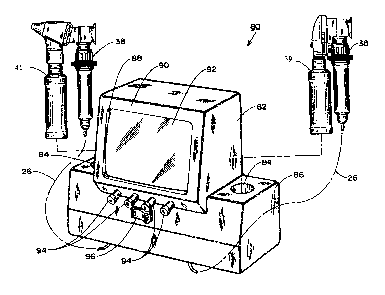
FIG. 1 is a perspective view according to one embodiment of the present
invention;
FIG. 2 is a bottom view of the apparatus according to Figure 1;
FIG. 3 is a perspective view according to another embodiment of the
invention;
FIG. 4 is a perspective view according to a third embodiment of the
invention; and
FIG. 5 is a bottom view of the apparatus according to Figure 4;
FIG. 6 is a block circuit diagram of one embodiment of the invention.
Detailed Description of the Invention
Referring to Fig. 1, there is shown an integrated video diagnostic center
embodying the teachings of the invention. The unit 10 has a housing 12 that is
suitable for wall mounting; however it is understood that the unit may be
placed on a table, or mounted on a roll-around cart, or the unit may be
attached
to a wall in a boom mount configuration. Preferably the housing 12 is
constructed of hard plastic or a similar material. The housing 12 has a recess
14 formed into the top surface 16. In the preferred embodiment, the recess 14
is substantially cylindrical in shape in order to accommodate and provide a
cradle for a rechargeable battery pack 18 of the type which is used with a
hand-
25, held medical diagnostic instrument, such as an otoscope, ophthalmoscope or
the
like wherein the battery pack also serves as the handle for the instrument.
The
recess 14 has an electrical connection integrated therein, which will be
explained in greater detail below. The housing 12 has formed therein a
convenient holder 20 for specula 22 that are used with the instrument. It will
be
2197484
4
understood that the housing may be formed to provide storage for other
accessories such as swabs or currettes.
Referring to Fig. 2, the bottom face 24 of the unit 10 is depicted. There
is an umbilicus 26 that is connected at the distal end to a video adapter 38
:c (shown in Fig. 4) which connects to a hand-held diagnostic instrument 39.
The
umbilicus 26 serves to relay the video information from the video adapter 38
to
the unit 10. The unit 10 houses electronic video processirig circuitry which
is
well known in the art that processes the video information for suitable use,
such as viewing a target area on a video monitor or for transmission of the
video data to various data storage instruments (not shown).
The bottom face 24 of the unit 10 also includes a calibration button 25.
The user of the apparatus focuses the hand-held diagnostic instrument 39 with
the video adapter 38 (Fig. 4) attached onto a suitable calibration surface
such as
a white sheet of paper. The user depresses the calibration button 25, which
when depressed provides an electrical signal to the video processing
circuitry,
thereby color calibrating the video signal to white. This calibration
procedure
ensures that any video information transferred to a video monitor or data
storage unit is of suitable quality. In addition to performing color
calibration,
the video processing circuitry may also compensate for back lighting on a
video
screen. For instance, the hand-held medical instrument, such as an otoscope
39, that is in use with the video adapter 38 may form an image on the video
screen that is circular due to the viewing area of the instrument 39. If the
image that is projected onto a video screen is not properl;y compensated for
back lighting, the image blooms on the video screen and is not of suitable
quality.
The video processing circuitry calibrates for both color and back lighting
when the calibration button 25 is depressed. The video processing circuitry
stores and maintains the calibration data in memory, even when the unit is
powered down. This feature eliminates the need for the user to calibrate the
unit each time the unit is used. Of course, one skilled in the art would
2197484
recognize that the calibration button 25 can be located at any convenient
location on the unit 10.
The bottom face 24 of the unit 10 also has a video output connection 28.
The video output connection 28 relays information from the video processing
5 circuitry which is contained within the housing 12 to an external monitor or
to
a video tape machine (not shown). A second video output connection 30 is
supplied to provide video information in the Y/C format for external uses.
There is also supplied in the bottom face 24 an electrical input connection 32
to
provide power to the unit 10. Electrical power from a power source is
transformed to provide 2.5v or 3.5v to the battery pack recharger as is
described below. In addition, electrical power is transformed to provide 12v
to
the video adapter 38.
Referring again to Fig. 1, the rechargeable battery pack 18 rests in the
recess 14. The battery pack 18 is provided with a female coupling 34 to
provide contact with the unit 10 by connecting to male coupling 36 which is
integral to the unit 10 in order to provide an electric current for
recharging. It
is understood that there are various configurations capable of performing the
recharging of the battery pack 18. For example, a battery recharger may be
surface mounted on the housing 12. The hand-held diagnostic instruments
include rechargeable batteries to provide power, for example, to operate a
light
source in the instrument. Typically, the recharging function provides charge
at
either 2.5 volts or 3.5 volts.
In use, the physician stores the hand-held medical diagnostic instrument
in the recess 14 of the housing 12 during which time the battery pack 18 is
charged. When the physician performs an examination that requires video
technology, the physician removes the battery pack 18, connects the
appropriate
medical diagnostic instrument to the battery pack 18, and connects the video
adapter 38 to the medical instrument 41 (Fig. 4). The physician will have a
video monitor 92 in use or the physician may provide the video image to a
remote location through telecommunications such as a modem (not shown).
2197484
6
Referring now to Fig. 3, there is shown an additional embodiment of the
present invention. The unit 40 has a housing 42 that is preferably formed from
hard plastic. The housing 42 has a recess 44 in the top surface 46 thereof to
accept and cradle a rechargeable battery pack 48 of a hand-held medical
diagnostic instrument, in this instance an ophthalmoscope is depicted. The
recess 44 provides a connection to the battery pack 48 in order to facilitate
recharging, similar to that shown in Fig. 1. An aperture 50 is formed in the
front face 52 of the housing 42. The aperture 50 is of sufficient size to
accommodate a small video monitor 54, preferably a 5 inch or greater diagonal
screen.
The unit 40 provides video output connections, a power supply
connection, a calibration button, and an umbilicus connecting the video
adapter
to the video processing circuitry contained within the housing 42 as shown in
Fig. 2. It is understood that the output connections, powei= supply
connection,
calibration button and umbilicus may be placed on other faces of the housing
42
to facilitate placement of the unit 40 in the most convenient location. The
preferred method is to wall mount the integrated video diagnostic center with
the various connections located on the bottom face.
The video information is relayed from the processing circuitry to the
video screen 54 where the physician and/or patient can view the target area.
This embodiment eliminates the need for a separate monitor in the examination
room and thereby achieves the objective of further reducirig the number of
units
in the examination room.
In some instances, the physician may prefer to utilize video adapters
with more than one type of hand-held diagnostic instrument, such as an
ophthalmoscope and an otoscope, and the embodiment depicted in Fig. 4
provides for the use of two. Of course, a plurality of instruments may be
provided for. The unit 80 has a housing 82 preferably formed of hard plastic
with two recesses 84-84 formed therein on the top surface 86. The housing 82
has an aperture 88 formed in the front face 90. A video nionitor screen 92 is
2197484
7
located in the aperture to provide the physician and patient with a view of
the
target area. There are also standard video control knobs 94 on the front face
90
of the unit 80 to allow the user to tune the monitor 92 to the desired picture
quality as is known in the art. The unit 80 has a three-way toggle switch 96
placed in the front face 90, the functionality of which will be described
below.
Referring now to Fig.'s 4, 5 and 6, the unit 80 and the bottom face 71
of unit 80 are depicted. The unit 80 provides a coaxial video output
connection
78, a Y/C video output connection 72, a power supply connection 74 and two
umbilica 76-76 connecting each video adapter 38-38 to the system bus video
processing circuitry 102 contained within the housing 82. When the physician
selects the middle position on the switch 96, the video unit is inoperative. A
selection of the right or left position on the switch 96 determines which of
the
respective video adapters 38 is operative.
There is a calibration button 77 located on the bottom face 71 of the unit
80. As described above, the calibration button 77, when depressed by the user,
sends an electrical signal to the video processing circuitry (Fig. 6). As
depicted in Fig.'s 4, 5, & 6, the unit 80 has two umbilica 76-76 leading to
video adapters 38-38 attached thereto which are controlled by the switch 96.
The calibration button 77 operates to calibrate the particular video adapter
which has been selected by the positioning of the switch 96. The calibration
information for the particular video adapter selected by the switch 96 is
stored
in the non-volatile memory of the video processing circuitry. Each of the
video
adapters can be separately calibrated and the calibration iriformation for
each is
stored in the video processing circuitry in non-volatile memory. Once each
video adapter is calibrated, the user need not recalibrate when switching from
one adapter to the other because calibration information relating to each
adapter
is maintained in memory and the video processing circuitiy is adapted to
utilize
memory depending upon the location of the switch 96.
Referring now to Fig. 6, there is shown a block diagrani of the video
processing circuitry. The imagers 112 and 114 contained within the video
2197484
8
adapters 38-38 relay information to the system bus 102 located within the
housing (indicated by dashed line in Fig. 6). The system bus 102 relays the
information to the microprocessing system 104 which contains a central
processing unit 106. An electronic switch 108, corresponding to mechanical
three way switch shown as 96 in Fig. 4, selects which of the two imager
signals will be relayed to the monitor circuitry 110 for display on the
monitor.
The electronic switch 108 also relays calibration data for the appropriate
imager
from the microprocessing system 104 to the monitor circuitry 110. It is
understood by one skilled in the art that there are various circuit
configurations
to accomplish the objective of displaying one or the other of' two incoming
video signals to a monitor.
In use, the physician selects which hand-held instrument to use and
removes it from the recess 84. The physician then selects the corresponding
video adapter 38 and connects an instrument, such as a otoscope 39 or an
episcope 41, to the video adapter 38. The three way switch 96 is moved to the
corresponding position and the video adapter 38 is activated. The physician
then performs the examination utilizing video capabilities. When completed,
the
three way switch 96 is returned to the center/off position, the video adapter
38
is removed, and the hand-held instrument 39 with the battery pack is returned
to the recess 84 for storage and charging.
While this invention has been explained with reference to the structure
disclosed herein, it is not confined to the details as set forth. For example,
the
shape of the housing is shown for illustration purposes. One of ordinary skill
in
the art will recognize that many housing shapes are possible. Also, the
battery
pack is shown as being recharged in a recess. One skilleci in the art will
recognize that the battery pack could be mounted on a recharger on the surface
of the housing. This application is intended to cover any modifications and
changes as may come within the scope of the following claims.
