Note: Descriptions are shown in the official language in which they were submitted.
~ 219827~
WO ~/06112 PCT~ngS/~279
Title: Antigenic peptides derived from the G protein of RSV
for type- and subtype-specific diagnosis of respiratory
syncytial virus (RSV) infection
FIELD OF THE INVENTION
This invention relates to the fields of peptide-based
diagnostics an~ vaccines in connection with diseases caused by
or related with respiratory syncytial virus (RSV) infection.
The invention involves a so far unidentified small,
independently folding, globular protein module between mucin-
like regions in the attachment protein G of RSV, and its use.
More specifically, the invention relates to the design of an
antigenic substance, preferably peptide-based, corresponding
to said protein module in the attachment protein G of RSV,
that can be used as a basis for e.g. a diagnostic assay.
Peptides corresponding to this independently folding globular
protein can also be incorporated in vaccines along with other
peptides to induce protective lmmune responses to the virus.
BACKGROUND OF THE INVENTION
RSV infections are a major cause of respiratory tract
disease in humans, cattle, sheep and goats (Stott and Taylor,
1985). The virus is classified within the Pneumovirus genus of
the Paramyxoviridae. Human respiratory syncytial virus (HRSV)
is the most important causative agent of bronchiolitis and
pneumonia in infants and young children. Approximately 100,000
children are hospitalized each year in the USA as a result of
RSV infection. A vaccine against the virus is not available
and development of a vaccine is third - subsequent to Malaria
and Human Immunodeficiency Virus - on the priority list of the
World Health Organization. In cattle, respiratory disease is
one of the most frequently recorded diseases. Recent reports
indicate that respiratory disease can account for up to 60% of
morbidity and for around 60% of mortality in feedlot cattle
(Healy et al., 1993, Edwards, 1989). Bovine respiratory
syncytial virus ~BRSV) infections are the major cause of
respiratory disease in calves resulting in high economic
losses.
W0~Kll2 ~9~19 rcT~n~u~79
Because different antigenic subgroups are described for
HRSV and BRSV (Johnson et al., 1987, Furze et al., 1994), it
is important to monitor the prevailing subgroups in a popu-
lation to choose a candidate vaccine of the right subgroup(s).
The virus has two major surface glycoproteins: the
attachment protein G and the fusion protein F. The G protein
is unique for RSV, it is highly variable between HRSV
subgroups (53% amino acid homology; Johnson et al., 1987), or
between HRSV and ungulate RSV ~30% amino acid homology; Lerch
et al., 1990). However, within the subgroups the amino acid
homology is much larger (80% or more within several HRSV-A
strains; Cane et al., 1991), 90% or more within several HRSV-B
strains (Sullender et al., 1991) and 90% or more within four
BRSV strains (Mallipeddi and Samal, 1993a). The G protein
shares neither sequence nor structural homology with other
attachment proteins of other Paramyxoviruses (Satake et al.,
1985, Wertz et al., 1985). In contrast to the attachment
proteins of other paramyxoviruses, G is shorter and lacks
hemagglutination or neuraminidase activity. RSV-G is a type II
membrane protein and contains about 60% carbohydrate by
weight. Approximately 20% of the carbohydrate moieties are N-
linked carbohydrates and 80% are O-linked carbohydrates which
are linked to the unusually high number of hydroxy amino acids
in the protein.
A number of diagnostic assays (reviewed by Welliver,
1988) are available for the detection of RSV. However, these
assays are based on whole virus or complete proteins that do
not (effectively) discriminate between subgroups of HRSV nor
between different~RSV types. secause the F protein is highly
conserved between all RSV types, a discriminating assay is
hard to design based on protein F and should therefore include
at least a part of the more variable G protein.
Empirical methods to determine the immunodominant site on
BRSV-G and HRSV-G showed that the immunodominant site of the
peptide was located within the C-terminal half of this peptide
(residues 174-188; Norrby et al., 1987). It has been suggested
WO96/06112 2 1 9 82 7g ~ PCT~ng5100279
that a 15-residue peptide (residues 174-188) could be used for
subtype-specific site-directed serology (Akerlind-Stopner et
al., 1990, Norrby et al., 1987).
The use of peptides as antigens in serological diagnosis
of infections has gained interest, because peptides are cheap
and easy to produce in a reproducible manner. However, the use
of peptides in routine diagnosis has so far been limited due
to lack of sensitivity.
SUMMARY OF THE INVENTION
The invention provides a peptide comprising an amino acid
sequence derived from protein G of a respiratory syncytial
virus, wherein said amino acid sequence has a length of from
~ about 28 to about 37 amino acid residues and is derived from a
- 15 region of said protein G which is located between two mucin-
like regions. Preferably, said amino acid sequence comprises
at least the amino acid residues Nos. 159-186, such as
comprising the amino acid residues Nos. 158-189 or Nos. 157-
193 of protein G (according to the numbering of protein G of
bovine respiratory syncytial virus) and has at most 4 amino
acid differences therewith. Preferably, said respiratory
syncytial virus (RSV) is selected from the group consisting of
bovine respiratory syncytial virus (BRSV), human respiratory
syncytial virus A (HRSV-A), human respiratory syncytial virus
B (HRSV-B), and ovine respiratory syncytial virus (ORSV).
Said amino acid sequence preferably comprises a member
selected from the group consisting of
HQDHNNFQTLPYVPCSTCEGNLACLSLC,
QQDYSDFQILPYVPCNICEGDSACLSLC,
PKDDYHFEVFNFVPCSICGNNQLCKSIC,
PNNDFHFEVFNFVPCSICSNNPTCWAIC;
more preferably a member selected from the group consisting of
NHQDHNNFQTLPYVPCSTCEGNLACLSLCHIE,
IQQDYSDFQILPYVPCNICEGDSACLSLCQDR,
KPKDDYHFEVFNFVPCSICGNNQLCKSICKTI,
KPNNDFHFEVFNFVPCSICSNNPTCWAICKRI;
WO~U~112 ~9 rCTnDD5~279
such as a member selected from the group consisting of
ENHQDHNNFQTLPYVPCSTCEGNLACLSLCHIETERA,
EIQQDYSDFQILPYVPCNICEGDSACLSLCQDRSESI,
~KKPKDDYXFEVFNFVPCSICGNNQLCKSICKTIPSNK,
NKPNNDFHFEVFNFVPCSICSNNPTCWAICKRIPNKK.
It is preferable that the peptide is capable of adopting
the tertiary structure of its counterpart in the corresponding
G protein.
This invention also relates to an antigenic substance, or
a precursor thereof, which allows discrimination between or
identification of different RSV types or subtypes, or allows
discrimination between or identification of antibodies against
different RSV types or subtypes, which antigenic substance or
precursor thereof comprises a peptide as defined herein.
A peptide, antigenic substance or precursor thereof as
defined herein may be used in diagnosis of RSV infections; or
in the prophylaxis of respiratory syncytial virus infections.
This invention also provides a diagnostic testkit for the
detection or identification of RSV types or subtypes, or anti-
bodies against RSV types or subtypes, which testkit comprises
a peptide, antigenic substance or precursor thereof as defined
herein, together with suitable means for detection. The
testkit preferably provides for an enzyme linked immunosorbent
assay, e.g. a blocking ELISA.
The invention also provides a method for the detection of
(antibodies against) RSV comprising contacting a sample of a
body fluid with a peptide, antigenic substance or precursor
thereof as defined herein, in a manner such that a complex
comprising said peptide, antigenic substance or precursor, and
an antibody directed against said peptide, substance or pre-
cursor can be formed, followed by detection of said complex.
Furthermore, the invention provides a pharmaceutical
composition for the prophylaxis of RSV infections comprising a
peptide, antigenic substance or precursor thereof as defined
herein, together with a suitable adjuvant or excipient for
administration to a mammal.
I
WO96/~112 2198279 ~ PCT~n95/00279
The invention also provides a method for the prophylaxis
of RSV infections comprising administering to a mammal a
composition as defined above, in an amount sufficient to
~ elicit an immune response against respiratory syncytial virus.
Furthermore, the invention provides a peptidomimeticum
which mimics a peptide as defined herein.
Another aspect of this invention is a method for inducing
antibodies against RSV types or subtypes comprising
administering to a mammalian host an antigenic substance or
precursor thereof as defined herein, together with a suitable
adjuvant and harvesting resulting antibodies or antibody
producing cells from said mammalian host.
An antibody directed agalnst a type or subtype of RSV
obtainable by the above method is also part of the invention.
Preferably, the antibody is a monoclonal antibody.
In another aspect, the invention provides a diagnostic
testkit for the detection of or the discrimination between
(antibodies against) subtypes or types of RSV comprising the
above antibody and suitable means for detection.
DETAILED DESCRIPTION OF THE INVENTION
Most peptides that have been used in serology represent
continuous epitopes. It is impossible to detect antibodies
against complex discontinuous epitopes using small linear
peptides and it is difficult to predict discontinuous epitopes
based on the amino acid sequence of a protein. In addition,
the antigenic surface of large globular proteins cannot be
mimicked accurately with a small linear peptide. We solved
this problem by predicting an independently folding region in
the G protein of RSV viruses that adopts a stable tertiary
structure while retaining its antigenicity. This prediction is
crucial for the correct design of a useful antigen.
RSV-G contains an unusual high amount of hydroxy amino
acids which are acceptor sites for O-glycosylation ~Johnson et
al., 1~37). We discovered that hydroxy amino acids like Serine
(S) ~nd Th~eonine (T) are clustered together with Proline (P)
WO gC~Kl12 ~9~ - PCT~n9~0027~
in two discrete regions of the G protein of RSV. These
~egions, enriched in serines, threonines and prolines (STP),
are common motifs that are heavily O-glycosylated and probably
adopt a stiff and extended conformation (generally reviewed by
Jentoft, 1990). Because such regions are the major constituent
of mucins (large polymeric molecules that form mucous gels),
the regions are called mucin-like regions. By definition, the
amino acid sequences in mucin-like regions comprise 25-40% of
serine or threonine residues (Jentoft, 1990).
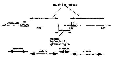
ne located the first mucin-like region in RSV-G as
extending from the transmembrane region until the conserved
double Pro1ss,1s6 and the second mucin-like region as extending
approximately from the conserved Pro1g4 until the C-terminus.
We defined the ectodomain of the G protein of RSV as a small
hydrophobic globular region which residés between two discrete
mucin-like regions. The proposed location of this small
central hydrophobic region of RSV-G (Fig. 1, 2), the possible
autonomous folding of this short sequence, its relatively
conserved nature, and the sparse potential glycosylation sites
therein make a peptide representing this short region a
promising candidate to be used as antigen for an immunoassay.
Comparative tests carried out with a peptide essentially
corresponding with the 15-residue peptide disclosed by Norrby
et al. (the peptide used in said comparative tests was a 16-
residue peptide composed of residues 174-189) have shown that
a 32-residue peptide corresponding to the central hydrophobic
region of HRSV-G reacted better as antigen in an ELISA than
said 16-residue peptide which represents only a part of said
central hydrophobic region. Probably, the 32-residue peptide
adopts a more native-like, complete structure. Tests with a
low sensitivity, as obtained when using the 15-residue peptide
of Norrby et al., are not suitable for performing sensitive
sero-epidemiological studies which is very important when
trying to discriminate between types or subtypes of RSV.
The invention provides an antigenic substance for discri-
mination between individuals infected with different subtypes
WO96/06112 21 982 79 ~" ~ PCT~n~51~279
of RSV. The antigenic substance is a peptide which corresponds
to an amino acid sequence of RSV-G, which is located between
the two mucin-like regions.
An antigenic substance according to this invention is to
be interpreted as any peptide-like or peptide-based substance
capable of inducing an immune response against RSV or recog-
nized by a serum containing antibodies against RSV. Precursors
of such antigenic substances are meant to read on comparable
peptide-like or peptide-based substances, which are not
immunogenic themselves but need for instance to be coupled to
a carrier to be able to induce an immune response or to be
recognized. Peptide-based or peptide-like substances are
intended to include anything with the function of the peptides
according to the present invention. This means that these
substances may be peptides themselves in which a number of
amino acid residues have been replaced or modified. It also
means that they may be fusion proteins for instance designed
to present the amino acid sequence of the peptides of the
invention on their surface. The definition also includes
peptidomimetics and anti-idiotype antibodies derived from the
peptides according to the invention.
In a preferred embodiment the invention provldes peptides
that can be used in diagnostic assays for detection of anti-
bodies directed against specific RSV types and subtypes (HRSV
and subtypes, BRSV and subtypes, ORSV and subtypes). Further-
more, these peptides can be incorporated in RSV vaccines.
The invention of the independently folding region in the
G-protein relates to all types of RSV. As a consequence, the
invention is not limited to the peptides specifically
disclosed herein, but extends to analogous peptides and their
derivatives in all types of RSV and all subtypes of these
viruses.
Preferred peptides to be used according to the invention
comprise at least the antigenic parts of the peptides given in
table l or derivatives thereof, their length being from about
28 residues ~p to about 37 residues.
WO ~K112 ~ ~9 ~q ~ rCT~n95100279
We have evaluated the applicability of the peptides in
diagnostics by the development of two different diagnostic
assays: an indirect ELISA and a blocking ELISA. Both of these
tests are type- and subtype-specific.
Other diagnostic assays can of course be easily designed
by the man skilled in the art.
These may of course be provided in any suitable format.
Assays can be performed in solution on solid phases, they can
be performed using any kind of label, such as enzymes, solid
particles, such as metal sols, or other sols, latex particles,
dyes, fluorescent substances or ~radioactive materials. They
even may be performed without labels, as can be done by
agglutination assays. The peptides can be used to detect
antibodies in for instance a fluid from a mammal, such as
blood, serum, urine, milk. Usually the antibody is bound by a
peptide according to the invention, which may be present on a
solid phase. Afterwards the complex of peptide and antibody
may be detected by a labelled reagent, which can be a labelled
antibody directed against human or bovine antibodies.
According to the invention the peptides can also be used
to obtain antibodies which are specific for RSV types and/or
subtypes. The peptides are administere~ to a mammal, usually a
rodent, in an immunogenic form and after one or more booster
administrations the serum from the animal is harvested and
antibodies can be purified therefrom. Alternatively, the
spleen of such animals may be removed to obtain antibody
producing cells. These can be changed, by fusion or trans-
formation, into cell lines producing monoclonal antibodies.
Assays based on (monoclonal) antibodies directed to RSV and
induced by the peptides according to the invention are
therefore also a part of the invention.
The peptides according to the invention can of course
also be used in vaccines to prevent infections with RSV. They
may be used in conjunction with other antigens or alone to
elicit an immune response against RSV. Usually the peptide has
to be coupled to a carrier to be presented in an immunogenic
WO96~K112 21 9 82 7~' ~ PCT~n~5n027~
form before administration to a host. Other ways of rendering
a peptide sufficiently immunogenic are known to the person
skilled in the art. Adjuvants are usually added to vaccines to
boost the immune response in a more aspecific manner.
The invention relates to a set of RSV diagnostic assays
based on peptides corresponding to the central region of the
RSV G protein. The regions are listed in Table 1 for BRSV,
ORSV, HRSV-B and HRSV-A. The length of the peptide to be used
in a diagnostic assay preferably is from about 28 to about 37
residues (Table 1). The minimal length of a suitable peptide
is dictated by the length of the module which lies between
Hislsg and Cysl~6. The maximal length of a suitable peptide is
dictated by the length of the module which lies between Pr0156
and Prolg4-
The region corresponds to a relatively conserved region
in a highly variable protein. Between different BRSV strains
an amino acid homology of 90% or more is observed, both in the
sequences corresponding to the underlined 32-mer peptide shown
in Table 1, and in the sequences of the 28-residue module
consisting of residues Nos. 159-186. Therefore the invention
relates to all peptides listed in Table 1 and all analogous
peptides with at most 4 amino acid differences within the 32-
mer or within the 28-residue module. Furthermore peptides
corresponding to yet to be sequenced subgroups of BRSV or ORSV
and their analogues with at most 9 amino acid differences are
also part of this invention.
The diagnostic assays based on the peptides can be used
to determine antibody levels in blood, serum, milk or other
body fluids.
The materials according to the invention can also be used
for incorporation in a vaccine.
wog6~ 9~~ ; rcrnnssou27s
F.x~m~l es
Structure analysis of RSV protein G
A detailed analysis of the primary structure of protein G
of RSV ~RSV-G) allowed a dissection of the protein in shorter
modules. In Figure 1, a schematic presentation is shown based
on this analysis of the primary structure. According to this
analysis, the protein comprises a cytoplasmic region, a trans-
membrane region, an elongated mucin-like region (37% Ser and
Thr), a central hydrophobic globular region, and a short
positively charged region within a second elongated mucin-like
region (38% Ser, Thr). The modular architecture demonstrateS
that the central hydrophobic globular region is positioned
between two mucin-like stalks, and limited approximately by an
N-terminal conserved double Pro1ss,1s6 and a C-terminal
consérved ProIg4 (Figure 1, 2). This schematic model suggests a
very important functional role for the central hydrophobic
region in protein binding because it may be the only exposed
protein domain.
The rest of the ectodomain of RSV-G iS mainly mucin-like.
The dense carbohydrate coat of the mucin-like regions is added
to the envelope protein by cellular enzyme systems and for
that reason the mucin-like regions are probably not very
antigenic.
The location of a small hydrophobic protein module
between two immunosilent mucin-like regions, the possibly
autônomous folding of this short sequence, the relatively
conserved nature, and the sparse potential glycosylation sites
make a peptide representing this short region a promising
candidate to be used as antigen for an immunoassay.
Peptide synthesis
Peptides were selected from the central hydrophobic
region of RSV-G that is located between the two mucin-like
regions. The central hydrophobic regions (residues 158 - 189)
of all cloned RSV types and subtypes were synthesized: BRSV-G
,
WO96/~112 21 9 82 j PCT~95/00279
(Lerch et al., 1990; WO 92/01471); ORSV-G (Mallipeddi and
Samal 1993b); HRSV-G type A (Wertz et al., 1985); and HRSV-G
type B (Johnson et al., 1987). Additionally, a peptide
corresponding to the immunodominant peptide (residues 174-189)
of HRSV-G type A was synthesized.
BRSV: acetyl-N H Q D H N N F Q T L P Y V P C S T C E G N L A C L S L C H I E-amide
ORSV: acetyl-I Q Q D Y S D F Q I L P Y V P C N I C E G D S A C L S L C Q D R-amide
HRSV-A: acetyl-~ P N N D F H F E V F N F V P C S I C S N N P T C W A . C K ~ I-amide
ace~yl-S I C S N N P ~ C W A ~ C K .~ l-amide
HRSV-B: acetyl-K P K D D Y H F E V F ~ F V P C S I C G N N Q L C K S I C K ~ I-amide
Synthesis of peptides was performed according to standard
procedures on an Applied Biosystems 430A synthesizer using
Fastmoc chemistry (Fields et al., 1991). Purified oxidized
peptide was obtained as follows: ~-mercaptoethanol reduced
peptide was slowly oxidized by dialysing against 1% NH4HCO3,
which was frequently refreshed, for three days. These peptides
were used as antigens in enzyme linked immunosorbent assays
(ELISA).
Serum samples and monoclonal antibodies
The following serum samples and monoclonal antibodies
were incorporated in the study.
Cattle. Negative field serum samples (N=40) were obtained
from 4 to 6 month-old calves which had no detectable
antibodies against BRSV for at least one month, using the F-
ELISA (Westenbrink et al., 1985). Sera were collected during
the summer season (Van der Poel et al., 1993).
In addition, negative serum samples (N=12) were obtained
from specific-pathogen-free (SPF) calves. The calves were
obtained by caesarean section, deprived of colostrum, and
reared in isolation.
WO ~112 ~9~ rCTnnD5l~279
12
BRSV-negative serum samples (N=4), containing antibodies
directed against either parainfluenza virus type 3 ~PI-3),
bovine herpesvirus 1 (BHVl), bovine viral diarrhea virus "
(BVDV) or mycoplasma, were also incorporated in this
investigation.
Field serum samples (N=102) that were positive in the F-
ELISA, were obtained from several Dutch farms with a history
of BRSV infection (Van der Poel et al., 1993). In the indirect
peptide-based ELISA (iG-ELISA), 102 sera were tested and in
the blocking peptide-based ELISA (bG-ELISA), 97 of these 102
sera were tested.
Paired serum samples ~N=152) from 76 different animals
were used to test for increases in antibody titer. Sera were
collected with one month interval in December 1990 and in
January 1991 at two different Dutch farms (Van der Poel et
al., 1993). Some of these sera (N=24) were used to distinguish
between reactivity against the BRSV-G peptide or ORSV-G
pept~ide.
Finally, we tested serum samples (N=6) that reacted non-
specifically in the F-ELISA.
Sheep. Sheep sera (N=3) positive for RSV in the F-EL~SA
were obtained from our internal sheep serum bank.
Human. Human sera (N=14), positive for HRSV specific
antibodies in a complement fixation test were obtained from
Dr. J.A. Melero of the National Centre for Microbiology
(Madrid, Spain). These sera were collected from patients, with
an age range of 7 months to 70 years, during the 1993-1994 RSV
epidemic.
Human sera (N=23) positive for RSV were obtained from Dr.
J.C. de Jong of the RIVM in Bilthoven, the Netherlands. These
sera were used in the tests to compare the 16-residue with
the 32-residue peptide as antigen.
~ Ahbit. Rabbits (N=3) were immunized with the supernatant
of HRSV-A (strain Long) and HRSV-B (strain 9320) infected
cells, respectively. Rabbits were vaccinated with 1 ml
WO~K112 219 ~ 7 ~ rCTnn~027g
13
Freund's complete adjuvant (FCA), mixed 1:1 with 1 ml of
supernatant.
MonoclonAl ~nt;~o~es. Production of a BRSV-G specific
monoclonal antibody (MAb 20) was performed as described
(Wensvoort et al., 1986). Balb/c mice were immunized
intraperitoneally with 100 ~g BRSV (Lelystad strain), grown on
embryonic bovine tracheal cells, mixed with FCA. The RSV-
specificity of MAb 20 was determined using an immunoperoxidase
monolayer assay (IPMA) as described (Wensvoort et al., 1986).
In this assay Vero cells infected with BRSV (strain Lelystad)
were used. Additionally, MAb 20 reacted in the BRSV-G peptide-
based iG- and bG-ELISA.
MAbs 2G and l9G specific for the G protein of HRSV A,
were obtained from Dr. J.A. Melero, National Centre for
Microbiology (Madrid, Spain). MAbs 26 and 30, specific for the
G protein of HRSV B and A, respectively, were a kind gift of
Drs. J. Furze and G. Taylor, AFRC Institute for Animal Health,
Compton, UK.
Comparison of antigenicity of different peptides
The single linear immunodominant region of BRSV-G as
determined by peptide binding studies (residues 174-185) and
the immunodominant peptide of HRSV-G described by Norrby et
al. ~residues 174-188) correspond to the C-terminal half of
the central hydrophobic region of RSV-G (residues 158-189). To
check whether the empirically determined immunodominant
epitope of HRSV-G type A (contained within the 16-residue
peptide 174-189) has the same antigenic characteristics as the
predicted antigenic site (the 32-residue peptide corresponding
to the central hydrophobic region 158-189), both peptides were
tested for their antigenicity in an iG-ELISA using the G-
peptide of HRSV subtype A as antigen. Four times more sera
were scored positive in the indirect G-peptide ELISA (iG-
ELISA) based on the 32-residue peptide than in the iG-ELISA
based on the 16-residue peptide (Table 2). Although the
peptide binding studies showed that the immunodominant site is
W09h~6ll2 ~9 '~ PcT~ngllo~79
contained in the 16-mer peptide (Norrby et al., 1987), the 32-
residue peptide corresponding to the central hydrophobic
region of HRSV-G type A as described in this study (Fig. 1) is
much more react-ive with human sera when compared with the 16-
residue peptide. Therefore, 32-residue peptides corresponding
to the central hydrophobic region of the G-protein of several
types and subtypes of RSV were used as antigen in immuno-
assays.
Respiratory syncytial virus specific F-ELISA
Test procedure. The RSV-specific indirect double antibody
sandwich assay, used as a routine diagnostic test in our
laboratory, was performed essentially as described previously
(Westenbrink et al., 1985), except that MAbs, instead of horse
anti-RSV serum, were used as capture antibody. In short,
microtiter plates coated with two MAbs (N~ 88953, ID-DLO,
Lelystad) directed against BRSV-F were subsequently incubated
with bovine RSV antigen, the test serum, rabbit anti-bovine
immunoglobulin peroxidase (Dakopatts, P159) and substrate
chromogen solution. Before use, and after each incubation
step, plates were rinsed six times with deionised water
containing 0.05% Tween 80. Dilutions of test sera and reagents
were made in ~high-salt~ ELISA-buffer (8.1 mM Na2HPO4, 2.79 mM
KH2PO4, 0.8 M NaCl, 2.68 mM KCl, lmM EDTA, 0.05% Tween 80,
pH 7.2) containing 4% horse serum. BRSV antigen stock solution
(N~ 88915, ID-DLO, Lelystad) was diluted 1:2 (100 ul/well) and
incubated during two hours at 37~C. Test sera were diluted
1:80 ~100 ul/well) and incubated for one hour at 37~C. Horse-
radish peroxidase ~HRPO) conjugated rabbit anti-bovine immuno-
globulin (Dakopatts, P159) was diluted 1:2000 (100 ul/well)
and incubated for one hour at 37~C. The substrate chromogen
solution consisted of 10 mM sodium-phosphate buffer (pH 6.8),
0.1 mM EDTA, 0.1% w/v 5-aminosalicylic acid, and freshly added
0.005% v/v H2O2. Incubation with substrate solution was
performed overnight at 4~C. Colour development was measured at
450 nm (Titertek Multlscan~. Absorbance values higher than two
WO ~K112 1 9 82 7g PCT~n951~27g
lS-
times the average background value of testsera in control
- wells without antigen, were considered positive. Although the
test has been developed for the detection of antibodies
specific for BRSV, antibodies against all other RSV types can
be detected with the test due to extensive F protein immune-
crossreactivity between RSV types and subtypes.
Indirect G-peptide ELISA (iG-ELISA)
Test proce~ure. The iG-ELISA was based on the test
procedure of the F-ELISA as described above with the following
modifications. The antigen was not caught but directly coated
to the plate. One hundred and fifty ng of crude oxidized
peptide was coated per well ~high binding capacity flat bottom
microplate, Greiner) in l00 ~l carbonate buffer pH 9.0, 4~C,
overnight. The optimal dilution of the peptide to coat the
ELISA plates was chosen in such a manner that maximum binding
was obtained as determined in a checkerboard titration. Test
sera, diluted l:5, and conjugate were incubated for one hour
at 37~C in "low-salt" ELISA buffer (8.1 mM Na2HPO4, 2.79 mM
KH2PO4, 0.5 M NaCl, 2.68 mM KCl, l mM Na2EDTA, 0.05~ v/v Tween
80, pH 7.2) containing 4% horse serum. Subsequently, the test
was performed as described above. The conjugates used in the
test were anti-bovine (1:2000), anti-sheep (l:l000), and anti-
human HRPO (l:l000) (Dakopatts). Absorbance values higher than
two times the average background value of testsera in control
wells without antigen, were considered positive.
iG-F.T.ISA
The reactivity of different panels of bovine sera in the
iG-ELISA was compared with the reactivity in a routine diag-
nostic F-ELISA (Fig. 3). By using the mean OD of all negative
sera (N=40) plus twice the standard deviation (% + 2SD =
0.062) as cut-off value for negativity, the relative
specificity of the iG-ELISA was found to be 0.98. Using this
cut-off value, the sensitivity of the test was determined
using 102 positive field serum samples from several Dutch
wog6~ 2 ~19:' ~ PCT~n95/~279
16
farms and was found to be 0.90 (92/102). Four different sera
containing antibodies against other microorganisms (BHV1,
BVDV, PI-3, mycoplasma) were all negative in the iG-ELISA
(data not shown). Six sera that reacted non-specifically in
the routine F-ELISA reacted also non-specifically in the iG-
ELISA (data not shown).
The low sensitivity compared to the routine F-ELISA may
be due to (i) a relative low antigenicity of BRSV-G in
comparison to that of BRSV-F, (ii) low antibody titers of some
animals, or (iii) some sera may be directed to another, yet
uncharacterized, subtype.
Blocking G-peptide ELISA (bG-ELISA)
Test procedure. This ELISA for measuring BRSV-specific
antibodies is based on blocking of the interaction of a BRSV-G
specific monoclonal antibody (MAb 20) with the coated peptide
by peptide-specific antibodies that may be present in the test
sample. ELISA plates were coated with 30 ng crude oxidized
peptide per well in 100 ~l carbonate buffer pH 9.0, 4~C,
overnight. The optimal dilution of the peptide to coat the
ELISA plates was chosen in such a manner that a near maximum
binding was obtained as determined in a checkerboard titration
and that the sensitivity of the test was maintained high.
Before use, and after each incubation step, plates were rinsed
six times with deionised water containing 0.05% Tween 80.
Plates were subsequently incubated with test serum diluted
1:2, a HRPO-conjugated monoclonal antibody specific for the G-
pepti~e (bovine RSV - MAb 20, ID -DLO, Lelystad) diluted 1:5000,
and substrate chromogen solution. Incubation with substrate
solution was performed overnight at 4~C. Test sera and
conjugate were incubated for one hour at 37~C in "low-salt"
ELISA buffer containing 4% horse serum.
Blocking percentages of each test sample was calculated
by using the optical density at 450 nm of "low salt" ELISA
buffer containing 4% horse serum as reference (- 0% blocking)
according to the following formula:
WO7RX1~2 ~79 '~t rcTnnJ~279
blocking percentage of test sample =
OD(~low sAlt" hllffer + horse serllm) - OD(test sA~le) x 100%
OD("low salt" buffer + horse serum)
hG-F.T.ISA
Blocking percentages of different panels of bovine sera
were compared in the bG-ELISA (Fig. 4). When the mean blocking
percentage of all negative sera (N=40) (X + 2SD = 42%) was
used as cut-off value for negativity, the relative specificity
was found to be 0.98. The relative sensitivity of the test as
determined using 97 of the 102 positive field serum samples,
was found to be 0.98 (95/97). Sera containing antibodies
against other microorganisms (BHV-l, BVDV, PI-3, mycoplasma)
were all negative in the bG-ELISA (data not shown). The six
sera that reacted non-specifically in the routine F-ELISA and
in the iG-ELISA, were tested in the bG-ELISA. One of these
sera blocked significantly (75%), suggesting that this serum
was positive for BRSV antibodies.
}i
Detection of RSV infection
An antibody titer rise ( 2 4x) in paired sera is normally
regarded as being the result of an infection or reinfection.
In 76 paired serum samples, seroconversion or at least a
fourfold titer rise was detected 42 times in the iG-ELISA and
32 times in the routine F-ELISA (Table 3). The difference in
frequency of titer rise seems to be related to the age of the
animals ~Table 3). The BRSV iG-ELISA was more sensitive in
detecting reinfections than the F-ELISA. The difference in
frequency of titer increases seemed to be associated with the
age of the animal. The iG-ELISA and the routine F-ELISA were
equally sensitive for detection of seroconversion in young
calves (age < l year). However, in older cattle (age more than
l year), antibody titer increases were detected more
frequently when the iG-ELISA was used. Therefore, the number
of reinfections may be underestimated when titer increases are
based on the F-ELISA. The different results of both assays may
WO ~K112 ~9~ ~ ' PCT~ng5/~27g
be explained by (i) a faster drop of antibody titers against G
compared with that of F after infection, or (ii) by the lower
antibody response against G as compared to that against F
after the first infection. Consequently, a reinfection may
induce a more pronounced increase in antibodies against G than
against F. Therefore, G-specific ELISAs, including the peptide
ELISAs described in this patent, may have the advantage over
ELISAs based on the F protein, that they better detect
reinfections with RSV.
Type- and subtyDe-sDec;ficity of peptide-F.LISA
RSV ELISAs based on whole virus contain antigenic
proteins which are very conserved. Therefore, such ELISAs are
not type- or subtype-specific, which means that these assays
do not distinguish between human RSV and ungulate RSV, and
certainly not between HRSV-A and HRSV-B or between BRSV and
ORSV. A dendogram was calculated based on the phylogenetic
relationship of RSV according to the amino acid sequence of
RSV-G and on the central hydrophobic region of RSV-G (Fig. 5).
Because RSV-G is highly variable between RSV types and
subt~pes, we investigated whether peptide-based iG-ELISAs were
able to recognize type-, or subtype-specific antibodies. The
reactivity of 14 sera of patients collected during the 1993-
l994 epidemic in Madrid showed that 13 sera reacted
specifically in the HRSV iG-ELISA and not in the BRSV iG-ELISA
(Table 4). Furthermore, 12 sera had a higher reactivity in the
HRSV-A iG-ELISA than in the HRSV-B iG-ELISA. Polyclonal rabbit
sera directed against HRSV-A or HRSV-B were tested for their
reactivity against the HRSV peptides. Table 5 shows that sera
of rabbits immunized with HRSV-A reacted only in the HRSV-A
iG-ELISA, and the serum of the rabbit immunized with HRSV-B
reacted only in the HRSV-B iG-ELISA. Furthermore, four
G-specific MAbs with known subtype-specificity tested in the
iG-ELISA did not show cross-reactivity ~Table 4). Previous
studies confirmed the subtype-specificity of an ELISA based on
Wo~UKIl~ 98279 ~ rCTnn~ ~ 279
19
~ the 15-residue peptide using some paired serum samples
(Akerlind-Stopner et al., 1990, Norrby et al., 1987).
. ORSV and BRSV are two ungulate RSV types which are
genetically equally distant compared to the distance between
HRSV subtype A and HRSV subtype B (Fig. 5). The genetic
distance based on the amino acid sequence of the central
hydrophobic region is slightly longer between ORSV and BRSV
than between HRSV-A and HRSV-B. Therefore, the subtype-
specificity of the RSV iG-ELISA can also be checked with RSV-
positive sera of sheep and cattle, which most likely can onlybe infected with ORSV and BRSV, respectively. Bovine sera of
24 different animals, collected at the same timepoint, and
reacting positively in the routine F-ELISA, were tested for
reactivity in the BRSV iG-ELISA and the ORSV iG-ELISA,
respectively (Table 5). The bovine sera reacted in the BRSV
iG-ELISA and not in the ORSV iG-ELISA. In addition, three RSV-
positive ovine sera reacted only in the ORSV iG-ELISA and not
in the BRSV iG-ELISA (Table 5).
Vaccine study
Two peptide vaccines were used to test the immunogenicity
of the peptide in calves and to examine whether vaccination
with the peptide could reduce or inhibit virus infection. One
calf was vaccinated once with the 32-residue BRSV-G peptide in
Freund's complete adjuvant (FCA), one calf was vaccinated once
with the 32-residue BRSV-G peptide coupled to Keyhole Limpet
Haemocyanin (KLH) in FCA, and a control calf was not
vaccinated. Antibody reactivity against the peptide was
monitored during the experiment (Fig. 6). Nine weeks after the
vaccination the animals were challenged nasally with 2 ml
virus (Odijk strain, TCID50: 103~8/ml). Before challenge, and
5, 7, 11 and 14 days after challenge lung washings were taken
from the calves. Cells in the washings were tested for the
presence of BRSV antigen, and virus titrations were performed
with the washings (Fig 7 a,b). The calf vaccinated with the
peptide conjugated to the carrier protein shows a considerate
WO ~K112 ~9S~ rCT~n~l0027~
protection against virus challenge. The protection is better
for the conjugated peptide compared with the unconjugated,
which seems to be assoclated with the antibody response
against the peptide (Figs 6, 7).
s
DESCRIPTION OF THE DRAWING:
Fi~ure 1:
a schematic representation of the primary structure of
RSV-G; Hatched box: transmembrane region (TM); dotted box:
positively charged region. The extent of the mucin-like
regions is indicated by upper arrows, which corresponds with a
high content of serine, threonine and proline (37-38% serine
and threonine content). ~= cysteine. The bottom of the figure
displays the general variablility in G (Sullender et al.,
1991, Cane et al., 1991, Mallipeddi and Samal, 1993a).
Figure 2:
Schematic structural model of RSV-G. The central
hydrophobic region (residues 158-189) is shown as a grey
ellipse. Mucin-like regions are shown with potential O-linked
glycosylation sites (short horizontal lines) and potential N-
linked glycosylation sites (branched lines). Hatched regions
correspond to the transmembrane region and the cytoplasmic
region.
Figure 3:
a. Reactivity of different sera in the BRSV iG-ELISA as
described in the test procedure.
b. Reactivity of different sera in the BRSV F-ELISA as
described in the test procedure.
F;gure 4:
Blocking percentages of different sera in the BRSV bG-
ELISA as described in the test procedure. Note that some
negative sera have blocking percentages below ~ero.
~ WO96/~112 ~D~g PCT~n~K100279
.
F1gllre 5:
Amino acid distances between (a) RSV-G proteins, and (b)
central hydrophobic regions of RSV-G (residues 158-189).
Phylogenetic analysis was performed with the neighbour-joining
method (Saitou and Nei, 1987) and the UPGMA method of
clustering in the PHYLIP package (Felsenstein, 1989).
F;gure 6:
Reciprocal of antibody dilution positive for the BRSV iG-
ELISA. Numbers on the x-axis indicate the number of weeks
after vaccination. The arrow indicates the time of challenge.
Figure 7:
a: Percentage of positive cells in lung washing of two
vaccinated and one unvaccinated calf on day 5 and day 7 after
challenge. Cells were stained using a BRSV-F specific MAb
conjugated with fluoresceine.
b: Virus titer in lung washing.
WO96~K112 ~9 PCT~n~5/00279
22
REFERENCES
Akerlind-stopner B., G. Utter,- M.A. Mufson, C. Orvell,
R.A. Lerner, and E. Norrby (1990). Subgroup-Specific Antigenic
Site in the G Protein of Respiratory Syncytial Virus Forms a
Disulfide-Bonded Loop. J. of Vir. 64, 5143-5148.
Cane P.A., D.A. Matthews and C.R. Pringle (1991).
Identification of variable domains of the attachment (G)
protein of subgroup A respiratory syncytial viruses. J. of
Gen. Vir. 72, 2091-2096.
Edwards J.A. (1989). The effect of stressors like rumen
overload and induced abortion on BRD in feedlot cattle. Agri-
Practice 10, 10-15.
Felsenstein J. (1989). PHYLIP, Phylogeny Inference
Package (Version 3.2). Cladistics 5, 164-166.
Fields C.G., D.H. Lloyd, R.L. Macdonald, K.M. Ottenson,
and R.L. Noble. (1991). HBTU activation for automated Fmoc
solid-phase peptide synthesis. Pept. Res. 4, 95-101.
Furze J., G. Wertz, R. Lerch, and G. Taylor (1994).
Antigenic heterogeneity of the attachment protein of bovine
respiratory syncytial virus. J. Gen. Virol. 75, 363-370.
Healy A.M., M.L. Monaghan, H.F. Basset, H.M. Gunn, B.K.
Markey, and J.D. Collins (1993). Morbidity an mortality in a
large Irish feedlot; microbiological and serological findings
in cattle with acute respiratory disease. Br. Vet.J. 149, 549-
560.
Jentoft N. (1990). Why are proteins O-glycosylated? TIBS
15, 291-294.
Johnson P.R., M.K. Spriggs, R.A. Olmsted and P.L. Collins
(1987). The G glycoprotein of human respiratory syncytial
viruses of subgroups A and B: Extensive sequence divergence
between antigenically related proteins. Proc. Natl. Acad. Sci.
USA 84, 5625-5629.
Lerch R.A., K. Anderson and G.W. Wertz (1990). Nucleotide
sequence analysis and expression from recombinant vectors
demonstrate that the attachment protein G of bovine
W09~6il2 ~27"~ rcrrns~/002~
23
respiratory syncytial virus is distinct from that of human
respiratory syncytial virus. J. of Vir. 64, 5559-5569.
Mallipeddi S.K. and S.K. Samal (1993a). Sequence variabi-
- lity of the glycoprotein gene of bovine respiratory syncytial
virus. J. of Gen. Vir. 74, 2001-2004.
Mallipeddi S.K. and S.K. Samal (1993b). Analysis of the
ovine respiratory syncytial virus (RSV) G glycoprotein gene
defines a subgroup of ungulate RSV. J. of Gen. Vir. 74, 2787-
2791.
Norrby E., M.A. Mufson, H. Alexander, R.A. Houghten and
R.A. Lerner (1987). Site-directed serology with synthetic
peptides representing the large glycoprotein G of respiratory
syncytial virus. Proc. Natl. Acad. Sci. USA. 84, 6572-6576.
Saitou N. and M. Nei (1987). The neighbor-joining method:
a new method for reconstructing phylogenetic trees. Mol. Biol.
Evol. 4, 406-425.
Satake M., J.E. Coligan, N. Elango, E. Norrby and S.
Venkatesan (1985). Respiratory syncytial virus envelope
glycoprotein (G) has a novel structure. Nucleic Acids Res. 13,
7795-7812.
Stott E.J. and G. Taylor ~1985). Respiratory syncytial
virus: brief review. Arch. Virol. 84, 1-52.
Sullender W.M., M.A. Mufson, L.J. Anderson and G.W. Wertz
(1991). Genetic diversity of the attachment protein of
subgroup B respiratory syncytial viruses. J. Virol. 65, 5425-
5434.
Van der Poel W.H.M., J.A. Kramps, W.G.J. Middel, J.T. van
Oirschot and A. Brand (1993). Dynamics of bovine respiratory
syncytial virus infections: a longitudinal epidemiological
study in dairy herds. Arch. Virol. 133, 309-321.
Welliver R.C. (1988). Detection, pathogenesis and therapy
of respiratory syncytial virus infections. Clin. Microbiol.
Rev. 1, 27-39.
Wensvoort G., C. Terpstra, J. Boonstra, M. Bloemraad and
D. van Zaane. 1986. Production of monoclonal antibodies
WO96~K112 ~9 PCT ~9S/~279
24
against swine fever virus and their use in laboratory
diagnosis. Vet. Microbiol. 12, 101-108.
Wertz G.W., P.L. Collins, Y. Huang, C. Gruber, S. Levine
and L.A. Ball ~1985). Nucleotide sequence of the G protein of
human respiratory syncytial virus reveals an unusual type of
viral membrane protein. Proc. Natl. Acad. Sci. USA. 82, 4075-
4079.
Westenbrink F., J.M.A. Brinkhof, P.J. Straver, J. Quak
and P.W. de Leeuw (1985). Comparison of a newly developed
enzyme-linked immunosorbent assay with complement fixation and
neutralisation tests for serology of bovine respiratory virus
infections. Res. Vet. Sci. 38, 334-340.
~, 219827g
WO 9C/06112 ~ ' PCTINI~5100279
U C
,~, 3,
o ~'
~, ~ ~
,¢ H 2~ ~ ~ I -- ~
U~ Z
~ v~ z,_, ~ 5 _
~ ~t H H
m
H
~ 01 ~ ~ ~ O O
C~ V ~ V
H H S O
o U~ U~ ~ ~ C5~ N
-~ V V V V O ~ . ~
a~ ~ ~ ~ E~ c~ U ~ C
~, z ~ Z Z Q, c Q) m
Z Z ~ U
C;
~ V V V C o
SE-l H H H ~, ~ E ,~,
~ z
~ ~ o ~ V ~ ~ C
Q Q a~
C , ~ ~, , C ~ I~ --
C C
~ ~ Z Z J S_ ~)
~ 1 3
'I E~ H > > _ ~ ~ C
U01 o~ G
~J Z a:C :C .~
Z
~: ~ ~ ~ C >1 U~
~ ~ a ~ z ,-- o
~1~ 01 01 ~ Z Ql 0 J~ h
~ U~ ~ O
~~ ~ 01 D~ ~1 ~I h
u z H
Z ~ ~ C,
-_1 a~
~1
C, ~ _
m
,~
~ X
.Cu~ O h ~)
~ m o h n~ ~
E' m o ~ Z ~ U~
WO961~ 2 ~9~ = PCr/NI~S/00279
26
TABLE 2. Reactivity of human sera (N=23) in the iG-ELISA based
on the 16- or 32-residue peptidea.
Reactivlty
Peptide +
______ _______
16-mer 3 20
32-mer 12 11
a = Peptides corresponding to HRSV-A G-protein as described
in the test procedure.
Sera were diluted 1:25.
TABLE 3. Frequency of rise in antibody titer ~ 2 4x), or
seroconversion of paired bovine sera in different ELISAs.
Frequency for different ELISAs
______________________________
Farm (No. of iG-ELISA F-ELISA
animals)
______________________________________________________________
(N=24) 13/24 9/24
16, age < 1 year ~N=26) 17/26 18/26
age > 1 year (N=26) 12/26 4/26
total (N=76) 42/76 31/76
. WO ~K112 2i98279 ~ PCT~n95100279
TABLE 4. Reactivity of human sera in the F-ELISA and the iG-
ELISASa
human serum RSV-F iG-ELISA iG-ELISA iG-ELISA
5 (epidemic ELISA HRSV-A HRSV-B BRSV
'94 Madrid)
487 >160 10 <5 <5
2369 >160 640 20 <5
2219 >160 320 80 <5
1740 ~ >160 10 5 <5
1484 >160 80 <5 <5
2377 >160 10 5 <5
2387 >160 40 10 <5
1092 >160 20 <5 <5
420 >160 20 <5 <5
199 <20 <5 <5 <5
2319 >160 10 5 <5
455 >160 20 20 <5
483 >160 40 <5 <5
453 >160 20 <5 <5
rabbit
serum
__________________________________
126 A N.Tb 320 <5 N.T
127 A N.T 320 <5 N.T
128 B N.T <5 40 N.T
MAbs subtype
_____________________________________________________
2 G A N.T 1280 <100 <100
l9G A N.T 2000 <100 <100
A N.T 6000 <100 <100
26 B N.T <100 800 <100
a Sera were two-fold diluted, starting with a 1:5 d lution for
polyclonal sera and 1:100 for MAbs.
b N.T = not tested.
WO ~K112 ~9~ PCT~n951~27g
28 .
TABLE 5. Reactivity of bovine and ovine sera in the iG-ELISAs
based on the BRSV-G peptide or the ORSV-G peptide.
-
serum BRSV G ORSV G
_______________________
<5
2 5 <5
3 10 <5
4 > 40 <5
> 40 ~5
6 > 40 <5
7 > 40 ~5
8 > 40 <5
9 > 40 <5
15 10 > 40 ~5
11 > 40 <5
12 5 <5
13 > 40 <5
14 > 40 <5
20 15 > 40 <5
16 5 <5
17 > 40 <5
18 5
19 > 40 <5
25 20 5 ~5
21 5 <5
22 > 40 <5
23 > 40 <5
24 > 40 <5
30sheep 1 <5 40
sheep 2 <5 10
sheep 3 ~5