Note: Descriptions are shown in the official language in which they were submitted.
WO 96/09011 , ° - 2 2 0 0 2 01 PCT/US95/11055
INSTRUMENTATION FOR PROXIMAL FEMORAL
COMPACTION BROACHING
BACKGROUND OF THE INVENTION
The present invention relates to
implanting prosthesis devices, and, more
particularly, to the preparation of the femur
for implanting a prosthetic hip replacement
device therein.
Bone disease and injuries to joints
often make it necessary or~desirable to replace
a natural joint with an artificial prosthesis.
One such replacement involves the fixation of a
prosthetic hip joint to a proximal end of the
femur. The femur contains a hollow
intramedullary femoral canal running through a
central axis thereof. When implanting the joint
prosthesis into the femur, it is desirable to
fix the prothesis to the femur in such a manner
that the stem of the prosthesis lies along the
central axis of the femur. In preparing the
,proximal end of the femur to receive the
prosthesis, the femoral head is first removed,
exposing the femoral canal. If the femoral
canal is large enough to accommodate the
prosthesis, the prosthesis is then inserted into
the canal and fixed to the femur either by an
interference fit or by cement fixation. In
certain circumstances, the femoral canal is not
wide enough to accommodate the prosthesis; thus,
the canal is enlarged by drilling or the like to
a size appropriate for receiving the stem of the
prosthesis at the proximal end of the femur.
The femur comprises hard cortical bone
surrounding softer porous bone, which lines the
femoral canal. Prior to and after drilling to
open the femoral canal for insertion of the
WO 96/09011 . ~ . 2 2 0 0 2 01 PCT~S95/11055,
2
prosthesis therein, the density of the porous
bone remains low at the wall of the femoral
canal. Due to the low-density canal wall,
fixation of the prosthesis to the femur may
deteriorate over time. '
SUMMARY OF THE INVENTION
An object of the present invention is to
provide a series of instruments for use in
preparing a femur for implantation of a
prosthesis device. The instruments are sized
and configured for sequential dilation of the
bone lining the femoral canal. The dilation
results in compacting bone, thereby defining
high density canal walls which increases the
fixation properties between the femur and
prosthesis. In accordance with the principles
of the present invention, this objective is
obtained by providing a kit of instruments for
preparing a femur for implantation 'of a
prothesis therein, the femur including a femoral
shaft having an exposed femoral canal therein
defined by sidewalls, the prosthesis being
constructed and arranged to be received within
the femoral canal. The kit includes at least
one initial type of compactor instrument having
a proximal portion and a distal broach portion,
the broach portion being constructed and
arranged to conform substantially to a proximal
end of the femoral canal so as to compact bone
in the sidewalls thereof without intentionally
removing bone from the femoral canal. A second
type of compactor instrument is provided which
also has a proximal portion and a distal broach
portion, the latter being constructed and
arranged to conform substantially to a shape of
WO 96/09011 . ~ - 2 2 0 0 2 01 PCTIUS95/11055
3
the prosthesis so as to further compact bone in
the sidewalls of the proximal end of the femoral
canal so that they conform substantially to the
shape of the prosthesis. A third type of
compactor instrument is provided and has a
proximal portion and an elongated distal broach
portion. The elongated broach portion is
constructed and arranged to extend into the
femoral canal and compact bone in sidewalls of
the femoral canal at a location distal to
previously compacted sidewalls of the proximal
end of the femoral canal.
In accordance with a further aspect of
the invention, a method is provided for
preparing internal walls of a cavity in bone for
receiving a medical device within the cavity.
The method includes the steps of selecting a
sizing instrument capable of compacting which
comprises the internal wall of the cavity;
compacting the bone which comprises the internal
wall, without intentionally removing bone from
the cavity, by inserting the selected sizing
instrument within the cavity; and continuing
compacting the bone until the sidewalls of the
cavity include dense, compacted bone resisting
migration or translocation of a medical device
placed therein.
Other objects, features and
characteristics of the present invention, as
well as the function of the related elements of
the structure, and the economics of manufacture,
will become more apparent upon consideration of
the following detailed description and appended
claims with reference to the accompanying
drawings, all of which form a part of the
specification .
WO 96/09011 ~ ' ~ 2 2 0 0 2 01 pCT~S95/11055 ,
4
BRIEF DESCRIPTION OF THE DRAWINGS
FIG. 1 is a perspective view of an
initial femoral compactor instrument for use in
compacting bone in the femur, provided in '
accordance with the principles of the present
invention;
FIG. 2 is a perspective view of a second
type of femoral compactor instrument constructed
and arranged to conform to'the configuration of
a prothesis to be inserted into the femur;
FIG. 3 is a perspective view of a third
or final type of femoral compactor instrument
for compacting bone deep within the femoral
shaft;
FIG. 4 is a partial schematic view of
the compactor instrument of FIG. 1, shown
inserted into the femoral canal and compacting
bone at a proximal end of the femur;
FIG. 5 is a view of the compactor
instrument of FIG. 2, shown in position to
. further compact bone at the proximal end of the
femur; and
FIG. 6 is a view of the compactor
instrument of FIG. 3, shown in position to
compact bone within the femoral shaft.
DEVILED DESCRIPTION OF THE PRESENTLY
PREFERRED EXEMPLARY EMBODIMENT
FIGS. 1-3 show a series of femoral
compaction instruments defining a kit for use in
preparing a femoral cavity or canal 10 (FIGS. 4-
6) of a femur 12 for insertion of a femoral
prosthesis device (not shown) into the canal 10.
FIG. 1 illustrates an initial femoral
compactor instrument, generally indicated at 14,
WO 96/09011 2 2 0 0 ~ 01 PCT~S95/11055
comprising a proximal portion 16 and a distal
broach portion 18. The proximal portion 16
terminates in an enlarged head 20 which is
constructed and arranged to be contacted by a
5 hammer or mallet for use in inserting the
instrument 14 into the femur, as will be
explained below. The proximal portion 16 is of
generally rectangular cross-section having a
width A of approximately 10 mm. The proximal
portion 16 merges with the~broach portion 18 via
curved segment 22. The broach portion 18 is
constructed and arranged to conform
substantially to a proximal end of the femoral
canal 10. Thus, as shown in FIG. 1, the broach
portion 18 is of generally triangular
configuration having a blunt, rounded tip 23.
The radius R of the tip 23 is approximately 5
mm. The broach portion 18 has a planar base
segment 24, approximately 70 mm in length, as
shown by dimension B. The base segment 24
merges smoothly with the rounded tip 23 at one
end thereof and merges smoothly with segment 22
at the other end thereof. The broach portion 18
also includes an upper segment 26 which extends
from the tip 23 towards the curved segment 22 at
an acute angle with respect to the base segment
24. The maximum height C of the broach portion
18 is approximately 25 mm, and the width D is in
the range of 2-4 mm. It is within the
contemplation of the invention to include a
plurality of initial instruments 14 with the
kit, each instrument 14 having slightly
increasing broach portion widths. For example,
one instrument 14 may have a broach portion
width D of 2 mm, another, 3 mm and yet another,
4 mm. The instrument 14 is used to compact bone
in the femoral canal 10, and the availability of
WO 96/09011 - ~ PCT/U995111055 i
6
a series of increasing widths for the broach
portion 18 assists in achieving the desired
dilation of the femoral canal 10, as will be '
explained more fully below.
The broach portion 18 is formed '
integrally with the proximal portion 16,
preferably from stainless steel or like material
suitable for medical purposes. As shown in FIG.
1, the broach portion 18 is solid, while in the
illustrated embodiment, the proximal portion 16
includes a plurality of spaced bores 28
therethrough which reduces the amount of
material required to manufacture the instrument
14, without diminishing its strength. Of
' course, the proximal portion 16 may be formed as
a solid member, if desired.
FIG. 2 shows a second type of femoral
compactor instrument, generally indicated at 30,
including a proximal portion 32 and a distal
broach portion 34. The proximal portion 32 is
identical to the proximal portion 16 of the
initial instrument 14 of FIG. 1. Thus, the '
description of the proximal portion 32 need not
be repeated here. The broach portion 34 is a
solid member constructed and arranged to conform
generally to the configuration of the prothesis
to be implanted into the femoral canal 10. In
the illustrated embodiment, broach portion 34 is
generally short and blunt, as compared to the
corresponding portion of instrument 14 of FIG.
1, so as to permit bone in a different portion
of the femoral canal 10 to be compacted. The
broach portion 34 has a maximum height C of
approximately 25 mm and a width D which tapers
from approximately 10 mm at the juncture between
the proximal portion 32 and the broach portion
34 to approximately 5 mm at the tip 38 of the
WO 96/09011 PCT/US95/11055
22_ 00201
broach portion 34. Portion 34 includes a curved
segment 40, an end segment 42, two tapered
segments 44, each extending from the end segment
.42, and an upper curved segment 46 merging with
the proximal portion 32. These peripheral
segments of the broach portion 34 are
constructed and arranged to compact bone in the
femoral canal 10. It is contemplated by the
invention to use instrument 30, after instrument
14 has been employed, to further compact bone in
preparing the femur for receiving the
prosthesis, as will become apparent below.
Further, it is contemplated to include a
plurality of these second type of instruments 30
with the kit, each such instrument 30 having
slightly increasing broach portion widths.
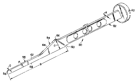
FIG. 3 shows a third, or final type of
femoral compactor instrument contained in the
kit. The third femoral compactor, generally
indicated at 50, is used preferably after
compaction has been performed with the second
type of instrument 30. As with the instrument
of FIG. 2, the proximal portion 52 of instrument
50 is identical to the proximal portion 16 of
the instrument of FIG. 1. As shown in FIG. 3,
the distal broach portion 54 of instrument 50 is
similar to the broach portion 18 of instrument
14, but further includes an elongation,
generally indicated at 55, to the distal broach
portion 54. The broach portion 54 of instrument
50 has an overall length B of approximately 160
mm and a maximum height C of approximately 25
mm. The elongation 55 has a length of
approximately 90 mm, a height F of approximately
9 mm, and a width D which tapers from
approximately 10 mm at one end thereof to
WO 96/09011 2 2 0 0 2 01 pCT~S95/11055.
~'~,, .,~
8
approximately 5 mm at the distal end 56 of the
broach portion 54.
As shown in FIG. 3, the instrument 50 '
,includes a planar base segment 58 spanning the
length of the broach portion 54. The base
segment 58 merges smoothly with the rounded tip
56 and also merges smoothly with the proximal
portion 52 at end 60 thereof. The broach
portion 54 also includes an upper segment 62
which extends generally pa~'allel to the base
segment 58 from the tip 56 towards the proximal
portion 52, a distance of approximately 90 mm.
A top segment 64 extends at an acute angle from
the upper segment 62 and merges with the
proximal portion 52 at end 66. These peripheral
segments are constructed and arranged to compact
bone deep within the femoral canal 10. The
broach portion 54 is solid and made of stainless
steel or similar materials. As with the other
types of instruments in the complete kit, it is
contemplated to include a plurality of the third
or final type, such instruments 50 having
slightly increasing broach portion widths.
With reference to FIGS. 4-6, the use of
the instruments of the kit to prepare a femur
for receiving a prosthesis will be described.
The first step in a procedure for preparing the
femur is to expose the proximal femoral neck and
shaft of the femur by removing the femoral head
(not shown). The orientation and direction of
the femoral canal 10 is then determined for
proper instrument placement. If the canal width
is narrow, or a cortical fit is desired, the
surgeon first drills the canal 10 in a reverse
drill operating mode to prevent removal of bone
from the canal 10. If such a drilling operation
is not possible, then a forward aggressive
WO 96/09011 ' PCT/US95/11055
2200201
9
drilling mode is used, alternated with reverse
drilling, so as to preserve bone fragments
within the canal for compaction along the
anticipated prothesis route. If the canal width
is larger than the prothesis to be used, then no
drilling is required, and the instruments
comprising the invention are sufficient to
prepare the femur for the prothesis placement.
When the instruments contained in the
kit are utilized to compact bone to prepare the
femur for the prosthesis implant, a 2 mm wide
initial femoral compactor 14 first is grasped at
the proximal portion 16 thereof and is pushed
and/or impacted with a mallet on its head 20 to
insert the broach portion 18 thereof into the
proximal portion 13 of the femoral canal 10,
such that the peripheral segments of the broach
portion 18, in particular, segments 24 and 26,
displace the softer bone 11 in the sidewalls of
the canal 10. The instrument 14 is then removed
from the canal 10. After compaction with the 2
mm width initial instrument 14, the 3 mm and the
4 mm width instruments 14 may be inserted
sequentially into the canal 10 to further
compact bone in the sidewalls thereof, if
desired.
Next, as shown in FIG. 5, the second
type of femoral compactor instrument 30 is
inserted into the femoral cavity 10 in a similar
manner as the initial compactor instruments)
14, and along the same orientation plane.
Instrument 30 is also pushed and/or impacted at
its head 20 so the peripheral surfaces of broach
portion 34, particularly segments 40 and 44,
compact bone in the sidewalls of the canal 10 so
that the compacted sidewalls correspond to the
shape 9f the prosthesis to be inserted into the
WO 96/09011 I2 2 0 0 2 01 p~T~S95/11055~
canal 10. It is within the contemplation of the
invention to customize the geometry of the
broach portion 34 of the second type of femoral '
compactor instrument 30 to correspond to the
5 geometry of the implant under consideration. '
Additional instruments 30 having slightly
increasing broach portion widths can be used
sequentially to dilate a portion of the canal
10, if desired.
10 Next, the final type of femoral
compactor instrument 50 is inserted into the
canal 10 in the manner described above, along
the same orientation plane as the previously
employed instruments. The elongated broach
portion 54 of instrument 50 creates compaction
of the softer bone 11 in the sidewalls deep
within the femoral canal at a location distal to
the previously compacted proximal portion 13 of
the femoral canal 10, as shown in FIG. 6. If
desired, additional instruments 50 having
slightly increasing widths can be used
sequentially to dilate a portion of the canal 10
deep within the femur.
After using the instruments of the kit
to compact bone of the femur, the femur is now
ready to receive a prosthesis (not shown)
therein. The procedure which has been described
for preparing the femur may be used when
implanting either a cemented or non-cemented
prosthesis.
The method described for preparing a
femur for insertion of a femoral component for
total hip replacement provides an effective '
means of compacting bone by the use of a series
of compaction instruments to produce dense
sidewalls of the canal 10 for receiving the
prothesis. Such density of the sidewalls of the
WO 96/09011 PCT/US95/11055
2200201
11
canal 10 ensures a degree of bone compactness
which creates high resistance to migration or
translocation of the prothesis inserted into the
prepared femur. The dense bone wall of the
canal 10 provides resistance to angular or
rotational stress of the prosthesis secured to
the wall of the canal. Thus, the use of the
instrumentation kit of the invention to compact
bone clearly offers significant advantages over
conventional femoral prosthesis insert
preparation, wherein either nothing is done to
the canal sidewalls, or wherein the canal is
prepared by removing bone via drilling.
While the invention has been described
~ in connection with what is presently considered
to be the most practical and preferred
embodiment, it is to be understood that the
invention is not limited to the disclosed
embodiment, but, on the contrary, is intended to
cover various modifications and equivalent
arrangements included within the spirit and
scope of the appended claims. .