Note: Descriptions are shown in the official language in which they were submitted.
2 ~ O ~ 4 3 4
WO 96/09398 PCTINL95100310
GeIle eo~tructs enco~ erop-protecti~ age~ts, as well as tr~n~formed pl~nts
cont~ining ~nd expressing sucb constructs, and methods of controllirtg plague o~ ",~
~nd p~thogens in erops.
The present invention relates to gene constructs suitable for ~ sillg agents
5 which protect a plant against plague org;~ni~m~ and pathogens.
Attack of plants by pathogens such as fungi, nematodes, insects, bacteria and
viruses, constitutes a considerable economic problem, in particular for large-scale
culture crops sueh as eorn, rice, beans, potatoes, tomatoes and grapes. Protection of such
plants by chemical means is highly undesired for environmental reasons.
A more effective and more acceptable way of protecting plants against attack
by plague org~ni.~m~ consists in making the plant resistant to the action of ~tt~cking
o~ islll~ by providing it with genetic information controlling the effect of such
or~ni~m~. As some recent examples of this type of plant protection have shown,
l~sisl~llce to viruses can be achieved by expression of viral coat protein (Beachy et al,
1990), or by expression of a single chain variable antibody fragment (scFcv) against the
Ca2~ binding domain of a viral coat protein (Tavladoraki et al, 1993). Resistance to
insects can be obtained by expression of Bacillus thuringiensis insecticidal crystal
proteins (ICP's) (Vaeck et al, 1987). Resistance to fungi can be achieved by expression
of chitin~e and/or glucanase (Alternaria longipes: US-A-4,940,840, Fusarium solani:
EP-A-440304, Botrytis cinerea and Rhizoctonia solani: Broglie et al, 1989, WO-A-90-
07001). Resistance to plant-parasitic nematodes can be achieved by using giant ccll
specific promoters in combination with barnase or anti-scnsc DNA techniques
(Opperman et al, 1994).
Major disadvantages of these methods are:
2s - The application is limited to a relatively small number of plague org~ni~ms. For
example, ICP's have so far only been effective against Lepidoptera, Diptera and
Coleoptera, because the binding domain of the ICP-toxin only recognises receptors
in the guts of insects belonging to these orders. In addition, each ICP-toxin (e.g.
CryLA(b), CryIC and CryIE) has its own specific spectrum with regard to the insect
species.
- There are strong indications that resistance by ICP's is not durable because of
mutations in the receptors (Van Rie et al., 1990; McGaughey and Whalon, 1992;
T~ hnik, 1994).
W0 96/09398 2 2 ~ PCT/NL95/0031
- These methods often only lead to partial resistance (viral coat protein, scFv, glucanase/chitinase) .
The objective of the present invention is to provide means and methods for
protecting plants, in particular agricultural crops, against plagues in a more effective and
s ecologically acceptable manner. This objective is met according to the invention, by
fusing monoclonal antibodies or parts thereof to toxins or enzymes having toxic
activities or to effective parts of these toxins or enzymes. The chimeric protein can be
made either by fusing the sequences encoding the respective parts of the chimera or by
chemically or biochemically linking the two parts of the chimeric protein together as for
o instance described in W0-A-9318162.
Thus, the invention relates to gene constructs comprising a nucleotide sequence
encoding an antibody or part thereof which is specific for a plague or~nislll orpathogen, and a nucleotide sequence encoding a protein which has a toxic effect on said
plague organism or pathogen. The invention furthermore relates to chimeric ploteills
consisting of an antibody or parts thereof, which is specific for a plague O~ lliSlll or
pathogen and a protein which has a toxic effect on a said plague ol~nisl.. or pathogen
and which has been constructed by chemically or biochemically linking the antibody (or
parts thereof~ to the toxic protein or enzyme (or parts thereof~. The terms "pathogen"
and "plague organism" are both used to denote any org;~ni~m~ or agents that affect the
20 growth, development or utility of a plant.
Monoclonal antibodies can be raised to almost any epitope or to almost any
molecular structure of a pathogen which is vulnerable to toxins or toxic enzymes (and
parts thereof), using the hybridoma technique (Kohler and Milstein, 1975). Single chain
antibodies can be prepared from monoclonal antibody producing hybridoma's by
25 established molecular techniques. In addition, single chain antibodies with affinities for
selected epitopes or molecular structures which are vulnerable to toxins or toxic enzymes
(or parts thereof~ can be obtained from phage display libraries (Hoogenboom et al. 1992;
Winter et al., 1994). A major advantage of this strategy is that the effectiveness of the
toxicity can be enhanced and furthermore that the specificity can be tuned to the target
30 or~ni~m~. With this strategy it is possible to construct fusion products which are active
against a single species or a range of species by selecting the applopliate epilopes. In
addition, it is possible to improve the durability of the resistance by a) selecting
antibodies against conserved epitopes and b) fusing the enzyme or toxin to two or more
antibodies (or parts thereof) having different receptors.
~ WO ~G~33g~ O 0 4 3 4 PCT/NI9SI00310
~ntibodies
Antibodies can be raised to structures of the plague organism which will
directly or indirectly lead to resistance or partial resistance when these antibodies are
fused to the aL)plo~liate toxin or enzyme. Examples of these structures are cellS membranes and cell walls, especially in the case of bacteria and fungi; or alimentary
tract structures, e.g. epithelial antigens, especially in the case of nematodes and insects;
or coat proteins, especially in the case of viruses. The antibodies are preferably single
chain antibodies.
Toxic proteins
0 Toxic proteins include all proteins that have a toxic effect on plague or~;~ni~m~
or pathogens, such as toxins and toxic enzymes.
Toxins that can be fused with monoclonal antibodies include the following:
- Rac;~1rl ~ thuringiensis insecticidal crystal proteins (ICP). ICP's or ~-endo-toxins are
a family of proteins produced during sporulation in the cytoplasm of B. thuringien-
1S sis. These proteins crystallize as parasporal inclusion, which are solubilized in the
insect larvae gut. The toxins are highly specific and effect lysis of gut cells of
susceptible insect larvae. Many B. thuringiensis strains producing ICP's with
different insect host spectra have been isolated. ICP's are classified according to
their specificity spectrum (Hofte and Whiteley, 1989).
Genes of various ICP's have been cloned and sequenced, including the gene
encoding CryL~(b) (Wabiko et al., 1986). CryL~(b) is produced as a 131 kDa
olo~ill which is activated by removal of an N- and a C-terminal ~ e~lide by
proteæes present in the insect gut. The mature toxin (65-66 kDa) comprises threedomains. The N-terminal (first) domain, which colll~ s several conserved
2s hydrophobic sequences, is assumed to form a pore in the apical membrane of gut
epidermis cells. The second domain is highly variable and presumably binds to a
receptor in the cell membrane. The C-tçrmin~l (third) domain also contains
conserved sequences.
- colicins (Pattus et al., 1990). Colicins are a family of plasmid encoded antibiotical
proteins, which kill bacteria closely related to the producing strain (generallyEscherichia coli). They are composed of structural domains, which exert dirrelelll
functions, such as receptor binding, translocation and killing. Based on their mode
of action colicins can be classified into two groups. The major group of colicins
wo 96,09398 -- 2 2 ~ ~ ~ 3 ~ PCT/NI9!j/0031~
causes permeabilisation of the cytoplasmic membrane, thereby destroying the
membrane potential. The C-terminal domain of these colicins form ion channels inartificial membranes. The other group of colicins causes enzymatic cleavage of
DNA or 16S rRNA.
s - thionins (Bohlmann and Apel, 1991); these are toxic for bacteria and fungi.
- cecropins (Fink et al, 1989), which are toxic for bacteria as they form pores in their
membranes.
- AaIT (Androctonus australis scorpion venom, which is a single polypeptide chain
of 70 amino acids, acting on sodium channels of insects, Gordon et al., 1984).
0 These are examples of toxins which interfere with cell membranes and are
particularly suitable for obtaining resistance against insects and nematodes.
Other toxins like ribosome-inactivating proteins may be suitable as well to
obtain resistance against plague org;~ni~m~.
Further examples are:
- Saporin (Stirpe 1983; Stirpe and Barbieri, 1986)
- Abrin A and C, (Wei et al. 1974; Lin et al. 1981)
- Melittin (26 amino acids long haemolytic peptide from bee venom)
- Gelonin (Stirpe et al. 1980)
- Momordin (Barbieri et al. 1980)
- rSLTAd7 (Shigella-Like Toxic Agent)
- T ectin~, highly specific carbohydrate-binding proteins from animal, plant or
microbial origin, which can be toxic as a result of binding to parts of the organism.
Toxic enzymes that can be fused to monoclonal antibodies include:
- glucanases, in particular ,13-1,3-gluc~n~es. These PR-2 proteins can be classified
in an alkaline form and acidic form. The alkaline form is produced after severalprocessing steps of the translation product. First the N-t~rrnin~l signal peptide is
cleaved off during transport to the endoplasmatic reticulum, and then the C-terminal
part is glycosylated and removed for transport over the vacuole membrane. For
glucanases both an intracellular and an extracellular form exist. The intracellular
form is extended by 3-25 aminoacids at the C-terminus. The sequence of intra-
cellular ,~-1,3-glucanase gene is disclosed in EP-A-440304. Glucanase genes fromother plants are also known, e.g. glucanase (and endochitinase) from maize (Nasser
et al. 1988). Effective destruction of cell wall glucans by endo-,3-1,3-glllc~n~cs
sometimes appears to require cooperation by exoenzymes such as exogluç~n~çs.
~ W096tO9398 - 2 2 0 ~ 4 ~ 4 PCT/NI95/003lO
- cllitin~es. These are PR-3 proteins comprising two domains and a hydrophobic
signal peptide which is absent in the active enzyme. Chitinases are subdivided in
three classes: class I, alkaline chitinases, are localised in the vacuole and contain a
cysteine-rich rlom~in, and a C-terminal sequence of 6 aminoacids that is removedJ 5 after translation and are involved in vacuolar talg~lillg; class II, acid chitinases, lack
a cysteine-rich domain and have a lower enzyme activity; they are localised in the
apoplast; class III, lysozyme-active cllitinzl~es, contain other conserved sequenrçs
than I and II. Plant chitinase was found to derive its chitinase activity from a 30
kDa monomer. For chitinases, as for glucanases, both an intr~cçllular and an
extracellular form exist. The intracellular form is extended by 3-10 ~minr-acids at
the C-terminlls. The catalytic centre is localised in the C-terminal part which is the
same in both forms. The sequence of intracellular chitinase gene is disclosed in EP-
A-440304. Chitinase genes from other org~ni~m~ are also known, e.g. bean endo-
chitinase, (DeBroglie et al. 1986).
C'hitin~e and ,~-1,3-glur~n~e are produced at an increased rate upon infection with
fungi (T~erticillium albo-atrum); Chitinase and ,B-1,3-gluc~n~e from tomato inhibit
growth of fungi in vitro (Young and Pegg, 1982, Young and Pegg 1981) and
probably also in vivo (Pegg and Vessey, 1973).
- lipase
- lysozyme
These are examples of enzymes which break down (parts of) the cell wall or
cell membranes. This strategy is particularly suitable for obtaining resistance against
bacteria and fungi.
In principle all enzymes which are able to lyse cells, damage the cell wall or
hllel~lt; with metabolic routes, replication, transcription, cell division, or interfere with
other essential functions to such an extent that the pathogen or plague organism will die
or will be severely hindered in growth, are suitable as part of the present constructs.
Con~tructs
Gene constructs may comprise nucleotide sequences encoding the complete
antibody molecule, the Fab part, the F(ab)2part, scFv part, bivalent scFv (diabody)
(Holliger, Prospero & Winter, 1993), minibody (Pack et al., 1993), or any other part
(like complementarity determining regions) which shows binding to the targets. In the
constructs according to the invention the antibody sequence is fused to a complete
sequence encoding an enzyme/toxin or to a part thereof which is still functionally active.
-220Q~4
WO 96/09398 PCT/NL95/0031
The chimeric protein consisting of an antibody (fragment~ and an enzyme/toxin can also
be obtained by chemical or biochemical linkage.
The antibody (fragment) and the enzyme/toxin (fragment) is fused directly or
using a flexible linker which does not intelr~l~ with the structure and function of the
s two proteins. Such flexible linkers are for instance those which have been used to fuse
the variable domains of the heavy and light chain of immunoglobulins to construct a
scFv, those used to create bivalent bispecific scFvs or those used in immunotoxins (see
Whitlow and Filpula, 1991; Kihlberg et al., 1993; Huston et al., 1992; Takkinen et al.,
1991). Linkers can also be based on hinge regions in antibody molecules (Pack and
0 Pliiclcthl-n, 1992; Pack et al., 1993) or on peptide fragments between structural domains
of proteins. When only a functional part of the toxin is to be conjugated to the antibody
(fragment) the linker present between two domains of the complete toxin itself could be
used. Fusions can be made between the enzyme/toxin and the heavy chain (fragment)
or the light chain (fragment) of the immunoglobulin at both the C and N tçrminlls. In
the case of a scFv fusion the variable domains can be in both the order VH-linker-VL
and VL-linker-VH.
The desired cellular location of the proteins can be achieved using the
o~liate targeting sequences. Proteins synthesized without targeting sequences stay
in the cytoplasm of the cell, whereas others are directed into the secretory pathway by
a signal peptide. When no other targeting signal is present, the latter proteins are
secreted by default. Additional targeting signals can be present to direct the proteins for
example to the vacuolar compartment of the cell or to retain them in the endoplasmic
reticulum (Chrispeels, 1991). Targeting signa!s to direct proteins to the chloroplast,
mitochondria, peroxisomes or nucleus have been described (Austen and Westwood,
2s 1991). An example of a targeting route is the secretion via endoplasmic reticulum and
golgi a~a~lus. Examples of signal sequences for secretion are described in Briggs and
Gierasch (1986), Firek et al., (1993), During et al., (1990) and Shirasu et al., (1988).
If the fusion protein has to be expressed in a heterologous organism for
production of the protein as such, it may be necessary to modify the gene construct in
order to improve ~ ssion because of the codon preference of this olg~ ll, to
remove mRNA instability motifs (e.g. AT regions, false splice sites) and polyadenylation
signals.
T~rget o~
Fungi, bacteria, nematodcs, insects, viruses and other plague org~ni~m~ or pathogens.
2 2 0 0 4 ~ ~
~ WO ~Cir~3g~1 PCT/NL95/00310
Pronloters
The fusions genes are expressed in plants under control of any type of promoter
which is active in plants. Examples are:
a) constitutive promoters such as the CaMV-35S (Kay et al., 1987)
5 b) tissue specific promoters such as described in Nap et al. (1993) (leave), De Almeida
et al. (1989) (leave, SSU-promoter), Nap et al. (1992) (potato tuber, patatin
promoter), Hendriks et al. (1991) (potato tuber), Guerche et al. (1990) (seed);
c) inducible promoters such as the 2' promoters (Langridge et al., 1994), wound
inducible promoters (Logemann et al., 1989; Suh et al., 1991) or chemically in~ ced
lo promoters (Williams et al., 1992).
T~ ,srO~ ation
Transformation can be done using any method which ensures a stable
integration of the chimeric gene in the plant genome in such a way that it can still be
transcribed. Examples of l~dll~ro~ ation are:
a) Agro~acterium t~lmefaciens mediated transformation (Horsch et al., 1985): based on
a natural transformation system in which the bacterium stably incorporates part of
a plasmid DNA (T-DNA) into the plant genome. The T-DNA includes the gene to
be expressed.
b) Microprojectile bombardment (Vasil et al., 1992): particles coated with DNA
penetrate the plant cell nuclei at high velocity where the DNA is integrated into the
genome by host recombination processes.
c) Tissue electroporation (D'Halluin et al., 199 ): under the influence of a strong
electric field DNA penetrates the plant cells and, after being transported to the
nuclei, it is incorporated into the plant genome by host recombination processes.
~)irect application
The gene constructs can also be used for plague control through external
application on crops which are to be protected. Such direct application can be achieved
in the form of ~rlmini~tering the expression product of the chimeric gene, i.e. the
immunotoxin comprising an antibody linked to a toxic protein. Another form can be by
applying an organism containing the immunotoxin as such or containing a gene encoding
the immunotoxin and capable of producing it. Suitable carrier Olg~ include
microor~ni~m~ such as bacteria (e.g. B. thuringiensis~, fungi, yeasts and viruses. The
ol~ may be alive or dead.
Thus the invention also relates to an immunotoxin colllplisillg an antibody
W096/09398 ~ ~ 0 ~) 4 3 ~ PCT/NL95/0031
linked to a protein which is toxic for a plague organism or pathogen, and which
immunotoxin is obtainable from an expression system as described above and can be
used for external protection of plants against plague org~ni~n-~. The protein may be
purified by known methods.
The invention also comprises org;~ni~m~ which contain such an immunotoxin
and o,~ni.ci"l~ which are stably transformed with the gene construct encoding the
immunotoxin. These org~ni~m~ can be used for e~rtrrn~l protection of plants against
plague or~;~ni~m~.
The invention further relates to pesticidal compositions co~ g an
lo immunotoxin as such or in encoded form, together with an acceptable carrier. The
compositions may also contain solvents, agents preventing the composition from
washing away, stabilisers, attractants, UV-absorbers, and the like. The invention also
relates to a process for protecting a plant against the action of a plague olg~llislll or a
pathogen, wherein the plant is externally treated with an immunotoxin as described or
an Ol~,~llislll contzlining the immunotoxin, or with a composition co~ "i"g it. Treatment
may be done by spraying and the like, by hand using any suitable equipment including
tractors, aircraft etc.
F~mple 1
Fusion of monoclon~l ~ntibodies or parts thereof with en~ymes h~vi~g toxic activities
Fusion of a plant chitinase and glucanase to a scFv derived from a monoclonal antibody
against a fungus (Verficillium dahliae)
The following steps are taken:
1) Antibodies against mycelium or purified cell wall components of Verticillium
dahliae are raised, and monoclonals are isolated.
2) cDNA sequences encoding antibody variable region are cloned to create a single-
chain Fv construct.
3) The functionality of bacterially expressed scFv is checked.
4) N- or C-tcrmin~l fusion between scFv and chitinase or ,~-1,3-gluc~n~e is
performed using a suitable linker, e.g. CBHI linker (Takkinen et al. 1991), and the
chimeric gene is inserted in an expression vector, e.g. pNemS or pNem6 (Figures
1 and 2), which is a derivative of vector pHenl (Hoogenboom et al., 1991).
S) Both binding activity (by ELISA) and enzymatic activity (bioassay) of b~ctr.ri~lly
(E coli) expressed fusion product are checked.
~ WO 9GJ~3~ 2 2 ~ 0 4 3 4 PCT/NL95/00310
6) The fusion gene is transferred, together with suitable lalgethlg sequences, to the
plant hdll~rollllation vector in between a promoter-termin~tion cassette.
7) Expression and functionality in plant cells is checked by a transient e~lGssion
assay.
8) The expression czlssettc with fusion gene and selection marker is transferred into the
plant genome by plant transformation.
g) Regenerated plants are screened for e~pleSSiOn of fusion product.
10) Activity of fusion protein in transgenic plants is checked through bioassay.The steps can be followed, with applo~liate adaptations of antibody production
and fusion with enzyme, for producing transgenic plants with resistance to Botrytis
cinerea, Fusarium oxysporum f.sp. radicis-lycopersisc~ and Phytophthora infestans.
Fusions with other proteins having toxic activities are also possible, e.g. with potato
lectine.
Fx~mple ?
Fusion of monoclon~l ~ntibodies or pzlrts thereof with to~cin~
Fusion of CryIA(b)_BT(29-607) to ~ scFv derived from a monoclonal antibody against
the gut of the insect Spodoptera exigua larvae.
The following steps are taken:
1) Antibodies against gut epithelial tissue of S. e~cigua larvae are raised, and monoclonals are isolated.
2) cDNA sequences encoding antibody variable regions are cloned to create a single-
chain Fv construct.
3) The functionality of bacterially expressed scFv is checked.
4) N- or C-terminal fusion between scFv and Cr~IA(b)_BT(29-607) is pelrolllled
2s using a suitable linker, e.g. CBHI linker (Takkinen et al. 1991), and the chimeric
gene is inserted in an expression vector, e.g. pNem6 (Figure 2), which is a
derivative of pSPORT1 (Gibco, BRL).
5) The binding activity of the bacterially (E. coli) expressed chimeric protein will be
analyzed by Western blot analysis, ligand blot assays in combination with
competition experiments (Bosch et al., 1994), on cryo-sections of midguts of insect
larvae (Martens et al., 1994) and on primary cultures of epithelial cells of insect
midguts (Baines et al., 1993). Insecticidal activity of the chimeric protein will be
checked by bioassays and lysing effect of the chimeric proteins will be followed
=-~2ao434
WO 96/09398 PCT/N195/0031j
using primary midgut cell cultures.
6) The fusion gene is transferred, together with suitable targeting sequences, to the
plant transformation vector in between a promoter-termination cassette for stable
rol.,lation. For transient expression the fusion gene will be cloned behind a
coll~liLulive promoter (i.c. CaMV-35S promoter) and a suitable termination cassette.
7) Expression and functionality in plant cells is checked by a transient e~,cssion
assay.
8) The expression cassette with fusion gene and selection marker is ll~n~re"~d into the
plant genome by plant transformation.
lo 9) Regenerated plants are screened for expression of fusion product.
10) The binding activity of the chimeric protein will be checked by w~ lll analysis and
ligand blot assays in combination with competition experiments, on cryo-sectionsof midguts of insect larvae (Martens et al., 1994) and on primary cultures of
epithelial cells of insect midguts (Baines et al., 1993). Insecticidal activity of the
1S chimeric protein will be checked by bioassays and lysing effect of the chimeric
proteins will be followed using primary midgut cell cultures.
Additionally N- and C-terminal fusions will be made between a scFv and
CryLA(b)_Bt(1-607); CryIA(b)_Bt(29-429); CryIA(b)_Bt(1-1155). Furthermore,
domain II (or part of it) of CryIA(b), which is thought to be responsible for receptor
binding, will be replaced by a scFv.
The steps can be followed, with a~plopliate adaptations of antibody production
and fusion with toxins, for producing transgenic plants with resistance to nematodes by
raising monoclonal antibodies against the intestine of the nematode.
Fx~mple 3
1) Balb/c mice were immunized with brush border membrane vesicles (BBMV's),
isolated from the midgut of Spodoptera exigua as described by Bosch et al., 1994.
The mice were immunized twice (with a four week interval) by subcutaneous
injection of BBMV's using the equivalent of 50 ,ug protein with the addition of
Freund's incomplete adjuvans. Four weeks after the last immunization a boost wasgiven with BBMV's (50 ~lg protein equivalent) injected intraperitoneally. Three days
later the spleen was removed and the fusion was carried out as described by Schots
et al, 1992.
- 2200~34
WO ~GI'~53~8 11 PCTINL95/00310
2) Antisera and monoclonal antibodies were checked for their ability to react with
epitopes present in BBMV's with ELISA according to standard procedures.
3) Western blot analysis was carried out to check the reaction pattern of the
monoclonal antibodies. 250 ~lg protein equivalent of BBMV's were separated on ans SDS-polyacrylamide gel (12%). The proteins were transferred to PVDF membrane
by western blotting.
4) In order to determine if the monoclonal antibodies could react with epitopes present
at the luminal side of the midgut of insects, cryosections of Spodoptera exigua
midguts were first incubated with the monoclonals, followed by incubation by a
lo second antibody reacting with mice antibodies and labelled with FITC.
S) In order to c~etermine if the monoclonal antibodies could react with epitopes at the
outside of the midgut and possibly at the luminal side of the midgut, primary
epithelial cell cultures of S. exigua midguts were prepared and incubated with the
monoclonal antibodies, followed by incubation with a second antibody reacting with
mice antibodies and labelled with FITC.
6) Single chain antibodies were isolated from hybridoma's producing monoclonals
which bound to epitopes present at the luminal side of the membrane and at the
outside of the midgut cells of S. exigua, according to standard procedures as, for
instance, described in (Johnson & Bird, 1991); (Huston et al., 1992).
F~:~mple 4
Fusion of colicin Npore-forming domain to a scFv derived from a monoclonal antibody
raised against the gut of the insect Spodoptera exigua larvae
The steps as in example 2 are followed, with the adaptation that in step 4 the
scFv is coupled to the N-t~rminn~ of the pore-forming domain of colicin N (C-terminal
region of the protein) (Pugsley, 1987), with in front of this domain the peptide fragment
which normally links the N-terminal part of the complete colicin N to its pore-forrning
domain, the latter peptide fragment serving as a linker between the two domains of the
- chimeric protein.
Fx~mple 5
30 Sprayable immunoe~7~yme formulation
Steps 1) to 5) of Example 1 are repeated, the antibodies being raised in step 1)against the mycelium or cell wall components of Botrytis cinerea. The fusion gene is
WO 96/09398 ~ 2 0 0 ~ PCT/NL95/0031Q~;
cloned into vector pNemS (Fig. 1). Upon induction with IPTG (isopropylthio~
galactosidase), the fusion protein is produced through ove~ lession. The fusion protein
(immunotargeted toxic enzyme) is then isolated, purified, and its activity against the
fungus Botrytis cinerea is checked through a bioassay, e.g. by incorporation in a suitable
s buffer cont~ining 0.1% Tween 20 as a wetting agent and spraying on a fungus culture
on an agar medium and then on a test plant previously infected with conidia of the
fungus. The fusion protein can then be formulated e.g. into a wettable powder orspraying powder and then be applied on crops threatened with the fungus.
Fx~mple 6
10 Sprayable immunoto~in forrnulation
The steps 1) to 5) of Example 2 are repeated. The fusion gene is cloned into
vector pNem6. Upon induction with IPTG, the fusion protein is produced by over-
e~pl~;ssion. The fusion protein (immunotoxin) is then isolated, purified, and its activity
against insect Spodoptera exigua checked by adding it to the artificial diet for this
15 insect. The fusion protein can then be formulated e.g. into a wettable powder or spraying
powder and then be applied on crops threatencd with the insect.
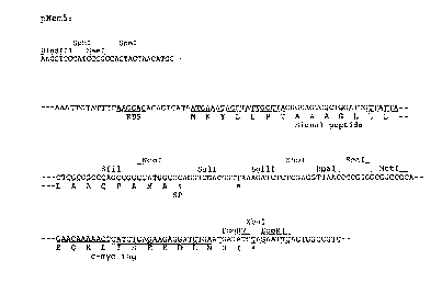
nescription of the fi~ures
Fig. 1 shows the nucleotide and partial aminoacid sequences of the vector
pNemS, a derivative of pHenl (Hoogenboom et al. 1991). The sequence shown was
20 cloned between the HindIII and EcoRI sites of pHenl. This sequence replaces the
multiple cloning site and gene III encoding a minor coat protein of phage Fd in pHenl.
In addition an extra multiple cloning site was introduced 3' of the HindIII site. Single
chain antibodies can, for instance be cloned between the SfiI and NotI sites or the SalI
and SmaI sites. RBS is a prokaryotic ribosomal binding site; the sequence encoding the
25 pelB signal peptide (signal peptide of pectate lyase of Erwinia carotovora) (Hoogen-
boom et al., 1991) and a c-myc tag are indicated and their amino acid sequences given.
Fig. 2 shows the nucleotide sequence of the pNem6 cloning vector. The
sequence shown was cloned in pSPORT1 (Gibco, Life Technologies) between PstI andSphI sites of the polylinker (S' and 3' ends respectively), in such a way that both the
30 original PstI and the SphI sites were dcstroyed. At the 5' end the sequence begins with
the nucleotide that was changed from G (last nucleotide of the original Pstl site~ to C
in order to disrupt PstI site. The last restriction site (AatII) of the polylinker of
~ W0 96/09398 2 2 Q 0 4 3 4 PCTII~I~5100310
pSPORT1 is indicated. Single chain antibodies can for instance cloned between the SalI
and the SmaI sites. RBS is a prokaryotic ribosomal binding site; the sequences encoding
<! the PelB signal peptide (signal peptide of pectate lyase of Erwinia carotovora)
(Hoogenboom et al., 1991) and a c-myc tag are indicated.
J
References
Austen, B.A., and Westwood, O.M.R. (1991) Protein targeting and secretion. IRL press
at Oxford University Press, Oxford UK. 85 pp.
Baines, D., Blowllw~lght, A., and Schwartz, J.L. (1993). J. Insect Physiol. 40, 347-357.
Barbieri et al. (1980). Biochem. J., 186, 443.
0 Beachy, R.N., Loesch-Fries, S., and Tumer, N.E. (1990). Ann. Rev. Phytopathol. 28,
451-474.
Bohlm~nn, H., and Apel, K. (1991). Ann. Rev. Plant Physiol. Plant Mol. Biol. 42, 227-
240.
Bosch, D., Schipper, B., van der Kleij, H., de Maagd, R.A., and Stiekema, W.J. (1994).
Bio/technology 12, 915-918.
Briggs, M.S., and Gierasch (1986). Adv. Protein Chem. 38, 109-180.
Broglie, K.E., Biddle, P., Cressman, R., and Broglie, R. (1989). Plant Cell 1, 599-607;
Chrispeels, M., (1991). Annu. Rev. Physiol. Plant. Mol. Biol. 42, 21-53.
D'Halluin, K., Bonne, E., Bossut, M., DeBeuckeleer, M., and Leemans, J. (1992). Plant
Cell 4, 1495-1506.
De Almeida, E.R.P., Gossele, V., Muller C.G., Dockx, J., Reynaerts, A., Botterman, J.,
Krebbers, E., and Timko, M.P. (1989). Mol. Gen. Genet. 218, 78-86.
DeBroglie et al. (1986). Proc. Natl. Acad. Sci. USA 83, 6820-24.
During, K., Hippe, S., Kreuzaler, F., and Schell, J. (199G). Plant Mol. Biol. 15, 281-
2s 293.
Fink, J., Boman, A., Boman, H.G., and Merrifield, R.B. (1989). Int. J. Peptide Protein
Res. 33, 412-421.
Firek, S., Draper, J., Owen, M.R.L., Gandecha, A., Cockburn, B., and Whitelam, G.C.
(1993). Plant Mol. Biol. 23, 861-870.
Gordon, D., Jover, E., Couraud, F., and Zlotkin, E. 1984. Biochem. Biophys. Acta. 778,
349-358.
Guerche, P., Tire, C., Grossi De Sa, F., De Clercq, A., Van Montagu, and Krebbers, E.
(1990). Plant Cell 2, 69-478.
WO ~Gi~9398 -- 2 2 0 0 4 ~ ~ PCT/NI95/003l~
14
Herl~1rik~, T., Vreugdenhil, D., Stiekema, W.J. (1991). Plant Mol. Biol., 385-394.
Hofte, H., and Whiteley, H.R. (1989). Microbiol. Rev. 53, 823-825.
Holliger, P., Prospero, T., and Winter, G. (1993). Proc Natl Acad Sci USA, 90, 6444-
6448.
s Hoogenboom, H.R., Marks, J.D., Griffiths, A.D., and Winter, G. (1992). Immunol Rev,
130, 41-68.
Hoogenboom, H.R., Griffiths, A.D., Johnson, K.S., Chiswell, D.J., Hudson, P., and
Winter, G. (1991). Nucleic Acids Res. 19, 4133-4137.
Horsch, R.B., Fry, J.E., Hoffm~nn, N.L., Eichholtz, N.L., Rogers, S.G., and Fraley, R.T.
0 (1985). Science 227, 1229-1231.
Huston, J.S., Mudgetthllnter~ M., Tai, M.S., McCartney, J., Warren, F., Haber, E., and
Opperrn~nn, H. (1997). Methods in Enzymology, 203, 193-200.
Johnson, S., and Bird, R.E. (1991). Methods in Enzymology, 203, 88-98.
Kay, R., Chan, A., Daly, M., and McPherson, J. (1987). Science 236, 1299-1302.
Kihlberg, B.M., Sjobring, U., Kastern, W., and Bjorck, L. (1993). J. Biol. Chem. 267,
25583-25588.
Kohler, G., and Milstein, C. 1975. Nature 256, 52.
Langridge, W.H.R., Fitzgerald, K.J., Koncz, C., Schell, J., and Szalay, A.A. (1994).
Proc. Natl. Acad. Sci. USA 86, 3219-3223.
Lin, J-Y, et al. (1981). Toxicon 19, 41.
Lo~çm~3nn, J., Lipphardt, S., Lorz, H., Hauser, I., Willmitzer, L., and Schell, J. (1989).
Plant Cell 1, 151-158.
Martens, J.W.M., Flipsen, J.T.M., Groffen, A.J.A., Vlak, J.M., and Bosch, D. (1994).
PhD thesis J. Martens. CIP-DATA bibliotheek, I~en Haag, 41-50.
2s McGaughey, W.H., and Whalon, M.E. (1992). Science 258, 1451-1455.
Nap, J.P., van Spanje, M., Dirkse, W.G., Baarda, G., Mlynarova, L., Loonen, A.,
Grondhuis, P., and Stiekema, W.J. (1993). Plant Mol. Biol. 23, 605-612.
Nap, J.P., Dirkse, W.G., Louwerse. J., Onstenk, J., Visser, R., Loonen, A., Heidekamp,
F., and Stiekema, W.J. (1992). Plant Mol. Biol. 20, 683-694.
Nasser W. et al. (1988). Plant Mol. Biol. 11, 529-538.
Opperman, C.H., Taylor, C.G., and Conkling, M.A. (1994). Science 263, 221-223.
Pack and Pluckthun, (1992). Biochemistry 31, 1581-1992.
Pack, P., Kujau, M., Schroeckh, V., Knupfer, U., Wenderoth, R., Riesenberg, D., and
Pliick~hlln, A. (1993). Biotechnology, 11, 1271-1277.
~ W096/09398 - 2 ~ D 0 4 3 4 PCT/NI95/00310
Pattus, D., Massotte, D., Wilmsen, H.U., Lakey, J., Tsernoglou, D., Tucker, A., and
Parker, M.W. (1990). Experientia 46, 180-192.
Pegg, G.F., and Vessey, J.C. (1973). Phys. Plant Pathol. 3, 371-382.
Pugsley, A.P. (1987), Molec. Microbiol. 1, 317-325.
J s Sang-Gon Suh, Stiekema, W.J., and Hannapel, D.J. (1991). Planta 184, 423-430.
Schots, A., Pomp, R., and Van Muiswinkel, W.B. (1992) Production of Monoclonal
Antibodies In: Techniques in Fish Immunology, Stolen et al. (Eds). SOS Publica-
tions, Fair Haven NJ, p. 1-18.
Shirasu, Y. et al. (1988). Hybridoma 7, 485-493.
lo Stirpe, F., et al. (1980). J. Biochem., 225, 6947.
Stirpe, F. et al. (1983). Biochem. J., 216, 617.
Stirpe, F., and Barbieri, L. (1986). FEBS Lett., 195, 1.
Tab~hnik, B.E. (1994). Annual Review of Entomology 39, 47-79.
Takkinen, K., T~llkk~nen, M.-L., Si~m~nn, D., Alfthan, K., Immonen, T., Vanne, L.,
Kaartinen, M., Knowles, J.K.C., and Teeri, T.T. (1991). Prot. Eng. 4, 837-841.
Tavladoraki, P. et al. (1993). Nature 366, 469-472.
Vaeck, M. et al. (1987). Nature 328, 33-37.
Van Rie, J. et al. (1990). Science 247, 72-74.
Vasil, V., Castillo, A.M., Fromm, M.E., and Vasil, I.K. (1992). Biotechnology 10, 667-
674.
Vigers, A.J., Roberts, W.K., and Selitrenikoff, C.P. (1991). Molecular Plant Microbe
Interactions, 4, 315-323.
Wabiko, H., Raymonds, K.C. & Bulla. L.E. (1986) DNA 5, 305-314). Wei, C.H., et al.
(1974) J. Biol. Chem. 249, 3061.
2s Whitlow, M., and Filpula, D. (1991). Methods 2, 97-105.
Williams, S. Friedrich, L., Dincher, S., Carozzi, N., K~.s~m~nn, H., Ward, E., and Ryals,
J. (1992). Biotechnology 10, 540-543.
Winter, G., Griffiths, A.D., Hawkins, R.E., and Hoogenboom, H.R. (1994). Ann Rev Immunol, 12, 433-455.
Young, D.H., and Pegg, G.F. (1981). Phys. Plant Pathol. 19, 391-417.
Young, D.H., and Pegg, G.F. (1982). Phys. Plant Pathol. 21, 411-423.
,~ ,, . . ~, !, ' . ' -