Some of the information on this Web page has been provided by external sources. The Government of Canada is not responsible for the accuracy, reliability or currency of the information supplied by external sources. Users wishing to rely upon this information should consult directly with the source of the information. Content provided by external sources is not subject to official languages, privacy and accessibility requirements.
Any discrepancies in the text and image of the Claims and Abstract are due to differing posting times. Text of the Claims and Abstract are posted:
| (12) Patent: | (11) CA 2200492 |
|---|---|
| (54) English Title: | METHOD AND APPARATUS FOR COLLECTING FRAGMENTS OF BONE TISSUE |
| (54) French Title: | PROCEDE ET APPAREIL POUR RECUEILLIR DES FRAGMENTS DE TISSU OSSEUX |
| Status: | Expired and beyond the Period of Reversal |
| (51) International Patent Classification (IPC): |
|
|---|---|
| (72) Inventors : |
|
| (73) Owners : |
|
| (71) Applicants : |
|
| (74) Agent: | BORDEN LADNER GERVAIS LLP |
| (74) Associate agent: | |
| (45) Issued: | 2004-08-31 |
| (86) PCT Filing Date: | 1995-09-20 |
| (87) Open to Public Inspection: | 1996-03-28 |
| Examination requested: | 2002-05-21 |
| Availability of licence: | N/A |
| Dedicated to the Public: | N/A |
| (25) Language of filing: | English |
| Patent Cooperation Treaty (PCT): | Yes |
|---|---|
| (86) PCT Filing Number: | PCT/SE1995/001065 |
| (87) International Publication Number: | WO 1996009079 |
| (85) National Entry: | 1997-03-19 |
| (30) Application Priority Data: | ||||||
|---|---|---|---|---|---|---|
|
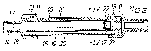
The invention relates to method and device for collecting bone tissue
fragments at a surgical operation in bone tissue occurring liquid
fluid being evacuated by suction from the operation site. In the method liquid
is allowed to pass during evacuation by suction into a
cylindrical sieve (19) through one open end thereof and through the sieve wall
from the inside to the outside thereof. A displaceable end
wall (22) disposed at the other end of the sieve is moved axially through the
cylindrical sieve in order that bone tissue fragments collected
on the inside surface of the sieve will be scraped off therefrom and deposited
outside the sieve from said one, open end thereof. The device
comprises a cylinder (10) having an inlet and an outlet at one end and the
other, respectively, which can be connected into a suction conduit.
The cylindrical sieve is located inside the cylinder, a space being provided
between the outside curved surface of the sieve and the inside
curved surface of the cylinder. One, open end of the sieve communicates with
the inlet of the cylinder while the outlet of the cylinder
communicates with said space. The end wall disposed at the other end of the
sieve can be displaced as a piston axially through the sieve.
L'invention concerne un procédé et un dispositif pour recueillir, pendant une opération chirurgicale, des fragments de tissu osseux apparaissant dans le fluide évacué par aspiration du site de l'opération. Selon ce procédé, le liquide évacué est introduit dans un tamis cylindrique (19) par une extrémité ouverte de celui-ci, pour traverser ensuite la paroi du tamis vers l'extérieur. Une paroi terminale (22) que l'on peut déplacer est prévue à l'autre extrémité du tamis et elle est déplacée axialement dans le tamis cylindrique pour recueillir les fragments de tissu osseux déposés sur la surface interne du tamis et les évacuer à l'extérieur du tamis, par l'extrémité ouverte. Le dispositif comprend un cylindre (10) ayant une entrée et une sortie aux deux extrémités respectives, pouvant être reliées à une conduite aspirante. Le tamis cylindrique est disposé dans le cylindre. Un espacement est prévu entre la surface courbe externe du tamis et la surface courbe interne du cylindre. Une extrémité (celle qui est ouverte) du tamis communique avec l'entrée du cylindre alors que la sortie du cylindre communique avec ledit espace. La paroi terminale prévue à l'autre extrémité du tamis peut être déplacée axialement dans le tamis, à la manière d'un piston.
Note: Claims are shown in the official language in which they were submitted.
Note: Descriptions are shown in the official language in which they were submitted.
2024-08-01:As part of the Next Generation Patents (NGP) transition, the Canadian Patents Database (CPD) now contains a more detailed Event History, which replicates the Event Log of our new back-office solution.
Please note that "Inactive:" events refers to events no longer in use in our new back-office solution.
For a clearer understanding of the status of the application/patent presented on this page, the site Disclaimer , as well as the definitions for Patent , Event History , Maintenance Fee and Payment History should be consulted.
| Description | Date |
|---|---|
| Time Limit for Reversal Expired | 2014-09-22 |
| Letter Sent | 2013-09-20 |
| Inactive: Office letter | 2007-01-23 |
| Inactive: Corrective payment - s.78.6 Act | 2007-01-15 |
| Inactive: IPC from MCD | 2006-03-12 |
| Inactive: IPC from MCD | 2006-03-12 |
| Inactive: IPC from MCD | 2006-03-12 |
| Inactive: IPC from MCD | 2006-03-12 |
| Grant by Issuance | 2004-08-31 |
| Inactive: Cover page published | 2004-08-30 |
| Inactive: Applicant deleted | 2004-08-23 |
| Inactive: Inventor deleted | 2004-08-23 |
| Inactive: Applicant deleted | 2004-08-23 |
| Pre-grant | 2004-06-15 |
| Inactive: Final fee received | 2004-06-15 |
| Notice of Allowance is Issued | 2004-04-15 |
| Notice of Allowance is Issued | 2004-04-15 |
| Letter Sent | 2004-04-15 |
| Inactive: Approved for allowance (AFA) | 2004-02-24 |
| Letter Sent | 2002-07-04 |
| Letter Sent | 2002-06-27 |
| Amendment Received - Voluntary Amendment | 2002-06-27 |
| Request for Examination Received | 2002-05-21 |
| Request for Examination Requirements Determined Compliant | 2002-05-21 |
| All Requirements for Examination Determined Compliant | 2002-05-21 |
| Reinstatement Requirements Deemed Compliant for All Abandonment Reasons | 2002-05-21 |
| Inactive: Entity size changed | 2002-04-24 |
| Letter Sent | 2002-02-21 |
| Inactive: Single transfer | 2002-01-10 |
| Deemed Abandoned - Failure to Respond to Maintenance Fee Notice | 2001-09-20 |
| Inactive: Single transfer | 1999-02-25 |
| Inactive: First IPC assigned | 1997-08-14 |
| Inactive: IPC assigned | 1997-08-14 |
| Inactive: IPC assigned | 1997-08-14 |
| Application Published (Open to Public Inspection) | 1996-03-28 |
| Abandonment Date | Reason | Reinstatement Date |
|---|---|---|
| 2001-09-20 |
The last payment was received on 2003-08-22
Note : If the full payment has not been received on or before the date indicated, a further fee may be required which may be one of the following
Please refer to the CIPO Patent Fees web page to see all current fee amounts.
Note: Records showing the ownership history in alphabetical order.
| Current Owners on Record |
|---|
| ASTRA TECH AB |
| Past Owners on Record |
|---|
| DAN LUNDGREN |

