Note: Descriptions are shown in the official language in which they were submitted.
CA 02202060 1997-04-07
R'O 96/11492 PCT/US95/11908
2
BACKGROUND OF THE INVENTION
1. Field of the Invention
This invention relates to a gas-detection
sensor and more particularly to a solid state mass
spectrograph which is micro-machined on a semiconductor
substrate, and, even more particularly, to a mass to
charge ratio filter for ion separation in the mass
spectrograph.
2. Description of the Prior Art
Various devices are currently available for
determining the quantity and type of molecules present
in a gas sample. One such device is the mass-
spectrometer.
Mass-spectrometers determine the quantity and
type of molecules present in a gas sample by measuring
the mass-to-charge ratio and quantity of ions formed
from the gas through an ionization method. This is
accomplished by ionizing a small sample and then using
electric and/or magnetic fields to find a charge-to-
mass ratio of the ion. Current mass-spectrometers are
bulky, bench-top sized instruments. These mass-
CA 02202060 2005-O1-21
3
spectrometers are heavy (100 pounds) and expensive.
Their big advantage is that they can be used to sense any
chemical species.
Another device used to determine the quantity and
type of molecules present in a gas sample is a chemical
sensor. These can be purchased for a low cost, but these
sensors must be calibrated to work in a specific
environment and are sensitive to a limited number of
chemicals. Therefore, multiple sensors are needed in
complex environments.
A need exists for a low-cost gas detection sensor
that will work in any environment. United States Patent
5,386,115, issued on January 31, 1995, discloses a solid
state mass-spectrograph which can be implemented on a
semiconductor substrate. Figure 1 illustrates a functional
diagram of such a mass-spectrograph 1. This mass
spectrograph 1 is capable of simultaneously detecting a
plurality of constituents in a sample gas. This sample gas
enters the spectrograph 1 through dust filter 3 which keeps
particulate from clogging the gas sampling path. This
sample gas then moves through a sample orifice 5 to a gas
ionizer 7
CA 02202060 1997-04-07
WO 96/11492 PCT/I1S95/11908
4
where it is ionized by electron bombardment, energetic
particles from nuclear decays, or in electrical
discharge plasma. Ion optics 9 accelerate and focus
the ions through a mass filter 11. The mass filter 11
applies a strong electromagnetic field to the ion beam.
Mass filters which utilize primarily magnetic fields
appear to be best suited for the miniature mass-
spectrograph since the required magnetic field of about
1 Tesla (10,000 gauss) is easily achieved in a compact,
permanent magnet design. Ions of the sample gas that
are accelerated to the same energy will describe
circular paths when exposed in the mass-filter 11 to a
homogenous magnetic field perpendicular to the ion's
direction of travel. The radius of the arc of the path
is dependent upon the ion's mass-to-charge ratio. The
mass-filter 11 is preferably a Wien filter in which
crossed electrostatic and magnetic fields produce a
constant velocity-filtered ion beam 13 in which the
ions are disbursed according to their mass/charge ratio
in a dispersion plane which is in the plane of Figure
1.
A vacuum pump 15 creates a vacuum in the mass-
filter 11 to provide a collision-free environment for
CA 02202060 1997-04-07
WO 96/11492 _ PCT/US95l11908
the ions. This vacuum is needed in order to prevent
error in the ion's trajectories due to these
collisions.
The mass-filtered ion beam is collected in an
5 ion detector 17. Preferably, the ion detector 17 is a
linear array of detector elements which makes possible
the simultaneous detection of a plurality of ions
formed from the constituents of the sample gas. A
microprocessor 19 analyses the detector output to
determine the chemical makeup of the sampled gas using
well-known algorithms which relate the velocity of the
ions and their mass. The results of the analysis
generated by the microprocessor 19 are provided to an
' output device 21 which can comprise an alarm, a local
display, a transmitter and/or data storage. The
display can take the form shown at 21 in Figure 1 in
which the constituents of the sample gas are identified
by the lines measured in atomic mass units (AMU).
Preferably, mass-spectrograph 1 is implemented.
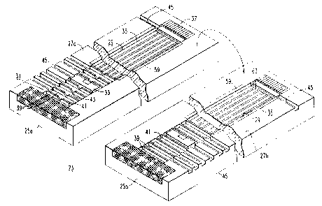
in a semiconductor chip 23 as illustrated in Figure 2.
In the preferred spectrograph 1, chip 23 is about 20 mm
long, 10 mm wide and 0.8 mm thick. Chip 23 comprises a
substrate of semiconductor material formed in two
CA 02202060 1997-04-07
WO 96/11492 PCT/US95/11908
6
halves 25a and 25b which are joined along
longitudinally extending parting surfaces 27a and 27b.
The two substrate halves 25a and 25b form at their
parting surfaces 27a and 27b an elongated cavity 29.
S This cavity 29 has an inlet section 31, a gas ionizing
section 33, a mass filter section 35, and a detector
section 37. A number of partitions 39 formed in the
substrate extend across the cavity 29 forming chambers
41. These chambers 41 are interconnected by aligned
apertures 43 in the partitions 39 in the half 25a which
define the path of the gas through the cavity 29.
Vacuum pump 15 is connected to each of the chambers 41
through lateral passages 45 formed in the confronting
surfaces 27a and 27b. This arrangement provides
differential pumping of the chambers 41 and makes it
possible to achieve the pressures required in the mass
filter and detector sections with a miniature vacuum
pump.
One of the methods utilized to determine the
nature of a molecular species is to determine its
molecular weight. This is not a unique property of a
molecule, since the same set of atoms which constitute
a molecule can be bonded together in a variety of ways
CA 02202060 1997-04-07
WO 96/11492 PCT/US95/11908
7
to form molecules with differing toxicities, boiling
points, or other properties. Therefore, in order to
uniquely identify a particular molecular compound, the
structure must be identified. A well-established
technique for determining the molecular structure of
molecules is the dissociative ionization of molecules
and then determining the quantity and mass to charge
ratio of the resulting ion fragments, also known as the
cracking pattern. The general technique is referred to
as mass spectroscopy.
To determine the mass to charge ratio of an
ion, a variety of methods are utilized which causes a
separation of the ions either by arrival at a detector
over a period of time, or by causing a physical
displacement of the ions. The number of detectors
simultaneously used determines the speed and
sensitivity of the device. Techniques which scan the
ion beam over a single detector are referred to as
mass-spectrometers and those which utilize multiple
detectors simultaneously are referred to as mass-
spectrographs. Mass-spectrographs can also be scanned
by utilizing an array which covers a subset of the full
range of mass to charge ratios; scanning multiple
CA 02202060 1997-04-07
WO 96/11492 PCT/US95/11908
8
subsets allows coverage of the entire mass range. In
order to provide a micro-miniature mass spectrograph,
there is a need for a micro-miniature mass separator
which can be used in that micro-miniature mass-
y spectrograph.
SUMMARY OF THE INVENTION
In order to utilize a detector array,
displacement of the various mass to charge ratio ions
in space is conventionally used. Time of flight
methods which separate the ions by arrival time at a
detector are typically single detector spectrometers.
For the present invention, physical separation in space
is utilized in order to take advantage of the
additional sensitivity gains through integration on an
array. Typically, magnetic and/or electrostatic fields
can be utilized to cause a separation of the ions in
space. Constant magnetic and electrostatic fields
cause a fanning of ions in physical space and are
amenable to the incorporation of detector arrays.
The mass filter of the present invention is
provided for use in a solid state mass spectrograph for
analyzing a sample of gas. The mass filter is located
CA 02202060 1997-04-07
WO 96/11492 PCT/US95111908
9
in a cavity provided in a semiconductor substrate. The
mass filter generates an electromagnetic field in the
cavity which filters by mass/charge ratio an ionized
portion of the sample of gas. The substrate has an
inlet through which the gas to be analyzed flows
through prior to reaching the mass filter. The mass
filter can be either a single-focussing Wien filter or
magnetic sector filter or can be a double-focussing
filter which uses both an electric field and a magnetic
field in two different regions of the ion trajectories
to separate the ions.
BRIEF DESCRIPTION OF THE DRAWINGS
A full understanding of the invention can be
gained from the following description of the preferred
embodiments when read in conjunction with the
accompanying drawings in which:
Figure 1 is a functional diagram of a solid
state mass-spectrograph in accordance with the
invention.
Figure 2 is a isometric view of the two halves
of the mass-spectrograph of the invention shown rotated
open to reveal the internal structure.
CA 02202060 1997-04-07
WO 96!11492 PCTlUS95/11908
Figure 3 is a schematic drawing of a first
presently preferred embodiment of the mass filter of
the present invention.
Figure 4 is a longitudinal fractional section
5 through a portion of the mass-spectrograph of Figures 1
and 2 showing a second presently preferred embodiment
of the mass filter of the present invention.
Figure 5, which is similar to Figure 4,
illustrates a variation of the embodiment of Figure 4.
10 Figure 6 is a schematic representation of the
mass filter of Figures 4 and 5.
Figure 7 is a graph showing the relationship
of the resolution and mass window width to the ion mass
for the mass filter of Figures 4, 5 and 6 for a device
with scanned electrostatic field and permanent magnetic
field.
Figure 8 is a graph illustrating the
relationship of the filter width in eliminating
cycloidal trajectories in the mass filter of Figures 4,
5 and 6.
Figures 9a and 9b are schematic drawings of a
third presently preferred embodiment of the mass filter
of the present invention.
CA 02202060 1997-04-07
WO 96/11492 PCT/US95/11908
11
DETAILED DESCRIPTION OF THE PREFERRED EMBODIMENTS
Three embodiments of the present separator are
provided which are miniaturizable and can cause
displacements of ion beams by tens of micrometers.
These separators can be incorporated into a
micromachined device with photolithographically defined
detectors to provide a small, compact gas sensor. The
three embodiments of mass filter 11 are the magnetic
sector shown in Figure 3, the Wien filter shown in
Figures 4 and 5, and the double-focussing filter shown
in Figures 9a and 9b. In all three embodiments, the
mass filter 11 is located at the mass filter section 35
of the cavity 29 shown in Figure 2.
Magnetic fields have been widely utilized to
separate ions according to their mass to charge ratio.
The separation is accomplished by passing a
monoenergetic ion beam with a defined cross section
between the poles of a magnet in a collisionless
environment. The interaction of the ion current with
the magnetic field imparts a force perpendicular to the
ion's velocity and the magnetic field lines which is
proportional to the product of the ion's velocity and
CA 02202060 1997-04-07
WO 96/11492 PCT/US95/11908
12
magnetic field strength, as represented in the
Maxwell's equation:
F = q * (v x B? ,
where F is the force vector, q is the charge possessed
by the ion, v is the velocity vector of the ion and B
is the magnetic field vector. If the ions are entering
the magnetic field monoenergetically, then the velocity
of the ion is proportional to the mass of the ion for
singly charged ions by the relationship:
v = [2 * q * K/mJ °~s,
where v is the velocity vector, K is the kinetic energy
of the singly charged ion and m is the mass of the ion.
For multiple charged ions, q, the charge on the ion
enters both relationships as shown.
A combination of the two relationships and the
use of uniform magnetic fields show that the ions
describe circles based on their mass to charge ratio.
The circular trajectories for a 90 degree sector magnet
design is:
r = q * B / (m * v) ,
where r is the radius which an ion having a charge, q,
mass, m, and velocity, v, will describe in a uniform
magnetic field, B. This results in a physical
CA 02202060 1997-04-07
R'O 96/11492 PCT/US95/11908
13
displacement of the ion according to its mass to charge
ratio, and an array can be utilized to collect the
dispersed ion spectrum. This system can also be
scanned by changing the magnetic field or the energy of
the ions.
A schematic of a magnetic sector mass filter
47 is shown in Figure 3. The detector array 49 is
situated perpendicular to the input 51 of the ion beam
direction for this 90 degree sector system. The
detector array 49 is situated on a line which is
slanted relative to the magnet pole face 53 due to the
focussing properties of the magnetic ffield. The ion
detectors 55 should be placed along the focal plane in
order to take advantage of the focussed ion beams to
obtain highest resolution for the system.
The mass range of the magnetic sector type
filter 47 is limited by the magnetic field strength and
the length of the pole face 53 in which the ions can
interact. Due to the small gaps obtainable in a
micromachined system, high magnetic fields can be
generated from permanent magnet materials. Mean free
path is also a consideration. In order to maintain a
collisionless environment, the mass filter 47 is
CA 02202060 1997-04-07
WO 96/11492 PGT/US95/11908
14
typically evacuated to low pressures. To obtain a mean
free path of one centimeter, pressures must be below
1 x 10'~ Torr. One centimeter for the mass filter is a
reasonable size to incorporate in a silicon
microelectronic fabrication. With this size
limitation, ion energies between 1 and 10 electron
volts, and magnetic field strengths of up to 0.8 Tesla,
the mass range of a magnetic section mass filter 47 is
from 1 amu to approximately 300 amu. The resolution of
such a system would be 1 amu at 300 amu. Higher ion
energies allow the system to scan wider ranges.
The magnetic sector type mass filter 47 is an
embodiment for a micro-miniature mass-spectrograph 1
which can be fabricated with standard silicon
photolithographic techniques. This enables
miniaturization and low power to expand sensing
applications using mass spectrometry techniques. For
high temperature applications, silicon carbide can be
utilized as an appropriate substrate, as well as other
etchable or machinable glasses and ceramics.
A more compact mass filter, known as a Wien
filter and shown in Figures 4 and 5, can be achieved by
placing a uniform electrostatic field perpendicular to
CA 02202060 1997-04-07
WO 96/11492 PCT/US95/11908
both the ion velocity vector and the magnetic field.
The electrostatic field can be polarized in this
situation so that the force exerted by the
electrostatic field opposes that exerted by the
5 interaction of the ion current and the magnetic field.
The force on the ion follows the relationship:
F = q*E + q* (v x B) ,
where F is the force vector, q is the charge on the
ion, E is the electrostatic field vector, v is the
10 velocity vector of the ion and B is the magnetic field
vector. For monoenergetic ions and uniform fields,
this causes one ion to travel down the centerline of
the filter undeflected with ions traveling slower
fanned to one side of the centerline and those
15 traveling faster to the other side. This permits a
straight through system to be fabricated with the ion
detection array at the end of the chamber, rather than
on the wall perpendicular to the initial ion trajectory
before it enters the mass filter.
The preferred embodiment of the Wien filter
utilizes a permanent magnet 57 which reduces power
consumption. As shown Figure 4, this permanent magnet
57 has upper and lower pole pieces 57a and 57b which
CA 02202060 1997-04-07
WO 96/11492 PCT/US95/11908
16
straddle the substrate halves 25a and 25b and produce a
magnetic field which is perpendicular to the path of
the ions. The orthogonal electric field for the Wien
filter is produced by opposed electrodes 59 formed on
the side walls 61 of the mass filter section 35 of the
cavity 29. As shown in Figures 2 and 4, additional
pairs of opposed trimming electrodes 63 are spaced
along the top and bottom walls of the mass filter
section 35 of the cavity 29. A spectrum of voltages is
applied to these additional electrodes to make the
electric field between the electrodes 59 uniform.
These additional electrodes 63 are made of non-
magnetic, electrically conductive material, such as
gold, so that they do not interfere with the magnetic
field produced by the permanent magnet 57. These
electrodes 63 are deposited on an insulating layer of
silicon dioxide 64a and 64b lining the cavity 29.
As an alternative to the permanent magnet 57,
the magnetic field for the Wien filter can be generated
by a magnetic film 65 deposited on the insulating
silicon dioxide layers 64a and 64b on the top and
bottom walls of the mass filter section 35 of the
cavity 29 as shown in Figure 5. In this embodiment,
CA 02202060 1997-04-07
R'O 96/11492 PGT/US95/11908
17
the electric field trimming electrodes 63 are deposited
on an insulating layer of silicon dioxide 66a and 66b
covering the magnetic film 65.
A second alternative Wien filter is shown in
Figure 6. In this schematic representation, the upper
magnet pole face is removed for clarity while lower
magnet pole face 57b is shown. The yoke of magnet 57
is provided outside the substrate of mass spectrometer
1. Opposed electrodes 63 and magnet pole faces 57 act
upon the ion beam to produce a series of ion
trajectories 66 which are received by detector array
17.
The Wien filter is the preferred embodiment of
the miniature mass filer 11. With permanent magnets
57a and 57b, the Wien filter offers a non-constant
resolution which depends on magnetic field strength,
ion energy and magnetic pole length. For 0.6 Tesla
magnets 57 and a pole length of 7.5 and 10 millimeters,
the resolution and mass window width is shown in Figure
7. The mass window width is limited by the need to
terminate cycloidal trajectories of ions with
velocities much different than the undeflected ion as
shown in Figure 8. This analysis indicates that a
CA 02202060 1997-04-07
WO 96111492 PCT/US95/11908
18
electrostatic field plate width of 1500 micrometers is
ideal and is the size of the Wien Filter. As shown in
Figure 8, for an ion of mass to charge ratio of 50
being undeflected in a 0.6 Tesla field, ions of mass to
charge ratios of 10 and 20 will fall very close in
physical space to where ion of mass to charge ration of
50 would land if the filter were unrestrictive in
width. With a half-width of 750 micrometers, these
ions would land and neutralize on the electrostatic
field plate, thereby, not appearing at the end of the
filter to be collected on the ion detector array.
Due to the ability to scan either the electric
or magnetic fields, the Wien filter can be utilized
over large mass ranges with practical resolutions. For
atmospheric gas sensing, molecules under 650 amu
molecular weight can be easily dispersed with a one
centimeter long magnetic field with a magnetic field
strength of greater than 0.4 Tesla.~ Higher magnetic
fields are required to obtain resolutions of one amu at
hundreds of amu.
Another embodiment of mass filter 11, known as
the double-focussing filter 67 and shown in Figures 9a
and 9b, separates ions according to their respective
CA 02202060 1997-04-07
WO 96/11492 PCT/US95/11908
19
mass to charge ratios through the use of electrostatic
and magnetic fields which act upon the same ion beam
over different regions of the ions' flight path. This
is commonly referred to as a double-focus mass
spectrometer, whereas, both the magnetic sector and
Wien filter are known as single focus mass
spectrometers.
In the double-focussing filter 67, the
electrostatic field is applied first in an
electrostatic filer region analyzer section 69 and then
the magnetic field is applied in a magnetic filter
region 71. Constant electrostatic fields by themselves
will not separate a monoenergetic beam according to its
mass to charge ratio, unless the ion beam already
possesses spatial dispersement of the ions according to
mass to charge ratio. An electrostatic field separates
ions according to their energies and then presents a
focussed, monoenergetic beam to the magnetic field.
This allows for higher resolutions, generally greater
than 1 amu at 5000 amu. Two most commonly used double
focussing mass spectrometers are shown schematically in
Figures 9a and 9b.
CA 02202060 1997-04-07
WO 96/11492 PCT/US95/11908
The use of a separate electrostatic analyzer
before the mass analyzer also has the advantage of
utilizing ion sources which produce ions with a
spectrum of energies, such as electrical discharges.
5 The electrostatic analyzer presents an ion beam whose
energies are of a narrow kinetic energy band. This
placement of an electrostatic analyzer between the ion
source and mass analyzer can also be used with the Wien
filter or the magnetic analyzer.
10 ~ The double-focussing filter is similar to the
wien filter discussed earlier, but requires the
fabrication of curved electrodes or segmented
electrodes to shape the electrostatic field to a curved
pattern. Pole shaping is required for the magnetic
15 field as well. Higher resolutions are possible with
this arrangement, but the total length is essentially
close to twice that required in the Wien filter. A
detector array 73 is placed at the end of the magnetic
filter region 71. Due to the need for precise shaping
20 of the fields in order to achieve the high resolutions,
the double-focussing filter 67 is more complicated than
either the magnetic sector or the Wien filter to
CA 02202060 1997-04-07
WO 96111492 PCT/US95/11908
21
fabricate, but can be fabricated using micromachining
techniques.
The miniaturization of the mass filter 11
requires the precise placement and sizing of the ion
optical apertures with respect to the mass filter
region 35. The ion optical apertures 9 determine the
size of the ion beam 13 and the acceptance angle of the
mass filter system 11. These determine the minimum
spot size achievable at the detector region 37 and,
therefore, the minimum displacement required to resolve
two closely spaced peaks. Silicon micromachining
allows the placement of micrometer size apertures
precisely between the ionizer region 33 and the input
to the mass filter 35. The use of a detector array 17
also requires that the ion optical control 9 occur
before the mass filter 11.
For the present design, a ten micrometer wide
aperture 9 is being used which translates to a beam
width 13 of twenty micrometers at the detector 17.
This means that the deflection required to resolve
peaks is on the order of twenty micrometers, which for
a one centimeter long magnetic field with strength
greater than 0.4 Tesla can be easily achieved.
CA 02202060 1997-04-07
WO 96/11492 PCTIUS95/11908
22
Therefore, the combination of the small size of the ion
optical aperture 9 and the precise placement of the
aperture 9 with respect to the mass filter region 35
permits the fabrication of small mass spectrographs 1.
The use of micromachining techniques makes this a
practical device to be fabricated at low cost and high
volume.
While specific embodiments of the invention
have been described in detail, it will be appreciated
by those skilled in the art that various modifications
and alternatives to those details could be developed in
light of the overall teachings of the disclosure.
Accordingly, the particular arrangements disclosed are
meant to be illustrative only and not limiting as to
the scope of the invention which is to be given the
full breadth of the appended claims in any and all
equivalents thereof.