Note: Descriptions are shown in the official language in which they were submitted.
CA 02202949 1997-04-17
-1-
REFLECTANCE SPECTROSCOPE WITH READ HEAD
FOR MINIMIZING SINGLY-REFLECTED LIGHT RAYS
Background of the Invention
The present invention relates to a spectroscope for performing tests on a
sample of
body fluid to be analyzed, and more particularly to a reflectance spectroscope
having a read
head for minimizing singly-reflected light rays.
It is useful for various medical diagnostic purposes to utilize a reflectance
spectroscope to analyze samples of body fluid, for example, to detect on
immunotest strips
or chemistry test strips the presence of blood in a person's urine.
Conventional reflectance
spectroscopes have been used to detect the presence of blood in a urine sample
disposed
on a reagent pad. Any blood present in the urine reacts with the reagent on
the reagent pad,
causing the reagent pad to change color to an extent which depends on the
concentration of
the blood. For example, in the presence of a relatively large concentration of
blood, such a
reagent pad may change in color from yellow to dark green.
One conventional reflectance spectroscope detects the concentration of the
blood by
illuminating the reagent pad and detecting, via a conventional reflectance
detector, the
amount of light received from the reagent pad, which is related to the color
of the reagent
pad. Based upon the magnitude of the reflectance signal generated by the
reflectance
detector, the spectroscope assigns the urine sample to one of a number of
categories, e.g. a
first category corresponding to no blood, a second category corresponding to a
small blood
concentration, a third category corresponding to a medium blood concentration,
and a fourth
category corresponding to a large blood concentration.
A prior art reflectance spectroscope has been provided with an optical system
in the
form of a read head in which a light bulb is disposed directly above the
reagent pad to be
tested and a reflectance detector is disposed at a 45o angle to the horizontal
surface of the
reagent pad. Light from that spectroscope passes through a first vertical
optical path from
the illumination source to the reagent pad and through a second optical path,
disposed 450
with respect to the first optical path, from the reagent pad to the
reflectance detector.
CA 02202949 1997-04-17
-2-
One problem with conventional reflectance spectroscopes is that light rays can
be
reflected from internal surfaces of the read head so that they are scattered
in unintended
directions, thus adversely affecting the accuracy of the spectroscope. Such
unintended
scattering of light rays may cause the reagent pad to be illuminated non-
uniformly; it may
cause areas adjacent the reagent pad to be illuminated which are not intended
to be
illuminated; and it may distort the distribution of light received by the
reflectance detector
from the reagent pad, thus adversely effecting the accuracy of the
spectroscope.
Summaryr of the Invention
The present invention is directed to a reflectance spectroscope with an
optical
illumination system that is designed with one or more optical paths which
prevent
substantially all singly reflected light rays from reaching the intended
destination. The
inventors have recognized that it is desirable, to obtain maximum accuracy for
the
spectroscope, to have light rays illuminate a reagent pad directly from an
illumination source
without reflection, and to have light rays from the reagent pad reach the
detection area
directly without reflection.
A reflectance spectroscope in accordance with the invention has a source of
illumination for generating light rays, a support member adapted to support a
reagent pad,
the support member having a position in which the reagent pad is illuminated
by the light
rays generated by the illumination source, a reflectance detector positioned
to receive light
rays from the reagent pad, and means for defining ari optical path in which
substantially all
singly-reflected light rays are prevented from reaching the intended
destination.
Where the optical path is between the illumination source and the reagent pad,
the
spectrometer may be provided with a housing having an aperture formed therein,
the
aperture being disposed between the illumination source and the reagent pad
and being
adapted to cause the light rays generated by the illumination source to
illuminate an area of
the reagent pad.
The means for defining the optical path may have a non-planar wall portion
comprising a first wall portion with a specular reflective surface disposed to
reflect
substantially all of the light rays generated by the illumination source which
reach the first
CA 02202949 1997-04-17
-3-
wall portion to an area which does not include the aperture, and a second wall
portion with a
specular reflective surface disposed to reflect substantially all of the light
rays generated by
the illumination source which reach the second wall portion to an area which
does not
include the aperture.
Where the optical path is between the reagent pad and the detection area, the
spectrometer may be provided with a non-planar wall portion comprising a first
wall portion
with a specular reflective surface disposed to reflect substantially all of
the light rays which
reach the first wall portion from the reagent pad to an area which does not
include the
detection area, and a second wall portion with a specular reflective surface
disposed to
reflect substantially all of the light rays which reach the second wall
portion from the reagent
pad to an area which does not include the detection area.
These and other features and advantages of the present invention will be
apparent to
those of ordinary skill in the art in view of the detailed description of the
preferred
embodiment, which is made with reference to the drawings, a brief description
of which is
provided below.
Brief Description of the Drawings
Fig. 1 is a perspective view of a reflectance spectroscope which may be used
to
perform various tests of a body fluid sample disposed on a reagent strip;
Fig. 2 is a perspective view of a reagent strip and a reagent tray used with
the
spectroscope of Fig. 1;
Fig. 3 is a cross-sectional view of a read head used in the spectroscope;
Fig. 3A is an enlarged view of a portion of the read head shown in Fig. 3;
Fig. 4 is a schematic view of a detector array used in the spectroscope; and
Fig. 5 is an enlarged view of a reagent pad and a number of illuminated areas
on the
reagent pad.
Detailed Description of a Preferred Embodiment
Fig. 1 illustrates a reflectance spectroscope 10 for performing various tests,
such as
urinalysis tests, on a reagent strip. The spectroscope 10 has an integral
keyboard 12 with a
CA 02202949 1999-06-11
- 4 -
number of entry keys 14 that may be depressed by the user. A visual
display 16 for displaying various messages relating to the operation
of the spectroscope 10 is disposed above the keyboard 12. Referring
to Figs. 1 and 2, the spectroscope 10 has a front face 17 with an
opening 18 formed therein in which a tray 20 for carrying a reagent
strip 22 is retractably disposed. The tray or support member 20 has
a central channel 24 and two side channels 26 formed therein, and
the central channel 24 is sized to conform to the shape of the
reagent strip 22.
The reagent strip 22 has a thin, non-reactive substrate 28 on
which a number of reagent pads 30 are fixed. Each reagent pad 30
is composed of a relatively absorbent material impregnated with a
respective reagent, each reagent and reagent pad 30 being associated
with a particular test to be performed. When urinalysis tests are
performed, they may include, for example, a test for leukocytes in
the urine, a test of the pH of the urine, a test for blood in the
urine, etc. When each reagent pad 30 comes into contact with a
urine sample, the pad changes color over a time period, depending
on the reagent used and the characteristics of the urine sample.
The reagent strip 22 may be, for example, a Multistix° reagent strip
commercially available from Bayer Corporation.
To perform urinalysis testing, the reagent strip 22 is dipped
into a urine sample to be tested so that all of the reagent pads 30
are immersed in the sample. After the side of the reagent strip 22
is blotted to remove excess urine, the strip 22 is placed in the
central channel 24 of the tray 20, and after the user presses one
of the start keys 14 to initiate testing, the tray 20 is
automatically retracted into the spectroscope 10.
A respective test is performed on each of the reagent pads 30
by illuminating a portion of the reagent pad 30 with white light
from a light source and then determining the color of the reagent
pad 30 based upon detection of light received from the illuminated
portion of the reagent pad 30 at an angle (e. g. 45°) from the upper
surface of the pad 30. After each test is performed, the tray 20
is repositioned relative to the light source so that the next
reagent pad 30 to be tested is illuminated. When the testing is
completed, the spectroscope 10 generates a record of the results,
which are displayed on the display 16 and/or printed on a strip of
paper 32 via a printer and/or sent to a computer.
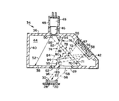
Fig. 3 is a cross-sectional view of an optical system, in the form
of a read head 34, for illuminating portions of the reagent pads 30,
and for detecting light from the reagent pads 30,
CA 02202949 1997-04-17
-5-
and a portion of the tray 20 on which the reagent strip 22 is disposed.
Referring to Fig. 3, the
read head 34 has a housing with a top wall 36, a bottom wall 38, a side wall
40, an angled
wall 42, a planar back wall 44, and a planar front wall (not shown) parallel
to the back wall
44. An illumination source in the form of a light bulb 46 is supported
directly above the
reagent pad 30 to be tested via a cylindrical housing portion 48 integrally
formed with the top
wall 36.
The lower spherical portion of the light bulb 46 has a concentrating lens
integrally
formed therein, and the lower spherical surface is acid-etched to provide it
with an uneven,
diffusing surface so that the shape of the bulb filament does not contribute
to non-uniformity
of the emitted light. When manufactured, the bulb 46 is dynamically fitted to
a ceramic base
49 when the bulb 46 is illuminated to ensure that the axial direction in which
bulb 46 emits
light is substantially parallel to the longitudinal axis of the ceramic base
49. The bulb 46
emits light through a circular aperture 50 formed in the top wall 36 to form a
cone of light
defined by a first edge ray 52 and a second edge ray 54.
The angled side wall 42 has a rectangular aperture 55 formed therein in which
a
rectangular detector array 56 is disposed. The detector array 56 has four
reflectance
detectors 57, 58, 59, 60 disposed therein (see Fig. 4), each of which is
composed of a
conventional colored or IR filter and a conventional silicon detector. Each
filter allows light
having a distinct wavelength to pass through so that each of the detectors 57-
60 is
responsive to light of a different wavelength range. The four wavelength bands
of the filters
are: 400-510 nm (nanometers) (blue); 511-586 nm (green); 587-660 nm (red); and
825-855
nm (infrared). Depending on the type of test being performed, one or more of
the detectors
57-60 may be used.
Light passes through a first optical path from the light bulb 46, through a
relatively
small rectangular aperture 62 formed in the bottom wall 38, to illuminate a
relatively small
rectangular area of the reagent pad 30 being tested. The reagent pad 30 may be
moved
relative to the aperture 62 so that different rectangular areas of the reagent
pad 30 are
illuminated.
Referring to Fig. 5, the illuminated areas may include a first area indicated
by a dotted
box 63, a second area indicated by a solid box 64, a third area indicated by a
dotted box 65,
CA 02202949 1999-06-11
- 6 -
and a fourth area indicated by a solid box 66. Although shown
slightly vertically offset in Fig. 5 so that each box can be
distinctly seen, the illuminated areas 63-66 are linearly offset
with respect to each other, and adjacent areas partially overlap
each other. A number of irregularly shaped areas 67 representing
non-hemolyzed blood cell fragments are also shown in Fig. 5.
Light passes through a second optical path from the illuminated
area on the reagent pad 30, through a first rectangular detection
aperture 68 having angled edges 69 formed in the bottom wall 38,
through a second rectangular detection aperture 70 having angled
edges 71, and through a rectangular aperture 72 formed in the angled
wall 42 to a detection area 73 (Fig. 4) in which the four detectors
57-60 are disposed.
The interior of the read head 34 is provided with an
irregularly shaped baffle 74 composed of a first planar wall segment
76, a second planar wall segment 78, and a zig-zag shaped wall
segment 80. The shape of the baffle 74 is designed to prevent
singly-reflected light rays from reaching the reagent pad 30 from
the light bulb 46 and to prevent singly-reflected light rays from
reaching the detector area 73 from the reagent pad 30.
All surfaces of the baffle 74 and all interior surfaces of the
housing walls 36, 38, 40, 42, 44 are shiny, specular surfaces so
that any light incident upon any surface at an angle of incidence
is reflected from that surface at an angle of reflection equal to
the angle of incidence. This may be accomplished by injection-
molding the read head 34 from a metal mold having highly polished
molding surfaces. The read head 34 is preferably formed of black
plastic so that only a small percentage of light, e.g. 5% incident
upon any of its internal surfaces is reflected. Consequently, any
light that undergoes at least two reflections from any interior
surfaces of the read head 34 is attenuated by at least 99.75%.
Referring to Fig. 3, the wall segment 76 has a non-planar wall
portion comprising a first wall portion with a specular surface 82
that is angled in a direction indicated by a dotted line 84, which
intersects the bottom wall 38, which constitutes a non-planar wall
portion which is a second wall portion with a specular reflective
surface, at a point just to the left of the aperture 62.
Consequently, any light rays emitted by the bulb 46 that impinge
upon the surface 82 are reflected to an area to the left of the
aperture 62. It should be noted that any such rays are reflected
at least twice (in actuality at least three times) before they can
pass through the aperture 62. It should also be noted that no light
CA 02202949 1999-06-11
- 7 _
can be reflected from the surface 82 and pass directly through the
aperture 62 without further reflection since the surface 82 is not
visible when the interior of the read head 34 is viewed from the
aperture 62.
The wall segment 78 has a specular surface 86 angled in a
direction indicated by a dotted line 88, which intersects the top
wall 36 at a point to the left of the circular opening 50 through
which light passes. Consequently, there is no direct path from the
light bulb 46 to the surface 86; therefore, any light that is
reflected from the surface 86 to the aperture 62 will have undergone
at least two (more than two in actuality) reflections from the
interior surfaces of the read head 34.
Fig. 3A is an enlarged view of a portion of read head 34 shown
in Fig. 3. Referring to Figs. 3 and 3A, the zig-zag wall segment
80 has angled surfaces 90-93, each of which is angled in a direction
indicated by a respective dotted line. Since all of the dotted
lines intersect the bottom wall 38 or the side wall 40 to the left
of the aperture 62, no light that impinges upon these surfaces 90-93
directly from the light bulb 46 can be reflected directly to the
aperture 62. The zig-zag wall segment 80 has two further surfaces
94, 95 (Fig.3) that are angled so that any light that impinges on
those surfaces directly from the bulb 46 is reflected exclusively
to the area of the bottom wall 38 to the right side of the aperture
62 .
The only surfaces from which light rays emitted by the bulb 46
can be singly-reflected and still pass through the aperture 62 are
the vertical walls of the aperture 62 itself . However, such singly-
reflected light rays constitute an insignificant amount of the total
light which passes directly from the light bulb 46 to the reagent
pad 30 without reflection. There is also a singly-reflected light
path from the bulb 46 to the walls 40 or 44 to the aperture 62. But
because the bulb 46 concentrates light in a forward direction within
the cone defined by rays 52 and 54, the amount of light going
through the aperture 62 from this path is insignificant.
The second optical path, from the reagent pad 30 to the
detector area 73 (Fig. 4), is generally indicated by a pair of
dotted line 96, 98. The side of the zig-zag wall segment 80 which
is disposed adj acent the second optical path has a plurality of non-
planar, specular surfaces 100, 101, 102 which are angled in a
direction indicated by a number of corresponding dotted lines (shown
CA 02202949 1999-06-11
- 7a -
in Fig. 3) which intersect the angled side wall 42 at a point to the
lower right of the detector area 73. Surface 101 forms a third wall
portion and surface 102 forms a fourth wall portion in zig-zag wall
segment 80. Consequently, any light rays that impinge upon
CA 02202949 1997-04-17
_$_
these surfaces 100-102 directly from the reagent pad 30 without reflection
cannot reach the
detector area 73 without at least one more reflection, and thus any such light
rays will be
attenuated by at least 99.75%.
The side of the zig-zag wall segment 80 which is disposed adjacent the second
optical path has a plurality of planar, specular surfaces 103, 104 (Fig. 3A)
which are angled
so that no light rays from the reagent pad 30 can reach the surfaces 103, 104
directly without
at least one reflection. Consequently, any light rays that impinge upon these
surfaces 103-
104 will already have undergone at least one reflection, and therefore any
such light rays
that eventually reach the detector area 73 will be reflected at least twice
and thus be
attenuated by at least 99.75%.
The wall surfaces 100 and 103 join at an edge 105, and the wall surfaces 101
and
104 join at an edge 106, the edges 105, 106 being substantially aligned with a
respective
edge of the detection area 73, and the edges 69, 71 of the detection apertures
68, 70 are
aligned with the edges of the detection area 73.
The configuration of the read head allows the read head to be made very small
while
maintaining small height sensitivity. Modifications and alternative
embodiments of the
invention will be apparent to those skilled in the art in view of the
foregoing description. This
description is to be construed as illustrative only, and is for the purpose of
teaching those
skilled in the art the best mode of carrying out the invention. The details of
the structure and
method may be varied substantially without departing from the spirit of the
invention, and the
exclusive use of all modifications which come within the scope of the appended
claims is
reserved.