Note: Descriptions are shown in the official language in which they were submitted.
CA 02206116 1997-0~-26
W O96/17546 PCTrUS95115567
SELF-EMISSION NONINVASIVE INFRARED SPECTROPHOTOMETER WITH
TEMPERATURE COMPENSATION
CROSS-K~-~ TO RELATED APPLICATIONS
This application is a continuation-in-part application
of U.S. Patent Application Serial No. 08/247,311, filed May 23,
1994, which is, in turn, a continuation-in-paYt application of
U.S. Patent Application Serial No. 08/010,634, filed January
28, 1993, now U.S. Patent No. 5,313,941.
R~R~ROUND OF THE lNv~NllON
Field of the Invention
The present invention relates to an instrument and a
method for noninvasively measuring the concentration of
glucose, dissoLved carbon dioxide, ethyl alcohol or other
constituents in a person's blood. In particular, the present
invention relates to an instrument and associated method for
monitoring the infrared absorption of such constituents in a
person's blood at long infrared wavelengths where such
constituents ha~Je strong and distinguishable absorption spectra
by measuring specific absorptions of the constituents of
interest within the infrared energy band emitted from the
person's body.
Brief DescriPtion of the ]?rior Art
Accord.ing to the American Diabetes Association, more
than 14 million people in the United States have diabetes,
though about half of them are not aware of it. Almost 750,000
people per year are diagnosed with diabetes, while
approximately 150,000 die from the disease or its complications
each year. Since people with diabetes are at risk for
CA 02206ll6 l997-0~-26
W O96/17546 PCTrUS95/lS567
-- 2
blindness, kidney disease, heart disease and stroke, they need
to control the disease by closely monitoring their blood
glucose levels and carefully controlling the intake of insulin
and glucose. Numerous home diagnostic devices have been
developed for this purpose.
Spectroscopic glucose monitoring techniques using
infrared light are known in the prior art and are the subject
of the present application. Prior art spectroscopic techniques
provide non-invasive solutions to the problem of measuring
glucose in the bloodstream. According to one techni~ue, near
infrared spectroscopy, light is passed through a finger or
suitable appendage for measuring glucose levels in vivo.
Unfortunately, this technique suffers from two sources of
inaccuracy: tissue interference and lack of specificity.
15 Moreover, while the near infrared wavelengths used are easily
and economically generated by light emitting diodes (LEDs) and
solid state lasers, they are not in a range specifically
absorbed by glucose. This lack of "fingerprint" absorbance and
interference from tissue pigment and condition render the
technique useless for accurate concentration determination but
possibly acceptable for trending if stability can be
maintained. Samples of prior art patents describing such
spectroscopic techniques are described below.
Kaiser describes in Swiss Patent No. 612,271 a
technique in which an infrared laser is used as the radiation
source for measuring glucose concentration in a measuring cell.
The measuring cell consists of an ATR measuring prism which is
wetted by the person's blood and an ATR reference prism which
is wetted with a comparison solution. CO2 laser radiation is
led through the measuring cell and gathered before striking a
signal processing device. A chopper placed before the
measuring cell allows two voltages to be obtained corresponding
to the signal from the sample and the reference prisms. Due to
absorption corresponding to the concentration of the substance
35 measured in the blood, the difference between the resulting
voltages is proportional to the concentration. Unfortunately,
the infrared laser used by Kaiser has the undesirable side-
CA 02206116 1997-0~-26
W O 96/17546 PCTrUS95/15567
effect of heating the blood, which may be harmful to the
person, and also does not overcome the effects of tissue
absorption. Alt.hough Kaiser suggests that heating of the blood
may be prevented by using extra-corporeal cuvettes of venous
5 blood and high blood flow rates, Kaiser does not describe a
noninvasive tec:~mique for measuring glucose concentration which
overcomes the eifects of tissue absorption or other sources of
error which are present in the portion of the infrared spectrum
were Kaiser makes his measurements.
March in U.S. Patent No. 3,958,560 describes a
"noninvasive" alltomatic glucose sensor system which senses the
rotation of polarized infrared light which has passed through
the cornea of the eye. March's glucose sensor fits over the
eyeball between the eyelid and the cornea and measures glucose
as a function of the amount of radiation detected at the
detector on one side of the person's cornea. Unfortunately,
while such a technique does not require the withdrawal o~ blood
and is thus "noninvasive", the sensor may cause considerable
discomfort to the person because of the need to place it on the
20 person's eye. A more accurate and less intrusive system is
desired.
Hutchinson describes in U.S. Patent No. 5,009,230 a
personal glucoFe monitor which also uses polarized infrared
light to noninvasively detect glucose concentrations in the
25 person's bloodstream. The amount of rotation imparted on the
polarized light beam is measured as it passes through a
vascularized portion of the body for measuring the glucose
concentration in that portion of the body. Although the
monitor descri.bed by Hutchinson need not be mounted on the
30 person's eye, the accuracy of the measurement is limited by the
relatively mini.mal absorption of glucose in the 940-1000 nm
range used by Hutchinson.
Dahne et al. in U.S. Patent No. 4,655,225 describe a
spectrophotometric technique for detecting the presence of
glucose using specially selected bands in the near infrared
region between llOo and 2500 nm Dahne et al. found that by
applying light at wavelengths in the 1000-2500 nm range
CA 02206116 1997-0~-26
W O96/17546 PCTrUS95/15567
acceptable combinations of sufficient penetration depth to
reach the tissues of interest with sufficient sensitivity may
be obtained for ascertaining glucose concentration variations
without the risk of overheating tissues.
Mendelson et al. in U.S. Patent No. 5,137,023 also
found that wavelengths in the near infrared range are useful
for noninvasively measuring the concentration of an analyte
such as glucose using pulsatile photoplethysmography. In
particular, Mendelson et al. describe a glucose measuring
instrument which uses the principles of transmission and
reflection photoplethysmography, whereby glucose measurement is
made by analyzing either the differences or the ratio of two
different near infrared radiation sources that are either
transmitted through an appendage or reflected from a tissue
surface before and after blood volume change occurs in the
systolic and diastolic phases of the cardiac cycle. The
technique of photoplethysmography can thus be used to adjust
the light intensity to account for errors introduced by
excessive tissue absorptions. However, despite the assertions
20 by Dahne et al. and Mendelson et al., the wavelengths in the
near infrared (below 2500 nm) are not strongly absorbed by
glucose yet are susceptible to interference from other
compounds in the blood and thus cannot yield sufficiently
accurate measurements.
Rosenthal et al. in U.S. Patent No. 5,028,787 disclose
a noninvasive blood glucose monitor which also uses infrared
energy in the near infrared range (600-1100 nm) to measure
glucose. However, as with the above-mentioned devices, these
wavelengths are not in the primary absorption range of glucose
and, accordingly, the absorption at these wavelengths is
relatively weak.
A more accurate glucose measuring technique which
monitors glucose absorption in its primary absorption range has
been proposed by two of the present inventors in U.S. Patent
35 No. 5,313,941. In that patent, Braig et al. disclose a system
for measuring glucose concentration in the middle to far
infrared range which provides improved glucose measurements
CA 02206116 1997-0~-26
W O96117546 PCTrUS95/15567
-- 5
despite the strong tissue absorption that typically attenuates
signals in the middle to ~ar infrared range. This is
accomplished by passing the long wavelength infrared energy
through a finger or other vascularized appendage as short
5 bursts or pulses of energy having a very low duty cycle and low
optical bandwiclth. The bursts are synchronized with systole
and diastole of the cardiac cycle so that only two pulses of
energy are senl_ per heart beat, one during systole and one
during diastol~. The absorption signals detected during
application ofl-he energy bursts are then used to calculate the
concentration of the blood constituents using a polynomial
equation. The description of the system and method as set
forth in U.S. E'atent No. 5,313,941 is hereby incorporated in
its entirety by reference.
Infrared emissions from bodies have been used to
determine the absolute temperatures of those bodies. For
example, some of the present inventors disclose a tympanic
thermometer in U.S. Patent No. 5,159,936 which measures the
absolute temperature of the person from the infrared energy
emissions of t:he tympanic membrane. However, such infrared
energy emissions have not previously been measured at
particular wavelengths to perform constituent absorption
measurements.
AccorcLingly, it is desired to extend the spectroscopic
techniques noted above to obtain absorption signals from
pulsing arterial blood and to provide more accurate
measurements of the concentration of glucose, ethyl alcohol and
other blood constituents by overcoming the problems caused by
interference fl~om tissues and the like. In particular, a
noninvasive blood constituent measuring device is desired which
uses long wavelength infrared energy emitted from the person~s
body for better absorption characteristics and improved signal
to noise ratic~s while also synchronizing long wavelength
infrared energy in those emissions with the cardiac cycle so
that very accurate, temperature compensated in vivo
measurements of the concentrations of such constituents in the
CA 02206116 1997-0~-26
W O96117546 PCTrUS95/15567
arterioles may be made. A method and device for such purposes
is described herein.
Sll ~ RY OF THE lNv~!iN-LloN
The above-mentioned limitations in prior art glucose
and other blood constituent measuring devices are overcome by
providing an instrument which noninvasively measures the
concentration of glucose and other blood constituents in a
person's blood by monitoring the infrared absorption of the
blood constituent in the blood at long infrared wavelengths
10 were such blood constituents have strong and readily
distinguishable absorption spectra. Preferably, the long
wavelength infrared energy emitted by the person's body,
preferably from a vascularized appendage such as the person's
arm, is used as the source of energy for the infrared
absorption measurement, which is made without injury,
venipuncture or inconvenience to the person. By adjusting the
absorption measurement to account for changes in the person's
internal temperature, improved accuracy in the glucose readings
has been achieved.
Since the person's tissue, water and bone are also
strong and variable absorbers of long wavelength infrared
energy, the signal to noise ratio in such a system could cause
serious errors in the blood constituent concentration
measurements. However, potential interference from these
sources is overcome in accordance with the present invention by
(1) synchronizing the optical transmission measurement with the
systolic and diastolic phases of the heart beat and using the
resulting expansion and contraction of the arterial walls to
isolate the measurement to only arterial blood, and/or (2)
using the infrared energy emitted by the person's arm (or other
vascularized appendage) which is not readily absorbed by the
skin or other tissue as the infrared energy source.
The present inventors have discovered that infrared
emissions generated as infrared radiation by a person's tissue
35 when radiated and partially reabsorbed by a blood constituent
such as glucose constitutes quantifiable information regarding
CA 02206116 1997-0~-26
W096tl7546 PCT~S95/15567
concentrations of those blood constituents. By using the
infrared emissions from the person's body as the source of
infrared energy, an infrared source is not necessary, which
greatly reduces the energy used and the system complexity, and
5 hence increases the portability of the device. Also, by
compensating Eor variations in the person's internal
temperature, imFIroved accuracy has been achieved.
Long wavelength infrared detectors typically have low
responsivities because of the attenuation of the signals by the
tissues. These problems are addressed by the device of the
invention by using infrared emissions from the person's arm or
other vascularized appendage for the blood concentration
measurement. l'hus, in accordance with a presently preferred
embodiment of tkle invention, it is unnecessary to apply high
energy infrared energy to the skin of the person, thereby
avoiding possible discomfort. Measurements also may be
synchronized with systole and diastole in accordance with the
invention so as to minimize the adverse interference effects of
tissue absorption. In such an embodiment, two or more
20 measurements typically are made per heart beat. An optical
plethysmograph or ECG may be used in accordance with the
invention to syn_hronize the measurements with the heartbeat.
However, synchroIlization is not necessary and may be eliminated
if it is desired that the spectrop~otometer respond to the
arterial, venous, and tissue glucose levels simultaneously.
The present invention thus relates to a noninvasive
infrared spectrophotometer and method thereof which measures
the concentration of at least one predetermined blood
constituent, such as glucose or ethyl alcohol, in a person's
blood using the infrared emissions from the person's arm or
other vasculari:zed appendage as the source of long wavelength
infrared energy In accordance with a preferred embodiment of
the invention, such a noninvasive infrared spectrophotometer
measures infrared absorption over a broad range of wavelengths
of at least 2.0~m which are emitted as heat by a person. Each
constituent to be measured readily absorbs infrared energy at
one or more of n wavelengths and minimally absorbs infrared
-
CA 02206116 1997-0~-26
W O 96/17546 PCTrUS95/15567
light at one or more other of the n wavelengths within that
range. Thus, infrared energy emitted by the person is absorbed
by the constituent to be measured, and by measuring this
absorption, the concentration of the constituent may be
determined. At least one infrared detector detects light at
the n wavelengths which has passed through, for example, an
arterial blood vessel of the person and been selectively
absorbed by the predetermined constituent(s). The infrared
detector outputs a detection signal for processing to determine
the constituent concentration. A temperature sensing device
for measuring the person's internal temperature at the arm or
other vascularized appendage is also used in adjusting the
constituent concentration measurement for temperature dependent
effects.
Synchronizing means may also be provided for
synchronizing the measurements with the systolic and diastolic
phases of the cardiac cycle of the person. Preferably, the
synchronizing means comprises a cardiac monitor and means
responsive thereto for controlling absorption measurements to
occur during the systolic and diastolic phases of the cardiac
cycle of the person. Alternatively, the plethysmograph signal
may be obtained from the infrared signal directly, thereby
eliminating the need for a separate plethysmograph. In this
embodiment, however, the infrared signal still would be gated
into systolic and diastolic components.
In addition, a chopper wheel may be used to convert
the measured signal to a high frequency in order to overcome
the low frequency noise in an HgCdT detector; however, other
detectors such as thermopiles do not require a chopper. Th~
chopped signal is then amplified and ~iltered and synchronously
demodulated to recreate the DC signal from the detector without
the added noise. The signal is then low pass filtered and
passed into the processor for calculation of the concentration
of the predetermined constituent(s) from the detection signal
35 to provide a concentration indication which is substantially
free of tissue absorption errors.
CA 02206116 1997-0~-26
W O 96/17546 PCTrUS95/15567
In a F~referred embodiment of a glucose monitor, the
detection wavelength is approximately 9.1 ~m while the
reference wavelength is approximately 10.5 ~m. In an
alternative el~odiment of a blood alcohol monitor, the
detection wave:Length is approximately 3.4 ~m and the reference
wavelength is approximately 4.8 ~m. Preferably, bandpass
analytical filters are also disposed between the arterial blood
vessel of the person and the infrared detector(s) for passing
infrared light in a narrow passband centered at such detection
and reference wavelengths.
Concentration of the predetermined constituent(s) is
calculated by forming a ratio R = (Sys Ll - Dias L1)/(Sys L2 -
Dias L2), where Sys L1 is a detected systolic phase signal at
the detection wavelength, Dias L1 is a detected diastolic phase
signal at the detection wavelength, Sys L2 is a detected
systolic phase ~:ignal at the reference wavelength, and Dias L2
is a detected diastolic phase signal at the reference
wavelength, and then solving the following equation:
C.C. = Cl + C2 * Ln(R) + C3 * [Ln(R)] 2 + C4 * [Ln(R)] 3 +
Cs * [L,n(R)]4,
where:
C.C. is the concentration of the predetermined constituent;
Cl - Cs are empi:r:ically determined calibration coefficients; and
Ln is a natural log function.
The above equation can be generalized for a system
using multiple cletection wavelengths and one or more reference
wavelengths by including cross-product terms in the polynomial.
Also, in the preferred embodiment of the invention,
the glucose measurement is compensated for temperature
30 variations in the person's body temperature by measuring the
temperature deep within the person's arm or other vascularized
appendage. From the measured temperature, Io is computed to
yield a value of the energy level emitted from within the
person's arm or other vascularized appendage before the glucose
absorbs any infrared energy. The resulting Io value is then
ratioed to the actual measured energy I for computing the
CA 02206116 1997-0~-26
W O96/17546 PCTrUS95/15567
-- 10 -
percentage of energy absorbed by the glucose. Discontinuities
in the ratio are also removed by converting the measured
voltages into watts of radiant power, adding the watts emitted
by the chopper blade, and then taking the ratio of that
quantity. Then, by treating the measured signals from the
person's arm or other vascularized appendage as the I value in
Beir's equation and ratioing these values to the measured Io
values using Planck's equation, Beir's law may be rewritten to
allow for compensation for temperature dependencies as:
I/Io = e~CLx,
where C is the glucose concentration, L is the pathlength from
where the infrared emissions originate to where they are
detected, X is an experimentally determined extinction
coefficient, I is the energy emitted by the arm, and Io is the
incident energy within the arm, defined from Planck's equation
as:
I o = T~ * ~ * C21
~5( e AT_ 1)
where TR is the transmission coefficient of the selected
filter, ~ is the emissivity of skin, C1 = 3.74 x 10⁴, C2 = 1. 438
X 104, T is the measured absolute temperature deep within the
20 body or arm, and ~ is the wavelength of the selected filter.
BRIEF DESCRIPTION OF THE DRAWINGS
The objects and advantages of the invention will
become more apparent and more readily appreciated from the
following detailed description of presently preferred exemplary
embodiments of the invention taken in conjunction with the
accompanying drawings, of which:
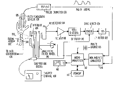
Figure 1 schematically illustrates a preferred
embodiment of a noninvasive infrared spectrophotometer in which
infrared energy emitted by the person is used as the infrared
energy source.
Figure 2 illustrates a photoplethysmograph for
detecting systole and diastole in the embodiment of Figure 1.
CA 02206ll6 l997-0~-26
W O96/17546 PCTrUS95/15567
Figure 3 illustrates a temperature sensing device for
determining the internal arm temperature for temperature
compensation of the glucose readings in accordance with the
invention.
Flgure 4 is a circuit diagram of the temperature
sensing circuit of Figure 3.
Figure 5 is a graph illustrating the ratio of target
wattage at 9.4 ~m divided by the target wattage at 10.5 ~m
before and after correction of the temperature dependent
effects and di<,continuity at zero.
DETATT~n DESCRIPTION OF THE PRESENTLY PREFERRED EMBODIMENTS
A noninvasive infrared spectrometer with the above-
mentioned beneficial features in accordance with the presently
preferred exemplary embodiment of the invention will now be
lS described with reference to Figures 1-5. It will be
appreciated by those of ordinary skill in the art that by
changing the wavelengths of infrared energy to be monitored for
absorption that other blood constituents such as ethyl alcohol,
carbon dioxide, urea, uric acid, lipids, creatinine, peptides,
cholesterol, hematocrit, drugs, and several other analytes can
be measured in accordance with the techniques of the invention.
Thus, the glucose monitoring device described herein in the
exemplary embod:Lments is for descriptive purposes only and is
not intended in any way to limit to scope of the invention.
25 All questions regarding the scope of the invention may be
resolved by referring to the appended claims.
As known by those skilled in the art, most covalent
bonds have characteristics and essentially invariant absorption
wavelengths so that the presence of an absorption band in the
infrared spectrum indicates the presence of a bond in the
molecule while the absence of an absorption peak guarantees the
absence of its corresponding bond. Hence, each compound or
blood constituent measured in accordance with the techniques of
the invention has a characteristic absorption spectrum in the
infrared range which may be used to establish one or more
detection and reference wavelengths for absorption measurement.
CA 02206116 1997-0~-26
W O96/17546 PCTrUS95/15567
- 12 -
~lucose measurement in the far infrared range will be described
herein as a presently preferred embodiment, although the
present invention also has particular utility as a blood
alcohol monitor in the middle and far infrared ranges.
The infrared spectra includes the near infrared
(approximately 1 to 3 microns), the middle infrared
(approximately 3-6 microns), the far infrared (approximately 6-
15 microns), and the extreme infrared (approximately 15-100
microns). As noted above, typical glucose and other
10 noninvasive blood constituent measuring devices operate in the
near infrared region where the absorption of infrared energy by
glucose and other blood constituents is relatively low.
However, the present inventors have found a technique whereby
absorption may be measured in the middle and far infrared
regions where glucose and other blood constituents have strong
and distinguishable absorption spectra while also minimizing
the adverse effects of tissue, water and bone absorption.
Glucose has strong characteristic spectra in the far
infrared above about 6 microns. As described by Mueller in W0
81/00622, glucose absorption may be measured using a detection
wavelength at approximately 9.1 ~m and a reference wavelength
at approximately 10.5 ~m. Similarly, ethyl alcohol has a
strong characteristic spectra in the middle infrared (3-4 ~m)
and in the far infrared (9-10 ~m). Accordingly, ethyl alcohol
concentration may be measured using a detection wavelength of
approximately 3.4 microns and a reference wavelength of
approximately 4.8 microns using differential absorption
calculation techniques.
Figure 1 illustrates a "self-emission~ glucose monitor
30 which noninvasively measures the concentration of glucose
and/or other constituents in a person's blood by monitoring the
infrared emission of glucose in the blood at long infrared
wavelengths near 10 microns. In other words, unlike prior art
analyzers or the analyzer described in U.S. Patent No.
5,313,941, the present embodiment of the invention does not
include an infrared source and instead utilizes the infrared
CA 02206116 1997-0~-26
W O96117546 PCTrUS95/15567
- 13 -
energy emitted by the person's blood and/or surrounding tissue
to perform the absorption analysis.
The inventors have found that in prior art "infrared
transmission" t:ype analy~ers only a small amount of signal
5 passes through the selected appendage. By contrast, in
accordance with the presently preferred embodiment, the
infrared source is no longer needed since the normal body heat
provides the source of infrared energy. However, analysis is
still performed at the longer wavelengths of infrared energy.
In particular, wavelengths between 2 and 12 microns are
particularly preferred because these wavelengths are naturally
emitted by the body as the wavelengths of heat, and because
molecular absorption in the 2-12 micron region is very molecule
specific. As noted in U.S. Patent No. 5,313,941, this region
contains the "fingerprint" region in analytical chemistry
because in this region each molecule has a distinct absorption
spectra.
This embodiment of the invention thus requires no
source of enerc~y other than the human body. No energy is
20 passed through ~n appendage, thereby eliminating any possible
danger associated with excessive radiation. Because it is
unnecessary to generate optical energy, the present embodiment
operates on less power, thus making this embodiment a more
likely candidale for portable operation.
As illustrated in Figure 1, the invention of the
preferred embodiment preferably includes an infrared detection
device which is designed to rest on the underside of a person's
wrist 100 for detection of infrared heat energy emitted from
the interior 102 of the person's arm. The detector includes a
1/4 inch aperture 104 which passes the infrared signal from the
interior 102 of the person's arm (or other vascularized
appendage) to a chopper 106 controlled by a chopper control
device 108 for converting the signal to a high frequency in
order to minimize the low frequency noise in the detector. The
signal then passes through a filter wheel 110 containing the
analytical and reference filters for making the absorption
measurements in accordance with the techniques described below.
CA 02206116 1997-05-26
W O96/17546 PCTrUS95115567
- 14 -
Analytical filter wheel 110 is controlled by microprocessor 112
to control detection of the absorption of the desired
constituents or the reference by infrared detector 114. In a
preferred embodiment, the filter wheel 110 changes filters
every 3 heartbeats.
Microprocessor 112 receives energy from an AC or DC
power source 115 which preferably permits the invention to be
miniaturlzed for use in a hand-held monitor. The calculated
concentration is then displayed on display 116 so that the user
10 may readily ascertain his or her glucose level, ethyl alcohol
level, and the like. Microprocessor 112 preferably further
includes memory 117 for storing sequences of such measurements
so that averaging and trending and the like may be conducted.
In a presently preferred embodiment, a cooled HgCdT
detector is used as infrared detector 114. The cryogenic
cooling of such a detector enhances the infrared signal since
infrared detectors generally respond to the difference in
temperature between the target (in this case the interior 102
of the person's arm) and the detector. However, with low noise
electronics and appropriate signal processing, room temperature
detectors such as the thermopile and pyroelectric types used in
the detector described in U.S. Patent No. 5,313,941 also may be
used in this embodiment.
Upon detection, the chopped and filtered signal is
amplified by preamplifier 118 and then filtered by bandpass
filter 119 to minimize noise before the signal is demodulated
by synchronous demodulator 120. Synchronous demodulator 120
demodulates the chopped signal to recreate the DC signal from
the detector, only now the noise is substantially reduced. The
signal is then low pass filtered by low pass filter 122 and
passed to multiplexer and A/D converter 124 for processing in
accordance with the techniques described below.
As with the monitor described in U.S. Patent No.
5,313,941, the monitor of this embodiment is designed to
analyze arterial blood metabolite levels. By monitoring the
cardiac pulse and synchronizing to that pulse, arterial blood
signals can be discriminated from other signals. In this
CA 02206116 1997-0~-26
W O96/17546 PCTnUS95/15567
- 15 -
embodiment, the in~rared detector 114 continuously measures the
infrared energy emitted from the interior 102 of the person's
- arm, for example. As with the monitor described in U.S. Patent
No. 5,313,9~1, in this embodiment an optical plethysmograph
5 monitors the person's pulse very near the infrared signal site.
As shown in Figure 1, the plethysmograph signal preferably is
taken from the person's middle (or ring) finger 126 by
plethysmograph ~3ensor 128 while the infrared signal is taken
from the underside of the person's wrist 100. The
10 plethysmograph signal is then applied to synchronous
demodulation electronics in a preamplifier of a pulse oximeter
or other cardiac monitor 130, which converts the silicon
detector OUtpllt into a useful signal. The resulting
plethysmograph signal is applied to multiplexer and A/D
converter 124 and then to microprocessor 112 for processing in
accordance with the techni~ues described below. The
plethysmograph signal is used to electronically gate the
infrared signal ~rom the underside of the wrist 100 into
systolic and cliastolic signals to help minimize tissue
absorption effects. Since veins and tissues generally do not
pulse, their contribution to the detected signal is cancelled
when the ratio is taken.
During operation, an LED of plethysmograph sensor 128
is located on the middle (or ring) finger 126 of the person,
thereby forming a visible or near infrared light source 200 as
shown in Figure 2. The LED 200 is pulsed by microprocessor 112
and LED driver circuits (not shown). The signal from LED 200
passes through the finger 126 and is detected by silicon photo
diode 202. As noted above, synchronous demodulation
electronics in a preamplifier of the cardiac monitor 130
convert the silicon detector output into a useful signal. The
LED 200, silicon photo diode 202, and preamplifier circuit of
the cardiac monitor 130 together constitute an optical
plethysmograph. As described in detail in U.S. Patent No.
5,313,941, the plethysmograph signal is used by microprocessor
112 to determine the phase of the cardiac cycle for controlling
CA 02206116 1997-0~-26
W O96/17546 PCTrUS95/15567
the gating of the infrared signal into systolic and diastolic
integrators within the software of microprocessor 112.
In an alternative embodiment to the embodiment of
Figures 1 and 2, the plethysmograph signal is obtained from the
infrared signal directly so as to eliminate the need for a
separate plethysmograph. In such an embodiment, the infrared
signal is still gated into systolic and diastolic components;
however, the plethysmogram is derived from the infrared signal
itself rather than a separate plethysmograph. Of course, other
techniques for monitoring the cardiac cycle maybe used. For
example, the cardiac monitor 130 may utilize an electro-
cardiogram for synchronizing to a characteristic feature of the
electrocardiogram.
On the other hand, those skilled in the art will
appreciate that synchronization to the cardiac cycle is not
strictly necessary and that the synchronization step may be
eliminated. Without synchronization, the spectrophotometer of
the invention will respond to arterial, venous, and tissue
glucose levels simultaneously, which may be desired in some
circumstances.
Thus, in accordance with the invention, if the
spectrophotometer is to be synchronized to the cardiac cycle,
the following steps are performed. In particular,
microprocessor 112 electively processes the plethysmograph
signal from photodetector 202 in order to determine systole and
diastole in the next cardiac cycle as follows:
1. A conventional plethysmograph signal is obtained
by photodetector 202, digitized by analog to digital converter
124 and recorded in memory 117 as pulse N-1. This is
accomplished by dividing the plethysmograph signal N-l into
sampling intervals having durations of approximately 0.1 to 10
msec. Preferably, the plethysmograph signal from photodetector
202 is sampled by analog to digital converter 124 every 1 msec.
2. A characteristic feature of the cardiac cycle
35 waveform is selected for purpose of synchronization.
Preferably, the dicrotic notch, which is a feature on the
CA 02206116 1997-0~-26
W O96/17546 PCTrUS95/15567
- 17 -
waveform of the cardiac cycle where a distinctive dip occurs as
a result of the closing of the ventricular valves in the heart,
is selected and labelled as time zero for cycle N-1. All other
1 msec intervals occurring after the dicrotic notch are
labelled as one, two, three, etc. until the next dicrotic notch
for the cycle N is found.
3. The waveform N-1 is then examined to find the peak
signal point lsystole) and the interval number (i.e., the
number of intervals or msec from the dicrotic notch) is stored.
4. The waveform N-1 is then ~mlned to ~ind the
minimum signal point (diastole) and the interval number is also
stored.
5. In cardiac cycle N, running in real time, the
dicrotic notch is again identified. The interval number stored
in step 4 for plllse N-1 is then counted from the dicrotic notch
to determine ~]le time interval anticipated to correspond to
diastole for cycle N. The infrared energy from underside of
the person's wrist 100 is then measured during a 2 millisecond
interval arou~cL diastole. At the end of this 2 millisecond
interval, the appropriate number of intervals is counted to
determine the time interval anticipated to correspond to
systole in cycle N. The infrared energy from the underside of
the person's w-cist 100 is measured again for approximately 2
millisecollds around systole.
6. The absorption signals developed by the infrared
detector 114 are digitized by analog to digital converter 124
and stored in memory 117 or another temporary register o
microprocessor 112.
7. In cycle N, the infrared LED plethysmograph signal
is again recorded and examined. If is determined that systole
and diastole occurred within approximately +/- lO msec of where
- they were predicted to have occurred during analysis of pulse
N-1, the long wavelength infrared data stored in memory 117 or
some other temporary register is then passed to the glucose
35 processing algorithm of microprocessor 112 for calculation of
the glucose concentration. However, if systole and diastole
did not occur within +/- 10 msec of where they were predicted
CA 02206116 1997-05-26
W O96/17546 PCTrUS95/15567
- 18 -
to have occurred in cycle N-1, the stored values are erased
from memory 117.
8. Steps 1-7 are then repeated until a number of
usable measurements have been made. The measurements may then
5 be averaged or the highest and lowest values thrown out so that
an accurate calculation of concentration may be made by
microprocessor 112 and displayed on display device 116.
Measurement of the infrared detection signal may be
synchronized with the heart beat as just described in order to
remove the effects of tissue and other "non pulsating"
interferants sometimes referred to as patient variations.
However, heart beats are not the same every time and vary from
individual to individual. These variations present a challenge
to calibration of an instrument. Accordingly, in order to
15 normalize the absorption readings and overcome the requirement
for individual calibrations, at least two long infrared
wavelengths are measured simultaneously during diastole and
systole as just described. As described above, for glucose the
analytical wavelength specifically absorbed by glucose is
20 preferably in the range of approximately 9.1 ~m, while the
reference wavelength is preferably in the range of
approximately 10.5 ~m, which is not absorbed by glucose.
Generally, glucose concentration is determined by forming a
ratio between the systolic and diastolic difference signals
25 measured at 9.1 ~m versus those measured at 10.5 ~m. More than
one reference and analytical wavelength may be used so that
multiple ratios are formed. The resulting arrays of numbers
are then operated upon by empirically determined calibration
coefficients. The resulting computation yields the
concentration of glucose in the person's arterial blood.
The general form of the mathematics used by
microprocessor 112 for calculating the concentration of a blood
component such as glucose from absorption signals generated at
two or more wavelengths in accordance with the techniques of
the invention will now be described.
In general, for a system of n+1 detection wavelengths
for detecting n blood constituents such as glucose, alcohol and
CA 02206116 1997-05-26
W O96/17546 PCTnUS95/15567
the like, where the systolic phase signal at wavelength n is
SYS LN and the diastolic phase signal at wavelength n is DIAS
LN, the concentration of the blood component (such as glucose)
being measured can be computed as a mathematical function of
5 SYS ~N and DIAS LN. For example, the component concentration
(C.C.) may be represented as:
C.C. = Fn (sYs LN, DIAS LN).
For a system u~,i.ng multiple (at least two) wavelengths where L1
- LN are analyti.cal wavelengths and LR is one or more referenc.e
lo wavelengths, then:
RN = (SYS LN - DIAS LN)/(SYS LR - DIAS LR); EQ. (1)
Of course, other mathematical forms of the ratio R may be used,
but in general, RN = FN (LN~ LR) -
The concentration of each blood constituent is then
a function of each ratio R for that constituent. For example,
glucose concentration (G.C.) may be calculated from a
polynomial equation of order p for a single detection and a
single reference wavelength as:
G.C. = C1 + C2 ~ Ln (R) + C3 * [Ln (R) ] 2 + C4 * [Ln(R)] 3 +
Cs * [Ln(R)] 4, EQ. (2)
where Cl-C~ are calibration constants, Ln is the natural log
function and p-.~.~. However, when plural detection wavelengths
and/or plural reference wavelengths are used, cross-product
terms would be added, resulting in the following generalized
equation:
x= (m-l ) y=p Z=P
C. C.n=B+ ~ y ( Y) ] ~ D~* [Ln (Rl ) *Ln (R2) . . . *Ln (R ) ~
x=l y=l z=l
EQ. (3)
where B, Cxyt and Dz are calibration constants, m is the total
number of analyt.ical and reference wavelengths (m ~= (n+1)) and
Ln in the natura.l log function.
_ _ _ _ _ . _ _ _ . . . .
CA 02206116 1997-0~-26
W O96/l7546 PCTrUS95/15567
- 20 -
Of course, other equations besides a polynomial
equation may be used by those skilled in the art to calculate
the concentration of the respective blood constituents. For
example, the systolic/diastolic ratios may be replaced with
their logarithms as in pulse oximeter computations.
Alternatively, the mathematical technique known as
"chemometrics" may be used to provide a multiple wavelength
spectral analysis.
As is apparent from the above, measurement of
concentrations by optical absorption involves making
measurements of absorbed light and applying some derivation of
Beir's law to compute the concentration, where Beir's law can
be stated as I = Io * e-C~, where C is the glucose
concentration, ~ is the pathlength from where the infrared
emissions originate to where they are detected, X is an
experimentally determined extlnction coefficient, I is the
energy emitted by the arm, and Io is the incident energy within
the arm, defined from Planck's equation as:
I O = TR * ~ * C12
EQ. (4)
20 where TR is the transmission coefficient of the selected
filter, ~ is the emissivity of skin, C1 = 3.74 x 10⁴, C2 = 1. 438
X 104, T is the measured absolute temperature deep within the
body or arm, and A is the wavelength of the selected filter.
Accordingly, the computation of C, the concentration of
interest, usually involves measuring I and IO and solving
Beir's equation for C.
However, in a system of the type proposed above in
which the energy emitted from within the body of a subject is
measured, an optical measurement of Io~ the incident energy, is
30 not possible because the energy can only be sensed after it has
passed through the skin and substance to be measured. However,
the present inventors have now discovered that Planck's
Equation may be used to compute the actual value of Ic based on
CA 02206116 1997-0~-26
.
W O 96/17546 PCTrUS95/15567
- 21 -
the temperature of the source o~ radiation, in this case, the
person's arm. As known by those skilled in the art, Planck's
equation givecs the radiant emittance of a "blackbody" at a
particular wavelength and temperature and can be written as:
W - Cl
A c2
A
EQ. (5)
where W = watls/cm2 per micron of wavelength, ~ = wavelength in
microns, T = absolute temperature in degrees Kelvin, Cl = 3.74
* 10~, C2 = 1.438 * 104, and W~ = radiant flux emitted per unit
area per unit increment of wavelength in watts/cm2-micron.
To correct for temperature dependent ef~ects, Io is
computed using Equation (4) to yield a value of the energy
level emitted ~rom within the target (person's arm) before the
glucose absorbs any energy. The computed value ~or Io is then
ratioed to the actual measured energy I for computation of the
15 percentage of energy absorbed by the glucose. By ratioing,
many of the terms, such as the pathlength L, effectively drop
out of the equation.
Thus, to compute Iol it is necessary to measure the
temperature deep within the person's body. For this purpose,
a temperature sensor 300 (Figure 3) may be placed, for example,
around the person's wrlst for measurement of the lnternal
temperature of the person's arm. As shown ln Figure 3, the
temperature sensor 300 may include a flexlble rectangular RTD
such as MINCO part #S386PD10Z36A whlch ls wrapped around the
25 person's wrlst 100 or forearm and covered wlth an insulatlng
elastlc foam 302, such as TRU-FIT model 405 Neoprene Wrlst
Support. As shown ln Figure 4, the temperature sensing element
(RTH) 301 ls e]ectrlcally connected in series wlth a precision
reslstor (R). Two voltage measurements Vre~ and Vr~h are made,
and a llnear equation ls used to determlne the temperature from
the voltage r.cltlo Vr~h/Vref. Once the temperature is known,
Equatlon (4) :i, used to compute Io in units of watts.
One of the problems assoclated with taking the ratios
of the 9.4 and 10.5 micron signals when applylng such
-
CA 02206116 1997-0~-26
W O96/17546 PCTrUS95/15567
techniques to the monitor of Figure 1 has been that the ratio
~'blew up" and became discontinuous as the signals approached
zero and/or went negative. Whenever the target is at a lower
temperature than the chopper wheel 106, the measured signal
5 would be less than zero. When the target temperature equals
the temperature of the chopper wheel 106, the signals will also
equal zero. On the other hand, when the target is warmer than
the chopper wheel 106, a positive signal is produced. As shown
in Figure 5, the ratio of the 9.4 signal to the 10.5 signal
results in a discontinuity at zero delta temperature. Since
the ratio is what is used to compute glucose concentration, it
is desired to remove this discontinuity.
To eliminate the discontinuity, the voltages measured
are converted into watts of radiant power and the ratio is
taken of that quantity. The conversion takes two steps:
1. Convert measured voltage to watts; and
2. Add the watts emitted by the chopper blade 106.
Step two is required because the system of the preferred
embodiment is only responsive to the difference between the
temperature of the chopper wheel 106 and the target temperature
as determined by temperature sensor 300.
Conversion of the measured voltage to wattage is
accomplished by calibrating the system experimentally. A
linear regression may be used to convert the voltage produced
25 by the detector to the wattage predicted by Planck's equation.
After the measured wattage is known, the wattage emitted from
the chopper wheel 106 is computed using Planck's equation.
Then, since the measured wattage equals the target-chopper
voltage, the chopper wattage is added to the measured wattage
to yield target wattage. Now, when the ratio of target wattage
at 9.4 microns is divided by the target wattage at 10.5
microns, the discontinuity is eliminated.
As shown in Figure 5, while the ratio discontinuity
is no longer a problem, the ratio shows a strong dependence
35 with temperature. Removing this dependence will allow for the
computation of more accurate glucose concentrations.
CA 02206116 1997-0~-26
W O96/17546 PCTrUS95/15567
The temperature dependence is caused by the shifts in
the peak and shape of the black body radiation curve with
temperature. Planck's Equation described this emission
spectra. As noted above, Planck's Equation and the measured
temperatures can be used to compute the theoretical Io value.
If the measure~ signals are then treated as the I value in
Beir's equation and ratioed to the Io values from Planck's
equation, Beir's law may be rewritten as I/Io = e~C~, as noted
in Equation (4) above.
To apply the theory, a measurement of the ratio may
be taken from a glucose solution soaked chamois. The target
temperature is swung over a wide range to test the temperature
correction to :lts fullest. Figure 5 shows the pre and post
correction rat'ios. The right scale is for the temperature
corrected ratic, while the left scale is for the "raw ratio".
As shown, the discontinuity at zero and the temperature
dependencies in the ratio have been eliminated. These
techniques are thus desixably used in the calculation noted
above to eliminate such potential errors in the computations.
The amplitude of the signal detected by the in~rared
detector 114 establishes the signal to noise ratio of the
entire system. The larger the signal, the lower the system
noise. Lower system noise leads to shorter integration times
and faster mea.surements. In accordance with the present
embodiment, the signal output by detector 114 has been measured
to be approximately 8 nV per mg/dl. The raw signal has been
measured at 65 microvolts with a pulse amplitude of 1~ of the
DC signal. Whi.]e those skilled in the art will appreciate that
such a signal is sufficient to make a meaningful measurement,
it is desirable to increase the pulse amplitude to improve the
signal to noise ratio. One such technique in accordance with
the invention includes inflating a blood pressure cuff in
synchrony with the glucose measurement.
As noted above, preferred embodiments of the invention
described herein are specifically designed to monitor glucose
which absorbs selectively near 9.1 ~m. However, those skilled
in the art will appreciate that by changing the wavelengths of
CA 02206116 1997-0~-26
W O96/17546 PCTrUS95/15567
.
- 24 -
infrared energy detected other bloodstream constituents such as
carbon dioxide, ethyl alcohol, urea, uric acid, lipids,
creatinine, peptides, cholesterol, hematocrit, drugs (all
absorbing in the 5-10 ~m band) and several other analytes can
5 be measured. Also, the dialysis fluid of kidney patients may
be monitored using the techniques of the invention.
The invention herein described offers both specificity
and noninvasive measurement, thereby making it acceptable for
use by anyone needing to measure or monitor his or her blaod
glucose level, ethyl alcohol level or other blood constituents
levels. Use of long wavelength infrared absorbance
measurements provide signals at the exact wavelengths absorbed
specifically and strongly by glucose or some other blood
constituent, while use of pulsed and cardiac synchronized
infrared energy bursts removes interference effects caused by
tissue absorption yet provides for a high energy infrared
signal without discomfort.
Although an exemplary embodiment of the invention has
been described in detail above, those skilled in the art will
readily appreciate that many additional modifications are
possible in the exemplary embodiment without materially
departing from the novel teachings and advantages of the
invention. For example, the present invention may be used to
measure other blood constituents such as those mentioned herein
25 by selecting one or more analytical wavelengths and one or more
reference wavelengths using techniques known to those skilled
in the art. Accordingly, these and all such modifications are
intended to be included within the scope of the invention as
defined in the following claims.