Note: Descriptions are shown in the official language in which they were submitted.
-
CA 02206600 1997-0~-30
WO g6/17948 1 ~ 5ll5gl4
METHODS, COMPOSITIONS AND APPARATUS FOR CELL TRANSFECTION
BACKGROUND OF THE lNv~NllON
This invention relates generally to genetic
engineering, and more specifically, to methods of
increasing transfection efficiency of target cells.
Genetic engineering technology is used routinely
to transfect cells. Transfection is the introduction of a
foreign gene(s) into a target cell and the incorporation of
that gene into a chromosome of the target cell. Once
inside the target cell, a functional foreign gene can
produce the RNA and protein product it encodes.
Transfection has diverse applications in fermentation,
research, agriculture, pharmaceuticals and medicine.
A particularly important application of
transfection is gene therapy. Gene therapy has the
potential to permanently treat diseases and deliver new
therapeutic proteins and RNA that currently cannot be used.
In gene therapy, a patient receives a functional foreign
gene which produces a product that affects the disease or
condition. Since the foreign gene can be stably
incorporated into the patient's genome, the foreign gene
has the potential to produce the product for the life of
the patient. The patient receives the foreign gene by
transfecting target cells ex vivo and A~m; n; stering the
transfected cells to the patient or the foreign gene can be
directly ~i n; stered to the patient and the cells
transfected in vivo.
A requirement of all transfection methods is that
the foreign gene gets into the target cell. Many
transfection methods have been developed but all can be
CA 02206600 1997-0~-30
WO 96117948 1 ~ sll59l4
classified as either direct or indirect methods. In direct
methods, a genetic engineer injects the foreign gene into
individual target cells using a microcAp;llary or
microprojectile. Indirect methods involve the target cells
passively or actively taking up the foreign gene. Indirect
methods are diverse and include, for example, pinocytotic
uptake of DNA-calcium phosphate and fusion of liposomes
with the plasma membrane of the target cell. A very
effective method is to use viral particles to infect the
target cell because, once inside the target cell, foreign
genes often express themselves at consistently higher
levels by this method than by other methods.
Viral particles are themselves quite diverse and
include DNA viruses, such as SV40, polyoma, adenovirus,
Epstein-Barr, vaccinia, herpes simplex and baculovirus, and
RNA viruses, such as tobacco mosaic virus, cucumber mosaic
virus, brome mosaic virus and retrovirus. Retroviruses are
particularly useful viral particles because, once inside
the target cell, these viruses lead to stable
transfections. Retroviruses which are replication-
incompetent appear well suited for gene therapy because, in
principle, these viruses do not produce any wild-type virus
and cannot infect other cells after infecting the target
cell. Replication-incompetent viruses are produced in so
called "packaging cells" because these cells "package" the
foreign gene into viral particles which can infect, but not
replicate.
The major problem with indirect transfection
methods is that they are inefficient at transfecting target
cells. Transfection efficiencies of 1-20% are achieved
but, for human target cells, the transfection efficiency is
at the lower end of the range. The transfection efficiency
is the number of target cells cont~;n;ng at least one copy
of the foreign gene divided by the total number of target
cells. Thus, most current indirect transfection methods
CA 02206600 1997-0~-30
WO 96117948 PCT/US9~;115914
waste large amounts of costly target cells, carriers and
foreign genes because only a small fraction of exposed
target cells is transfected. This inefficient method
~ particularly limits development of gene therapy because
gene therapy requires many transfected target cells. In
certain circumstances, higher transfection efficiencies are
possible but often heroic measures are needed to achieve
them. For example, bone marrow target cells can be
cultured for several weeks with repeated exposures to
retroviral particles. Such methods are not practical
because of expense, complexity or incompatibility of the
target cells and particles. There is a need for a more
efficient, easy-to-use, generally applicable transfection
method.
Contact between target cells and viral particles
is essential for transfection to occur. Generally,
indirect viral transfection occurs by culturing target
cells with viral particles suspended in the cell culture
medium. All indirect transfection methods are based on
random contact between viral particles and target cells.
Typically, the culture is gently agitated during
transfection and suspended viral particles contact target
cells by chance. Although specific target cells can be
selected for transfection using various techniques, the
contact between target cells and viral particles in these
methods remains a random event. Methods for selectively
transfecting target cells include bridging antibodies
between viral and target cell antigens and chemically
modifying particles for specific target cell receptors.
Current indirect transfection methods are therefore limited
to rAn~o~ contact between viral particles and target cells.
Increasing the concentration of viral particles
increases contact between viral particles and target cells.
However, the viral particle concentration that can be used
CA 02206600 1997-0~-30
WO 96/17948 PCT/US95115914
for transfection is limited because the proportion of
infectious viral particles decrease as the viral particles
are concentrated. Various methods are used to concentrate
retroviral particles including polyethylene glycol
precipitation, sucrose gradient centrifugation, pelleting
by centrifugation, aqueous two-phase systems, ammonium
sulfate precipitation, and hollow fiber ultrafiltration.
A measure of viral particle concentration is titer that,
for replication-incompetent retrovirus, is typically about
104 to 105 colony forming units (CFU)/ml. Viral particle
concentration limits the transfection efficiency of current
viral transfection methods by limiting the contact between
viral particles and target cells.
Current indirect transfection methods require
chemical additives to transfect target cells. Chemical
additives allow viral particles to enter target cells more
easily. Chemical additives include, for example, polybrene
and protamine sulfate. In current methods, chemical
additives are required because particle-target cell contact
is so infrequent it is necessary to m~;m;ze the number of
particles that enter target cells once contact occurs.
Without chemical additives, even the relatively low
transfection efficiencies achieved by current methods would
not be possible. Chemical additives are undesirable for
gene therapy because the chemical additives pose a
cont~min~tion concern.
Another problem with current methods is that some
target cells cannot be transfected because the particles
cannot contact the target cells in culture. ~or example,
hematopoietic stem cells (HSCs), a prime target for gene
therapy applications, are often grown in cell culture in
association with accessory cells (stromal cells). The HSCs
position themselves between stromal cells and the cell
growth support, and become physically inaccessible to
retroviruses in the cell culture medium. HSCs cannot be
CA 02206600 1997-0~-30
WO96117948 PCT~S9S/15914
transfected by retroviru~es because the stromal cells block
retroviral access to the HSC. A method is needed which
allows particles to contact target cells even though the
~ target cells are covered over by accessory cells.
Besides contact limitations, low transfection
efficiencies can result from cell culture inhibitors that
limit target cell growth. Retroviruses require dividing
target cells to transfect. Packaging cell culture
supernatant contains growth inhibitors that reduce target
cell growth. Since the target cells must divide for
transfection to occur, inhibitors reducing target cell
growth reduce transfection efficiencies. Using current
methods, it is difficult to remove inhibitors in the
packaging cell culture supernatant from the replication-
incompetent retroviruses.
Clearly, there is a need for new transfection
methods that improve the efficiency of target cell
transfection. New transfection methods are needed which
increase particle-target cell contact without adversely
effecting particle infectivity and do not require chemical
additives to transfect target cells. Further, new
transfection methods are needed to transfect target cells
that are not normally accessible in culture and to remove
growth inhibitors from transfecting cultures. These needs
are particularly acute in the field of gene therapy. The
present invention satisfies these needs and provides
related advantages as well.
SUMMARY OF THE lNV~llON
This invention provides a method of transfecting
target cells by particles comprising depositing the
particles on a cell growth support and contacting the
CA 02206600 1997-0~-30
WO 96/17948 PCIIUS95115914
target cells with the particle-loaded cell growth ~upport.
In one embodiment of the method, the particles are
retroviral particles. Another embodiment further comprises
cryopreserving or lyophilizing the particle-loaded cell
growth support prior to contacting target cells.
The invention also provides a composition
comprising particles capable of transfecting target cells
localized on a filter, membrane filter, cell culture
surface or tissue engineering material in an amount
effective for increasing the transfection efficiency of
target cells compared with that achieved by particles
suspended in liquid. In one embodiment, the particles are
retroviral particles. Another embodiment is a frozen
and/or lyophilized particle-loaded cell growth support in
which the particles are in an amount effective for
increasing the transfection efficiency of target cells
compared with that achieved by particles suspended in
liquid after freezing and/or lyophilizing.
The invention also provides an apparatus
comprising particles contained in a liquid, a cell growth
support and means for moving the liquid toward the cell
growth support. In one embodiment, the means for moving
the liquid comprises a container having a porous cell
growth support allowing passage of the liquid through it.
Another embodiment is where the means for moving the liquid
comprises a container having a solid cell growth support
allowing liquid to pass over it. Still another embodiment
comprises target cells contained in a liquid, a particle-
loaded cell growth support and a means for causing the
target cells to move toward the particle-loaded cell growth
support.
CA 02206600 1997-0~-30
WO 96/17948 PCT/US95/15914
BRIEF DESCRIPTION OF THE DRAWINGS
Figure 1 is a schematic representation of
particles undergoing Brownian motion, inactivation and
cellular absorption.
Figure 2 shows the number of colony forming units
(CFU) as a function of (A) retroviral concentration, (B)
initial density of target cells, and (C) depth of the
liquid layer above the target cells.
Figure 3 shows infectivity decays for retroviral
particles in: (A) in suspension, (B) on a cell growth
support due to stagnant loading, and (C) on a cell growth
support due to filtration deposition.
Figure 4 depicts an embodiment of the invention
in which particles are deposited on a cell growth support
by flowing the fluid cont~;n;ng the particles through a
porous cell growth support.
Figure 5 depicts an embodiment of the invention
in which viral particles are deposited on a cell growth
support by connecting a chamber producing viral particles
to a chamber cont~; n; ng a porous cell growth support.
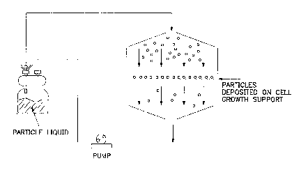
Figure 6 depicts an embodiment of the invention
in which viral particles are deposited on cell growth
supports by connecting a container producing viral
particles to a chamber cont~;n;ng a porous cell growth
= 25 support in which medium is recycled by means of a pump.
CA 02206600 1997-0~-30
WO 96/17948 PCTIUS95/15914
DET=A-TTl~n DESCRIPTION OF T~E lNv~NlION
The invention provides a new method that
dramatically increases the transfection efficiency by
increasing the contact between particles and target cells.
The contact is increased by localizing particles on a cell
growth support and directing target cells to contact the
particle-loaded cell growth support. As broadly claimed,
the method comprises two steps. First, the particles are
deposited on the cell growth support by various means such
as filtration or absorption. Second, the target cells are
directed to the particle-loaded cell growth support by
various means such as gravity se~ tation or filtration.
Localizing the particles on the cell growth support
increases the contact between particles and target cells,
lS which increases the transfection efficiency compared with
that achieved by particles in suspension. Moreover, this
method retains the infectivity of viral particles.
The method has broad application as any
transfection particle can be used. As used herein, the
term "particle~ means the combination of any carrier and
any foreign gene(s) used to transfect target cells.
Carriers include, for example, viruses, liposomes,
spheroplasts, red blood cell ghosts, colloidal metals,
calcium phosphate, DEAE Dextran and plasmids. Viral
carriers include DNA viruses, such as SV40, polyoma,
adenovirus, Epstein-Barr, vaccinia, herpes simplex,
papilloma particles and baculovirus, and RNA viruses, such
as tobacco mosaic virus, cucumber mosaic virus, brome
mosaic virus and retrovirus. An embodiment of the
retrovirus is a replication-incompetent retrovirus
including, for example, replication-incompetent
retroviruses produced by packaging cell lines ~2, ~AM,
PA12, PA317, PG13, Clone 32, GP+E-86, ~CRIP, ~CRE, D17-C3,
DSN, DAN, PHF-G, Isolde, Q2bn/Q4dh. As used herein "viral
CA 02206600 1997-0~-30
Wos6/17948 PCT~S95/15914
particle" means a particle cont~; n i n~ a viral carrier and
any foreign gene.
Any foreign gene includes any gene(s) wanted for
transfection and ~ ry nucleic acid sequences. The
foreign gene and/or ~llxiliAry nucleic acid sequences can be
either DNA and/or RNA. ~ ry nucleic acid sequences
are any nucleic acid sequences necessary or which improve
transfection, expression and/or detection of the foreign
gene. All~ ry nucleic acid se~uences include, for
example, expression elements, promoters, e~h~ncers and
homologous recombination sequences.
Particles are assembled using any method
appropriate for the carrier and foreign gene. Diverqe
methods of assembly are known including, for example,
simple precipitation of the foreign gene as DNA with
calcium phosphate, encapsulation of the foreign gene within
liposomes, adsorption of the foreign gene on colloidal
metal particles or genetically engineering the foreign gene
into the genome of viruses. Commonly used particles
suitable for this method and their methods of assembly are
described in many genetic engineering articles including,
for example, Kaufman, R.J. "Vectors Used for Expression in
M~ lian Cells~ in Methods in Enzymology Gene
Expression Technology Ed by D.V. Goeddel, Pub. by Academic
Press, Inc., San Diego, 185:487-511 (1990), Keown, W.A. et
al., "Methods for Introducing DNA into Mammalian Cells," in
Methods in Enzymology~ Gene Expression TechnoloqY Ed by
D.V. Goeddel, Pub. by Academic Press, Inc., San Diego,
185:527-537 (1990), Kriegler, M. Gene Transfer and
Expression A Laboratory Manual, Pub. by W.H. Freeman and
Company, New York, pp 3-81 (1990), Davision, A.J. and
Elliott, R.M. Molecular Virology, A Practical Approach,
IRL Press at Oxford University Press, Oxford, (1993) pp
171-198.
CA 02206600 1997-0~-30
WO 96/17948 1 ~ 35ll59l4
The method has broad application also because any
target cell can be used. As used herein, the term "target
cell" refers to cells that can be infected by a particle
and contact the particle-loaded cell growth support.
Target cells include procaryotic and eukaryotic cells.
Eukaryotic cells include plant, insect and animal cells.
An embodiment are cells useful for gene therapy, either for
human or ~ni~l use, and include, for example, bone marrow
cells, lymphocytes, fibroblasts, keratinocytes,
hepatocytes, endothelial cells, neurons, muscle cells, and
epithelial cells. Preferred human gene therapy target
cells include HSCs. Examples of target cells and their use
in gene therapy are discussed in several articles
including, for example, Mulligan, R.C. "The Basic Science
of Gene Therapy" Science 260:926-932 (14 May 1993),
Bluestone, Mimi, "Genes in a Bottle," Bio/Technoloqy
10:132-136 (February 1992), Krauss, J.C. "Hematopoietic
stem cell gene replacement therapy," Biochimica et
Biophysica Acta. 1114:193-207 (1992), and Verma, Inder, M.
"Gene Therapy" Scientific American 84:68-74 (1990). One
can readily employ other particles and target cells not
named but known to the art.
As used herein, the term ~cell growth support"
means any sterile material on which particles can be
deposited ex vivo and is compatible with subsequent ex vivo
and/or ln vivo transfection of target cells. An embodiment
of the cell growth support is disposable. Examples of cell
growth supports include filters, membrane filters, cell
culture surfaces, and tissue engineering materials. A
filter is any porous material capable of collecting
particles in suspension and supporting target cells. An
embodiment of the filter is that at the bottom surface of
a cell culture cup such as COSTAR TRANSWELL insert, FALCON
CLYCLOPORE, cell culture insert, NUNC ANOPORE and
polycarbonate TC inserts, or ~TTTTPORE MILLICELL insert.
Filters can be made of any material compatible with target
CA 02206600 1997-0~-30
WO96/17948 PCT~S95/15914
11
cell transfection, including for example, polyethylene
terephthalate, polystyrene or polycarbonate. Filters also
can be coated with any material compatible with target cell
transfection including for example, collagen or polycation.
Polycations include, for example, polybrene, protamine or
polylysine. Filters can be treated in any manner
compatible with target cell transfection including, for
example, plasma discharge. Any filter with a pore size
capable of supporting target cells can be used, especially
a pore size of approximately 0.l ~m to about 2.0 ~m
diameter. A membrane filter is a thin filter made of
synthetic or natural materials. An embodiment of the
membrane filter is made of nitrocellulose or other
cellulose esters. An embodiment of the membrane filter is
dialysis membrane made of either synthetic or naturally-
derived materials such as intestinal segments. See, for
example, Grass, G.M. and S.A. Sweetana, "In Vitro
Measurement of Gastrointestinal Tissue Permeability Using
a New Diffusion Cell" Pharmaceutical Research 5(6):372-376
(1988). A cell culture surface is a solid surface to which
particles can be localized and on which target cells grown.
An embodiment of the cell culture surface is made of glass
or plastic which may or may not be coated or treated as
above. An embodiment of the plastic cell culture surface
includes those dishes, plates, flasks, bottles and hollow-
fiber cell culture systems produced by various
manufacturers for growing cells including COSTAR, NUNC and
FALCON. Tissue engineering materials are materials designed
to replace or create new tissues, products or functions in
an animal. Tissue engineering materials are diverse and
include synthetic materials, natural materials and
combinations of both. Tissue engineering materials can be
porous or solid and can be designed to remain permanently
or temporarily in an ~n;m~l after implantation. Examples
of tissue engineering materials include hollow fibers,
synthetic polymer scaffolds, microcapsules, sheaths, rods,
discs, dispersions and feeder layers. Applications using
CA 02206600 1997-0~-30
WO 96/17948 ~ 95ll59l4
12
tissue engineering materials include, for example, hip
replacements, organ transplantations and skin replacement
in burns. One application of tissue engineering materials
is to provide a substrate on which to grow cells ex vivo to
replace damaged or missing tissue in the animal. See, for
example, Langer, R. and J.P. Vacanti, ~Tissue Engineering"
Science 260:920-926 (14 May 1993).
The first step of this method is to deposit
particles on the cell growth support. Particles can be
deposited by any means which localizes particles to the
cell growth support and preserves the ability of the
particles to transfect target cells. Examples of
deposition methods include, but are not limited to,
adsorption, absorption, non-covalent chemical bonding,
centrifugation, aerosol dispersion, electrophoresis or
combinations thereof. Adsorption collects particles on the
surface of a cell growth support from fluid cont~ining the
particles. An example of adsorption deposition is
filtration adsorption which is the collection of particles
on a filter or membrane filter as fluid cont~; n; ng the
particles flows through the filter or membrane filter.
Adsorption from a stagnant liquid or liquid agitated by
l~m; n~r lateral or circular fluid motion is specifically
excluded. Non-covalent chemical bond deposition collects
particles by binding particles to chemical binders attached
to the cell growth support. The chemical binders include,
for example, polycations, antibodies, and adhesion
molecules and may or may not be covalently attached to the
cell growth support. Polycations include, for example,
polybrene, protamine sulfate, and polylysine. Antibodies
include, for example, antibodies or fragments thereof which
bind retrovirus antigens, especially the env antigen.
Adhesion molecules include, for example, collagen and
fibronectin polypeptides or derivatives thereof.
Centrifugation collects particles by accelerating particles
through the liquid and onto a cell growth support. An
CA 02206600 1997-0~-30
WO 96117948 PCT/US9!i/lSgl4
13
example of centrifugation deposition is spinning at
sufficient speed a container contA;n;ng a solid cell growth
support and liquid cont~;n;ng particles such that the
particles collect on the solid cell growth support.
Aerosol dispersion collects particles by applying very
small micro-liquid droplets cont~;n;~g particles onto a
cell growth support. An example of an aerosol dispersion
deposition is a pressurized system including an aerosol jet
that sprays particles on a cell growth support.
Electrophoresis collects particles by electro-plating
particles onto a cell growth support. A particle-loaded
cell growth support is cell growth support on which
particles have been deposited.
Particles are deposited on the cell growth
support in an amount effective for increasing the
transfection efficiency of target cells relative to that
achieved by particles in suspension. The 'amount
effective" is a particle density greater than that
resulting from particle adsorption due to Brownian motion.
In preferred embodiments, the amount effective for the
claimed invention is a particle density approximately 2, 5,
10, 100, or 1000 fold greater than that resulting from
adsorption due to Brownian motion. An estimate of the
m~; mllm particle density due to Brownian motion of
approximately 1. 6 X 10⁵ to 1. 6 X 10⁶ particles per cm2 can
be calculated for particles having a short half-life. The
calculations assume particles with an initial concentration
of 107 or 108 particles per cubic centimeter (cc), a
diffusion coefficient of 4.5 x 10⁻⁸ cm2 per sec and a half-
life of 5 hours. The calculation boundary conditions are
that the initial concentration is the concentration at time
zero, there is an exponential decay of virus in the
suspension and complete and instantaneous adsorption of the
particles occurs at the surface. The calculated estimate
is a maximum density whereas the actual density may be
less. To achieve a particle density greater than that due
CA 02206600 1997-0~-30
WO 96117948 PCI'IUS95/15914
14
to Brownian motion, the liquid flow rate towards the cell
growth support must result in a Peclet number greater than
unity. The calculations are based on established methods,
for example, R.B. Bird, W.E. Steward and E.N. Lightfoot,
Transport Phenomena, Wiley & Sons (1960). For the
definition of Peclet number see, for example, B.O. Palsson
in Activation Metabolism and Perfusion of the Heart, S.
si~ n and R. Beyar, Eds., Marinus Nijhoff Pub., pp. 594-
596 (1987).
"Localized" as used herein means the particles
are in physical contact with the cell growth support and
accessible to target cells. The number of particles on a
cell growth support can be assayed by s~An~Ard methods
currently used to assay viral titers including, for
example, radioactivity assays and electron microscopy
particle counting assays and/or calculated based on
diffusion or mass transport theory. Transfection
efficiency can be assayed using stAn~Ard CFU assays.
Optimizing these parameters is routine work not requiring
undue experimentation. Comparisons between particles on
the cell growth support and in suspension are made using
the equivalent conditions, for example, the same target
cell concentration or density, age of target, temperature
and transfection time.
In the second step of the method, target cells
contact the particle-loaded cell growth support. For ex
vivo transfection, target cells are "directed" to contact
the particle-loaded cell growth support. Any means of
directing target cells to the particle-loaded growth
support can be used while transfection can occur. For
example, target cells can direct themselves to the
particles by naturally attaching to a cell growth support.
In another embodiment, the target cells are directed
gravitationally or centrifugally onto the particle-loaded
cell growth support. In another embodiment, the target
CA 02206600 1997-0~-30
WO96/17948 PCT~S95115914
cells are directed by filtering target cells onto the
particle-loaded porous cell growth support as fluid
contA;n;ng the target cells flows through the porous cell
growth support.
Although st~n~rd transfection culture conditions
are generally used, a significant advantage of the present
invention is that ch~mic~l additives are not required for
transfection. Until now, indirect transfection methods
required chemical additives for transfection. Chemical
additives, such as polybrene and protamine sulfate,
increase the proportion of particles entering target cells
after particle-target cell contact. Chemical additives,
however, are not always desirable and, for gene therapy,
pose a contAm;n~tion concern. In contrast, the broadly-
claimed invention does not require chemical additives
because excellent transfection efficiency results without
them. The broadly claimed method results in frequent
particle-target cell contact that eliminates the need for
m~;m-l particle entry into target cells after contact.
The frequent particle-target cell contact of the claimed
invention compensates for the infrequent entry of particles
into target cells that occurs without chemical additives.
Thus, the current transfection method offers, for the first
time, the option of transfecting target cells without
chemical additives. Other st~n~rd transfection culture
conditions, such as culture media, temperature and
accessory cells, used to transfect target cells by
particles in suspension are also used in the method of
particles deposited on a cell growth support. See, for
example, Cassel, A. et al, "Retroviral-m~ ted gene
transfer into CD34-enriched human peripheral blood stem
cells" Experimental Hematology 21:585-591 (1993), Kaufman,
R.J. "Vectors Used for Expression in Mammalian Cells" in
Methods in EnzYmology, Gene Expression Technoloqy Ed by
D.V. Goeddel, Pub. by Academic Press, Inc., San Diego,
185:487-511 (1990), Keown, W.A. et al., "Methods for
CA 02206600 1997-0~-30
WO 96/17948 ~ /15914
16
Introducing DNA into Mammalian Cells," in Methods in
Enzymoloqy, Gene Expression Technology Ed by D.V. Goeddel,
Pub. by Academic Press, Inc., San Diego, 185:527-537
(1990), Kriegler, M. Gene Transfer and Expression A
Laboratory ~AnllAl, Pub. by W.H. Freeman and Company, New
York, pp 3-81 (1990), Davision, A.J. and Elliott, R.M.
Molecular ViroloqY A Practical APProach, IRL Press at
Oxford University Press, Oxford, (1993) pp 171-198.
The term "liquid" as used herein is any free
flowing, nongaseous or non-solid material compatible with
particles and/or transfection of target cells. Different
liquids can be used at different stages of the transfection
method. For example, the liquid used for depositing
particles need not be the same as the liquid used during
transfection. Liquids include, for example, buffered and
osmotically controlled solutions, any cell culture medium,
and supernatant from packaging cells. As discussed above,
the liquid may include a chemical additive for improving
transfection such as a polycation. Examples of polycations
include, polybrene, or protamine sulfate, present at
approximately 4 to 10 ~g/ml. See, for example, Cornetta,
K and W.F. Anderson, ~Protamine sulfate as an effective
alternative to polybrene in retroviral-mediated gene-
transfer: implications for human gene therapy," J.
Virological Methods, 23:187-194 (1984).
Inhibitors in the supernatant contA;n;ng the
particles can be removed before the target cells contact
the particles. This capability offers significant
advantages for replication-incompetent retrovirus
transfection. Specifically, growth inhibitors present in
the supernatant from packaging cells can be removed before
the target cells contact the replication-incompetent
retrovirus. Some retroviral packaging cell supernatants
inhibit the growth of certain target cell types. For
example, commonly-used packaging cell line supernatants,
CA 02206600 1997-0~-30
WO 96117948 1 ~ Jb~;ll59l4
17
such as from PA317, ~CRIP, and PG13, inhibit growth of
primary human cell lines. Target cell growth is required
for retroviral transfection and, therefore, removing growth
- inhibitors increases target cell growth and transfection
efficiency. Inhibitors are removed by depositing the
particles on the cell growth support, removing the
supernatant and replacing the supernatant with a liquid
which does not contain the inhibitor and which is
compatible with transfection. Methods of removing
inhibitors include, for example, adsorbing or absorbing the
particles onto the cell growth support and washing the
particle-loaded cell growth support with a liquid not
cont~ining the inhibitors. See, for example, Paul, R.W. et
al. "Increased viral titer through concentration of viral
harvests from retroviral packaging lines," Human Gene
Therapy, 4:609-615 (1993).
Localizing particles on cell growth supports
allows transfection of target cells that would be difficult
or impossible to transfect by current methods. Many target
cells require co-cultivation with accessory cells in order
for the target cells to grow. For some target cell types,
target cells must grow between the cell growth support and
the accessory cells and the particles cannot contact the
target cells because accessory cells block access to the
target cells. However, a particle-loaded cell growth
support contacts the target cell because the target cell is
growing on the cell growth support. An example of target
cells having this property is the HSCs. HSCs can be more
efficiently transfected by particle-loaded cell growth
supports than by particles suspended in liquid because the
target cells contact the cell growth support.
After ex vivo transfection, the target cells can
be used in the culture or removed from the culture.
Various st~n~rd sterile techniques can be used to remove
target cells from the culture including, for example,
CA 02206600 1997-0~-30
WO 96/17948 1 ~ gS/15914
18
trypsin digestion to release attached target cells,
agitation and aspiration of media cont~;n;ng transfected
cells and gravitationally or centrifugally removing the
target cells from the cell growth support. An example of
a method for removing transfected cells from the culture is
to wash the culture with phosphate buffered saline, add
trypsin and EDTA, incubate, suspend and remove target
cells, inactivate trypsin and resuspend target cells in
growth medium. Cells transfected by this method can be
used for any purpose including use in fermentation,
research, agriculture, ph~rm~ceuticals and medicine. For
gene therapy, an embodiment of the application is to
~m; n; ster the transfected target cells to a patient to
treat a disease or condition.
The in vivo transfection of target cells is
achieved by implanting a particle-loaded tissue engineering
material into an An; m~ 1 . The physical contact of the
particle-loaded tissue engineering material with the body
tissue places the particles in contact with the target
cells. Selecting the site of implantation directs the
particles to specific tissues or organs. In one
embodiment, target cells attach to the particle-loaded
tissue engineering material ex vivo and the assembly of
target cells, particles, and tissue engineering material is
implanted into an ~n; r- 1 . In another embodiment the tissue
engineering material with attached particles is implanted
directly into an ~nir-l to transfect target cells in vivo.
See, for example, Langer, R. and J.P. Vacanti, "Tissue
Engineering" Science 260:920-926 (14 May 1993), especially
page 924, third column, third full paragraph and page 925,
first column, first full paragraph and Mulligan, R.C., "The
Basic Science of Gene Therapy," Science 260:926-932 (14 May
1993), especially page 931, first column, first full
paragraph.
CA 02206600 1997-0~-30
WO96117948 PCT~S95115914
19
The particle-loaded cell growth support can be
cryopreserved for later use. A particle-loaded cell
growth support is cryopreserved using stAn~rd methods
which preserve the ability of the particles to transfect
target cells. St~n~rd cryopreservation methods used for
viral particles in suspension can be used, for example,
freezing at -1~C/min. and storing the frozen particle-loaded
support at -70~C or colder. An embodiment is to add a
cryoprotectant to the particle-loaded cell growth support
before freezing, such as glucose, sorbitol or gelatin.
Just before use, a frozen particle-loaded cell growth
support is thawed using st~n~Ard methods used for thawing
viral particles, for example, rapidly thawing at 37~C.
Infectivity is always reduced because of the freezing and
thawing process. Cryopreservation and thawing is generally
adequate if transfection is retained after freeze/thaw. In
comparing transfection efficiencies from cryopreserved
particles attached to cell growth supports and suspended in
liquid, substantially the same freezing, storage and
thawing conditions must be used for both. In an
embodiment, frozen particle-loaded cell growth supports are
envisioned for commercial sale. See, for example, Elliott,
R.M. Molecular Virology, A Practical Approach, IRL Press
at Oxford University Press, Oxford, (l993) pp l7l-l98.
The particle-loaded cell growth support can also
be dehydrated and preserved dry for later transfection use.
A particle-loaded cell growth support is dried using any
method that preserves the ability of the particles to
transfect target cells. Drying methods include, for
example, lyophilizing or air drying. Lyophilizing can be
achieved by adding cryoprotectants, quickly freezing,
freeze-drying and storing the particle-loaded cell growth
support. Cryoprotectants include, for example, glucose,
sorbitol and gelatin. Rapid freezing can be achieved, for
example, in a dry-ice acetone bath. Lyophilization can be
achieved in st~n~rd freeze-drying apparatus such as, for
CA 02206600 1997-0~-30
WO 96117948 PCI'IUS95/15914
example, a MODEL FREEZEMOBIL LYOPHILIZER. Lyophilized
material can be stored at low temperature, 4 ~C or colder,
prior to rehydration and transfection.
Furthermore, apparatuses that allow easy-to-use
particle deposition on cell growth supports are developed.
The apparatus comprises particles in a liquid, a container
which has a cell growth support and means for causing the
liquid to move through or over the cell growth support. In
one embodiment, the liquid flows through a container having
a porous cell growth support, for example, filters having
pore sizes ranging from about 0.1 ~m to about 2.0 ~m, to
which the particles attach. The means for moving the fluid
through the cell growth support include gravitational,
centrifugal, vacuum and pumping means. In an e~oAim~nt~
particles in the fluid are produced by a separate particle-
producing container which is operationally connected to the
container which has the cell growth support. The particle-
producing cont~; ner can contain a cell culture that
produces particles, such as a viral particle producing cell
20 line. Specifically, such cell cultures could be done in
suspension, on suspended microcarriers or on solid
surfaces. In an embo~;m~t, the container that has the
cell growth support has a porous filter or membrane filter
and the fluid flows from the particle-producing container
to and through the container which has the filter or
membrane filter cell growth support. In one example, the
means for liquid flow is a liquid-absorbing matrix,
contacting one side of the porous cell growth support,
which pulls the fluid through the filter or membrane filter
by capillary action. A liquid-absorbing matrix is any
sterile material that can draw liquid through the porous
cell growth support. Examples of liquid-absorbing material
include sponges, cloth, and paper. In another example, the
means for liquid flow is any means that applies a partial
vacuum to one side of the porous filter or membrane filter.
A means for applying a partial vacuum includes any method
CA 02206600 1997-0~-30
WO 96/17948 1 ~ 9!illS914
21
that can produce a vacuum, for example, an aspirator,
roughing pump, peristaltic pump or combinations thereof.
In another embodiment, the particles contA;ne~ in the
fluid are recycled, after exiting the container which has
the porous cell growth support, back to the container which
has the porous cell growth support.
For clinical use, a fully-automated gene transfer
system is envisioned having the reproducibility and
documentation required by the United States Food and Drug
Administration. The system would allow automated
deposition of particles on cell growth supports, target
cell contact, transfected target cell purification and
target cell harvesting as needed for gene therapy
applications.
The following examples are intended to more-
clearly illustrate aspects of the invention, but are not
intended to limit the scope thereof.
EXAMPLE I
Contact between particles and target cells is
necessary for gene transfer to take place. Particles
suspended in a liquid move by random motion, called
Brownian motion. A typical prior art transfection method is
to overlay attached target cells with a liquid cont~;ning
particles. In this method, particle-target cell contact is
flln~A~?ntally limited by Brownian motion, a measure of
which is diffusivity. Moreover, even if the liquid is
gently agitated, contact r~m~;ns limited by Brownian motion
because of l~mi nar flow. The transfection efficiency in
these methods is expected to be directly proportional to
the concentration of particles in suspension, the
concentration of target cells, and increase with the
duration of time that the target cells are exposed to
transfection particles.
CA 02206600 1997-0~-30
WO 96/17948 ~ 3~ll59l4
22
These expectations are verified experimentally.
For the results of figure 2, African green monkey-derived
CV-l cell line (ATCC# CCL 70) is transfected by a murine
retroviral particles produced by a ~CRIP derived producer
cell line carrying the LacZ gene for B-galactosidase.
Figure 2a shows that for a fixed number of target cells and
transfection time, the transfection efficiency is directly
proportional to the concentration of viral particles.
Similarly, Figure 2b shows that for a given viral particle
concentration and transfection time, the transfection
efficiency is directly proportional to the density of
target cells.
Particles lose their ability to transfect target
cells over time. The loss occurs for various reasons
including, for example, particle disintegration. The half-
life measures the time in which half of the particles in a
population are lost. In suspension, the net distance that
particles can travel is limited by particle diffusivity and
half-life. For particles in suspension having a short half-
life, particles can only travel a very short distance.This distance is critical for transfection because only
particles within it can transfect target cells. The
critical distance can be estimated based on flln~mental
laws that govern Brownian motion as well as be
experimentally determined. For retroviral particles, the
critical distance is calculated to be about 380 to 440 ~m.
The calculation is based on retroviral particles traveling
for about one half-life, about 4.5 to 6 hours long, and a
diffusion coefficient of about 4.5 x 10⁻⁸ cm2/sec. After
four retroviral half-lives, corresponding to 760 to 880 ~m,
approximately 93% of the retroviral particles cannot
transfect target cells. For calculations see, for
instance, E.L. Cussler, Diffusion Mass Transfer in Fluid
Systems, Cambridge University Press, Cambridge ~1984).
CA 02206600 1997-0~-30
WO 96/17948 P~~ 59l4
23
Experimentally, the critical distance that
retroviral particles can move by Brownian motion and
transfect target cells can be shown to be less than about
500 ~m. In figure 2c, three depths of liquid contA;ning
equivalent concentrations of retroviral particles are
placed on top of adherent target cells for different
lengths of time. The three depths of liquid are 520, 832,
and 1559 ~m, and are denoted in figure 2c by different data
point symbols. Figure 2c shows that all three depths have
essentially the same transfection over time, showing two
important points. First, transfection does not increase
after about 12 to 15 hours or approximately 2 to 3
retroviral half-lives. Second, retroviral particles more
than 520 ~m away from the target cells do not significantly
transfect cells because greater depths of liquid do not
increase transfection. Therefore, the critical distance
that retroviral particles can diffuse and transfect target
cells approximately 520 ~m or less. Current methods
typically use a liquid depth of about 3000 ~m (3 mm) above
the target cells for transfection. These methods, thus,
waste the vast majority of retroviral particles because the
retroviral particles beyond the short critical distance
never transfect the target cells.
EXAMPLE II
Figure 3 shows the decay of retroviral particles:
(A) suspended in liquid; (B) on a cell growth surface
deposited by adsorption from stagnant liquid; and (C) on a
cell growth support deposited by filtration adsorption.
The half-lives for the three cases are 4.8, 4.2 and 4.6
hours, respectively.
The viral particles are replication incompetent
retroviral particles from a PA317/pMFG packaging cell line
and the target cells are NIH 3T3 murine fibroblast cells
(ATCC~ CRL 1658). Particles are deposited onto porous cell
CA 02206600 1997-0~-30
W O 96/17948 PC~rrUS9511S914
24
growth supports (COSTAR TRANSWELL insert, 0 .4 ym pore
diameter PE filter, cat. no. 345 0). Filtration particle
deposition is by vacuum filtering the fluid cont~in;ng the
particles through the porous cell growth supports using a
water aspirator. Stagnant particle deposition is done by
overlaying the porous cell growth support with fluid
cont~ining the particles for 2 hours. An equivalent
particle fluid volume cont~ining an equivalent number of
particles as for the particles in suspension is used to
particle-load the porous cell growth supports. An
equivalent number of target cells are used in both groups,
approximately 30,000 per insert, and allowed to naturally
attach to the cell growth supports. The same s~n~Ard
transfection fluid, including 4 ~g/ml polybrene, and
conditions are used for transfecting both groups. The
target cells are transfected for 24 hours.
The data of figure 3 has at least three important
ramifications. First, it shows retroviral particle half-
life is short and, as discussed above, the short half-life
severely limits the distance in which these particles can
travel in solution.
Second, figure 3 shows retroviral particles
deposited on cell growth supports by filtration deposition
have the same half life as particles in suspension or
deposited on cell growth supports by adsorption from
stagnant liquid. The half-life remains the same despite
the fact that an increased number of particles are
localized on the cell growth support by filtration
deposition. Generally, increasing retrovirus
concentration significantly reduces the retrovirus half-
life. The claimed invention, unlike prior art viral
concentration methods, produces the unexpected result that
locally concentrating retroviral particles on a cell growth
support do not reduce infectivity. See, for example,
Mulligan, R.C., "The Basic Science of Gene Therapy,"
CA 02206600 1997-0~-30
WO 96117948 r~.l/U~ s9l4
Science 260:926-932 (14 May 1993), see especially page 926,
last paragraph bridged to page 927.
Third, filtration deposition results in a Peclet
number exceeding unity. As a result, the effective amount
of particles deposited on the cell growth support by
filtration deposition is greater than that from stagnant
liquid. Particle-loaded cell growth supports made by
filtration deposition and stagnant liquid adsorption have
an initial transfection efficiency of 46% and 24%,
respectively.
EXAMPLE III
Table 1 shows the transfection efficiencies of
particles in suspension and particles deposited on a cell
growth support by filtration deposition. Approximately 24%
and 54% of the target cells are transfected by the
particle-suspension and particle-loaded cell growth support
methods, respectively.
The viral particles are replication incompetent
retroviral particles from a PA317/pMFG packaging cell line
and the target cells are NIH 3T3 murine fibroblast cells
(ATCC# CRL 1658). Particles are deposited onto porous cell
growth supports (COSTAR TRANSWELL insert, 0.4 ~m pore
diameter PE filter, cat. no. 3450) by vacuum filtering the
fluid contA;n;ng the particles using a water aspirator. An
equivalent particle fluid volume contA; n; ng an equivalent
number of particles as for the particles in suspension is
used to particle-load the porous cell growth support. An
equivalent number of target cells are used in both groups,
approximately 30,000 per insert, and allowed to naturally
attach to the cell growth supports. The same stAn~Ard
transfection fluid, including 4 ~g/ml polybrene, and
conditions are used for transfecting both groups. The
target cells are transfected for 24 hours. Transfection
CA 02206600 1997-05-30
WO 96/17948 ~ 35lls9l4
26
efficiency is the number of transfected cells divided by
total target cells times 100.
TABLE 1
TRANSFECIION METHOD TRANSFECTION EFFICIENCY (% of
TOTAL CELLS)
Particles in ~ H~
Experiment 1 23.2
Experiment 2 24.5
Average 23.9
10 P~ lc loaded cell growth support
Experiment 1 47 5
Experiment 2 59.4
AVERAGE 53-5
EXAMPLE IV
Table 2 shows transfection efficiency and
recovery of retroviral particles deposited on a porous cell
growth support before and after a freeze/thaw cycle.
Approximately 68% of the transfection efficiency is
recovered after freeze/thaw.
The retroviral particles are produced by the
PA317/pMFG retroviral packaging cell line. The retroviral
particles are suspended in st~n~rd cell culture fluid to
which 4 ~g/ml polybrene is added. The porous cell growth
support used is a COSTAR TRANSWELL 0 .4 ~m PE membrane
25 insert (COSTAR cat. no. 3450). The retroviral particles
are deposited on the inserts by vacuum deposition using a
CA 02206600 1997-0~-30
WO96/17948 PCT~S95/15914
27
water aspirator. Particle-loaded cell growth supports are
frozen by and stored in a -80~C freezer (REVCO) and thawed
24 hours later in 37~C cell culture medium. The recovery is
- the transfection efficiency after the freeze/thaw cycle
divided by the transfection efficiency before the
freeze/thaw cycle.
TABLE 2
EXP~.RTNI~.NTTRANSFECTIONTRANSFECTION % RECOVERY
EFFICIENCY EFFICIENCY
BEFORE AFTER
HAW FREEZE/I HAW
(% of TOTAL (% of TOTAL
CET~T~q) CELLS)
7.8 5.1 66
1 0 2 53.4 30.1 56
3 24.5 19.7 81
AVERAGE 68
EXAMPLE V
Tables 3 and 4 show the transfection efficiencies
of particles in suspension and particles deposited on cell
growth supports by filtration deposition with and without
the chemical additive, polybrene. The particle-loaded cell
growth supports without a chemical additive result in
transfection efficiencies as good as or better than that
- 20 achieved by prior art methods with a chemical additive.
This result is unexpected because the claimed invention is
the only method that does not require a chemical additive
for transfection.
CA 02206600 1997-0~-30
WO 96/17948 1 ~ 5/l59l4
28
The viral particles are replication incompetent
retroviral particles from a PA317/pMFG packaging cell line
contA;n;ng the lacZ gene and the target cells are NIH 3T3
cells. Particles are deposited as in Example III. An
equivalent number of target cells are used in all groups
and allowed to naturally attach to the cell growth
supports. For those groups contAining polybrene, 4 ~g/ml
polybrene is added to the DMEM with 10% FCS transfection
liquid used for all groups. The same stAn~Ard transfection
conditions are used for all groups.
The transfection efficiency is assayed by either
of two methods, flow cytometry or microscopic counting.
For flow cytometry, the FDG stain identifies infected from
non-infected target cells and cells analyzed using stAn~Ard
flow cytometer counting methods. For microscopic counting,
the X-gal blue stain identifies infected from non-infected
target cells and cells analyzed using stAn~Ard microscopic
cell ~A~inAtion and counting methods. The results
obtained by flow cytometry and microscopic counting are
shown in Tables 3 and 4, respectively. The results are
expressed as transfection efficiency.
TABLE 3
TRANSFECTION PARTICLES IN PARTICLE-LOADED
LIQUID SUSPENSION (~ OF CELL GROWTH
TOTAL CELLS) SUPPORT (% OF
TOTAL CELLS)
WITH POLYBRENE 16.8 30.0
WITHOUT POLYBRENE 1.9 19.8
CA 02206600 1997-0~-30
WO 96117948 PCI~/USg511S914
29
TABLE 4
TRANSFECTION PARTICLES IN PARTICLE-LOADED
LIQUID SUSPENSION (~ OF CELL GROWTH
TOTAL CELLS) SUPPORT (% OF
TOTAL CELLS)
WITH POLYBRENE 23.7 41.4
5 WITHOUT POLYBRENE 2.4 33.7
EXAMPLE VI
Figure 6 is a schematic diagram of an apparatus
used to particle load cell growth supports. The apparatus
includes a first container in which particles are deposited
on a cell growth support. The first container includes a
clamp which allows the container to opened and closed for
inserting an removing cell growth supports. The container
including clamp is made from machinable plastic, glass and
metal parts, including, for example polycarbonate,
polysufone and stainless steel, which can be sterilized.
The clamp forms a hermetic seal when closed which allows
liquid to flow through the container. The container
dimensions allow the cell growth support to fit within the
container. The cell growth support is a pre-sterilized,
porous, tissue culture well plate insert available from,
for example, COSTAR, ~TTTTPORE and NUNC. The first
container is operationally connected to a pump and a second
container cont~;ning particles in liquid. The pump is a
peristaltic pump which allows sterile liquid pumping. The
peristaltic pump is capable of producing sufficient
pressure to pump the fluid from the second container and
through the insert. The second container can be opened and
=
CA 02206600 1997-0~-30
WO 96/17948 PCI~/US9S/15914
subsequently hemetrically sealed. The second container can
be made of the same materials as the container cont~; n i ng
the insert. The second container contains sufficient
liquid to particle load the insert. The first and second
containers and pump are connected by tubing which can be
sterilized, for example, TYGON tubing. The apparatus,
first and second containers, pump and tubing, is sterilized
prior to use using conventional sterilization methods such
as an autoclave and alcohol wipe down. Liquid cont~; n; ng
particles and inserts are placed in the apparatus in a way
which preserves the sterility of the liquid, inserts and
apparatus, such as, for example, in a tissue culture hood
using sterile technique.
All articles cited herein are expressly
incorporated herein by reference.