Note: Descriptions are shown in the official language in which they were submitted.
CA 02208446 1997-06-20
WO 96/20013 PCT/GB95/03008
Title: Improvements in or Relating to Endometrial Function
Field of the ~nvention
This invention relates, inter alia, to a method of altering one or more characteristics of
mammalian endometrial tissue.
.
Background of the Invention
Endometrial physiology
Two major events are required for the embryo to become established in the mammalian
uterus; firstly, the preparation of the endomelrium so thal it is receptive to the presence
of a blastocyst which can then implant and acquire nutritive support through the formation
of the placenta; and secondly, the modification of myometrial activity which must become
quiescent and thereby allow the blastocyst to become resident within the uterine cavity
without the danger of expulsion. Both these events are controlled by the action of the
hormones of pregnancy. of which oestrogens and progesterone are particularlv important.
These steroid hormones act on the endometrium and myometrium through their receptors
which are located in the nucleus of tar_el cells Once activated, the steroid-nuclear
receptor complex interacts with specific regions within the DNA to stimulate, repress or
de-repress genes that code for proteins and polypeptides such as enzymes or growth
factors .
The initiation of implantation is brought about by a cascade of biochemical and biophysical
changes. Adhesion molecules (e.g. C~\~ I05) have been implicated in the early stages
ol attachment of the blastocyst to the wall of the uterus. Afterwards, the blastocyst and
endometrium adopt various stratagems to improve intimacy between fetal and maternal
~ tissues. In ungulates~ trophoblast cells which form the outermost layer of the blastocyst
migrate into the uterine epithelium with which they subsequently fuse. Cell migration is
carried a step further in women because it is not only isolated or specific cell types that
migrate but large areas of trophoblast which insinuate between the uterine epithelial cells.
In order to do this, some of the trophoblast cells fuse together to form a syncytium The
SUBSTITUTE SHEET (RULE 26)
CA 02208446 1997-06-20
W 096/20013 PCT/GB95/03008
process is very rapid and the embryo becomes established in the uterine tissues without
much apparent degeneration in the uterine epithelium. In some species the process of
implantation is delayed, either to await environmental cues which ensure the young are
born at favourable times of the year, or by physiological factors such as lactation so that
the mother has finished weaning the previous litter before the next pregnancy becomes
fully established.
The preparation of the uterus for implantation is regulated by the secretion of ovarian
hormones. The transport of the fertilised egg through the oviduct has to be precisely
timed so that it arrives in the uterus at the correct time of development and when the
uterus is in a fit condition to receive it. Under most conditions the uterus is hostile to the
embryo, more hostile in fact than some other areas of the body. The epithelial lining of
the uterus is, under most conditions, resistant to ~tt~hment and invasion by trophoblast
and it is only under very precise hormonal states that this resistance is relaxed.
In mice and rats nnm~ted ~nim~lc do not have a full oestrous cycle because they do not
form a normal secretory corpus luteum which produces increasing amounts of
progesterone. If mating occurs at oestrus, a time when high levels of oestrogens are
secreted by the ovarian follicle from which the ovum is shed, a corpus luteum will form
in the place of the ruptured follicle, rising concentrations of progesterone are then
secreted, implantation occurs and pregnancy progresses (length, 21 days). If an infertile
mating occurs, similar events occur except that the corpus luteum only lasts about 11 days
and pseudopregnancy is curtailed.
The cellular and biochemical changes that take place in the endometrium have been most
thoroughly studied in the mouse and the rat, though information about these aspects in
women has increased substantially in recent years. The endometrium in all species is made
up of three main tissues - luminal epitheliurn, glandular epithelium and stroma. Cell
proliferation occurs at different times in the three tissues. T l~min~l cells proliferate just
before oestrus (proestrus~~under the influence of the rising levels of oestrogens produced
by the follicles in the ovary. By day 1 of pregnancy (day of copulation plug in rodents)
they have ceased division but then undergo a second, though smaller, burst of activity on
CA 02208446 1997-06-20
WO 96t20013 PCT/GB95/03008
day 3. Gl~n~ r cells show most activity on day 4 and then decline. C,tromal cells do
not proliferate until day 4 but thereafter, under the influence of progesterone, they reach
high levels of proliferation by day 5. In women, less is known of these changes which
presage the process of implantation but there appears to be peak proliferation in epithelial
cells during the follicular phase of the cycle and in stromal cells during the luteal or
secretory phase, as in the mouse and rat.
The purpose of endometrial cellular proliferation is not fully resolved. It is believed to
prepare the endometrium for implantation by increasing the number of cells that will serve
a nutritive and secretory function (glandular epithelium) and that participate in the very
early stages of placentation (deci.1..~1i7~tion). As a prerequisite of succes~ful implantation,
cell mitosis may progress towards cellular differentiation and therefore plays a crucial role
in the early events of the establishment of pregnancy. Evidence in support of this role is
the endometrial production of growth factors (mitogens), cytokines and nuclear oncogenes.
Many of these compounds are produced in increased concentrations in response to ovarian
hormones acting through their receptors.
Amongst growth factors, much attention is currently given to epidermal growth factor
(EGF), heparin binding epidermal growth factor (HBEGF), amphiregulin and
insulin-binding growth factors (IGF-I and IGF-II). Evidence for the importance of the
local (paracrine) action of at least one of these growth factors, amphiregulin, has been
provided by recent ~elhllents in mice. Inhibition of the implantation-specific and
progesterone-regulated gene for amphiregulin was achieved by the anti-progestin, RU486,
and this resulted in the prevention of implantation (Das et al. Molecular Endocrinology
9, 691-705, 1995).
Amongst the cytokines, leukaemia inhibitory factor (LIF) and colony-stimulating factor
(CSF), which are also produced by the mouse uterus at the time of implantation, have
been found from gene knockout studies to be indispensable, demonstrating that their
removal is incompatible with implantation and normal placentation (Stewart et al. Nature
359, 76-79, 199~; Pollard et al. Developmental Biology 148, 273-~83, 1991).
CA 02208446 1997-06-20
Wo 96/20013 PCT/Gs95/03008
Amongst the nuclear oncogenes, levels of cjun and c-fos (which are early indicators of
gene transcription) increase in the uterus after oestrogen ~-lmini~tration, and are inhibited
by progesterone.
Important differences exist between various species in the extent of trophoblast invasion
at the time of implantation. In women, the early trophoblast is highly invasive whereas
in pigs, which have a non-invasive form of implantation, the endometrial epithelium is
never breached throughout the three month gestation period. Failure of implantation in
both these species is high, reaching about 60 and 30%, respectively. The reasons for this
high rate of failure are complex and incompletely understood. In women, about half the
loss is attributable to genetic abnorrnalities but in pigs, as in other ungulates where the loss
is also high, genetic defects only account for a few percent of the total.
After implantation failure in women a fall in progesterone secretion causes bleeding, as
at the end of the normal menstrual cycle; this does not occur in most other ~nim"l.c.
Disorders of menstruation, as well as of implantation, are common. In addition menstrual
bleeding, either as a consequence of sequential hormonal therapy, or in conjunction with
continuous combined hormone replacement therapy or progestin-only long-acting
contraceptives, is a significant cause of ill-health in women. The underlying reasons for
this bleeding are the focus of many current studies into biochemical (e.g. prostaglandins,
enzymes, polypeptides and proteins, vasoactive compounds such as platelet-activating
factor PAF, and vascular endothelial growth factor VEGF) and cellular mech"ni~m~ (e.g.
migrating cells homing to the uterus that produce immunosuppressive compounds).
Current underst"n~ing of reproductive processes largely centres on the control of steroid
hormone production and the actions of these hormones on their target tissues. However
paracrine and autocrine factors are increasingly seen to be key mediators of reproductive
function, albeit interacting with steroids (Benton, 1991 Current Opinion in Cell Biology
3, 171-175; Rozengurt, 1992 Current Opinion in Cell Biology _, 161-165; Tartakovsky
et al., 1991 Developmental Biology 146, 345-352; Robertson et al., 1992 Current opinion
in Immunology 4, 585-590; Smith, 1994 Human Reproduction 2, 936-946; and
Tabibzadeh, 1994 Human Reproduction 2, 947-967). The clearest example of this is seen
CA 02208446 1997-06-20
Wo 96/20013 PCT/GB95/03008
in the ovariectomi~ed mouse. In this model the uterus undergoes marked growth inresponse to a single dose of estradiol. This effect can be blocked by anti TGFa antibody
(TGF is "transforming growth factor") suggesting that the mitogenic effects of estrogen
in this tissue are mediated by TGF~x (Nelson et al., 1992 Endocrinology 131, 1657-1664).
Consequently medical intervention in Gynaecology is largely based on
steroidal/antisteroidal regulation of the uterus (Yen & Jaffe, 1991 in "Reproductive
Endocrinology", Eds. Yen, Jaffe & Benton, Pub. WB Saunders, Philadelphia; Baird, 1993
British Medical Bulletin 49, 73-87). Despite the undoubted success of this approach, no
conceptual advances in contraceptive technology have arisen for 20 years, no means
identified to improve implantation, no advances made in promoting placental growth and
development and no new approaches found to treat benign gynaecological disease
(menstrual dysfunction and fibroids).
A number of publications have been made in relation to the use of "gene transfer" in
m~mm~ to alter the genotype of at least some cells in a certain tissue or tissues. In
particular, it is known to attempt "gene therapy" of humans by the introduction into
recipients of nucleic acid sequences, with the aim of overcoming a genetic deficiency in
the recipient by the expression of polypeptides encoded by the introduced nucleic acid
sequences. Gene therapy trials have been conducted, for example, in which DNA
sequences (incorporated within viral vectors) were introduced into the airways of cystic
fibrosis patients, so as to alter the phenotype of at least some of the epithelial cells lining
the patients' respiratory tract. Thus far, there have been no published attempts to
introduce DNA into the m~mm~ n endometrium, despite the availability of suitabletechniques therefor.
Summary of the Invention
In one aspect, the invention provides a method of altering one or more characteristics of
at least some of the cells of the reproductive tract of a m~mm~ n individual by the
introduction into said cells of a nucleic acid.
In a second aspect the invention provides a composition comprising nucleic acid, for use
CA 02208446 1997-06-20
wo 96/20013 PCT/Gs95/03008
in altering one or more characteristics of at least some of the cells of the reproductive tract
of a m~mm~ n individual.
In a third aspect the invention provides for use of a composition comprising nucleic acid
for altering one or more characterstics of at least some of the cells of the reproductive
tract of a m~mm~ n individual.
In a fourth aspect the invention provides for use of a composition comprising nucleic acid
in the preparation of a substance for altering one or more characteristics of at least some
of the cells of the reproductive tract of a m~mm~lian individual.
In a fifth aspect the invention provides a method of maKing a composition for use in
altering one or more characteristics of at least some of the cells of the reproductive tract
of a m~mm~ n individual, comprising mixing a nucleic acid with a physiologicallyacceptable carrier substance.
The present invention can in no way be considered as an obvious extension of genetic
therapy techniques already known to be at least partially ~lccessful when applied to the
lungs of cystic fibrosis patients. Inherited genetic disorders are not thought to be
responsible for any of the known diseases of the endometrium, so there would have been
no incentive for those skilled in the art to apply gene therapy techniques to the
endometrium. Further, the epithelium of the endometrium is of a different type (cuboidal,
derived from coelomic epithelium) compared to lung epithelium {which is stratified) and
therefore could not have been predicted to behave in an analagous manner. Moreover, at
least in primates, there is cyclical shedding of the endometrial epithelium which would
tend to cause the loss of any transfected cells. Finally, the inventors have found that there
was no transfer of the introduced DNA into the organs of the mother, nor into the placenta
of the embryo, either of which might have occurred and could have caused practical
difficulties.
Typically the nucleic acid is introduced into a m~mm~ n female (preferably a woman)
and, in particular, into the endometrial cells thereof. Desirably the nucleic acid is
CA 02208446 1997-06-20
Wo 96/20013 PcT/Gs95/03008
introduced into the gl~n~ r epithelium of the endometrium. The nucleic acid may
encode a polypeptide which is already naturally synthesiced by the cells into which the
nucleic acid is introduced, such that the level of expression of that polypeptide is increased
via a gene dosage effect. Alternatively the method can be used to induce the cells to
express a polypeptide not previously synthesised in those cells. The polypeptide could,
for example, be an "artificial" recombinant polypeptide which does not exist in nature,
such as a chimeric polypeptide comprising, wholly or in part, functional domains from two
or more different proteins.
The nucleic acid is preferably DNA~ but one could seek to introduce RNA (either sense
or non-sense strands). An antisense molecule could be used to inhibit or otherwise
interfere with the expression of a polypeptide in the cells into which the nucleic acid is
introduced. The nucleic acid sequence introduced may be antisense RNA, or may be a
DNA sequence directing the synthesis, intracellularly, of antisense RNA. Another way
of achieving such inhibition is to introduce into the cells a sequence directing the synthesis
of a ribozyme, which will then specifically cleave the mRNA needed to synthesise the
polypeptide whose expression is sought to be inhibited.
The present inventors have found that the time of a~lminictration of the nucleic acid
(relative to the stage of the reproductive cycle) greatly affects the efficiency of uptake of
the nucleic acid. The inventors have found that, in general, in order to obtain the
optimum degree of uptake of the atlminictered nucleic acid it is necessary for the
~tlminictration to be made in the period following ovulation, up to and including the day
on which there is a peak of progesterone level in the blood. The progesterone level
normally peaks at around a similar time to the point at which an embryo, if present in the
uterus, could become implanted.
Thus, for example, the inventors have found that maximal uptake of ~t~minictered DNA
by mouse endometrium occurs at day ~-3 in the cycle ~with day 1 taken as the day on
which a vaginal plug is first detected). In humans, ovulation typically occurs at day 14
of the cycle, and implantation is generally reckoned to occur in the mid-luteal phase
(although the exact time is poorly defined in humans).
CA 02208446 1997-06-20
W 096/20013 PCT/GB95/03008
The nucleic acid may be ~-iminictered in a naked form, or may be bound or associated
with other substances (e.g. liposomes). Conveniently the nucleic acid is introduced into
the cells of the recipient m~mm~l by simple transfection (with or without liposomes),
which has been found by the present inventors to be surprisingly effective, without the
need for the sequence to be introduced within a viral vector. Nevertheless, viral vectors
may be desirable, especially those which may be targeted to certain cell types (e.g. as
disclosed in WO 93/20221).
The nucleic acid will conveniently be introduced as part of a construct (e.g. a plasmid,
cosmid or the like), which construct will advantageously comprise a promoter, operable
in a m~mm~l, to cause transcription of at least part of the introduced nucleic acid. The
promoter may be constitutive or, more preferably, inducible so as to allow greater control
of expression of the introduced sequence.
In one particular method performed in accordance with the invention, introduction of a
nucleic acid molecule into the endometrial cells of an individual m~mm~ n female allows
for the up- or down-regulation of the fertility of the individual. The invention may
particularly be used to provide a method of contraception for companion ~nim~l~ (e.g. cats
and dogs) to prevent unwanted litters. In other embodiments the invention provides a
method of improving the fertility of livestock species, such as pigs, cattle, sheep and the
like.
Preferably the nucleic acid is introduced into the reproductive tract via the vagina, which
avoids the need for invasive surgical techniques. However, if necessary, the nucleic acid
could be introduced by means of surgical techniques directly into the reproductive tract
(e.g. into the uterus). The invention offers the possibility of altering one or more
characteristics by the introduction of one or more of a very large number of different
nucleic acid sequences.
In one embodiment, the sequence introduced into the reproductive tract cells directs the
expression (preferably at high levels) of an effective portion of a cytokine or growth factor
(an effective portion is that part of the molecule which retains the biological activity
-
CA 02208446 1997-06-20
W 096/20013 PCT/GB95/03008
particularly associated with the whole e.g. binding to a specific ligand etc.). Examples
of such polypeptides which might be expressed by the introduced sequence include, but
are not limited to, the following: interleukins, leukaemia inhibitory factor (LIF), vascular
endothelial growth factor (VEGF), epidermal growth factor (EGF), heparin-bindingepidermal growth factor (HBEGF), insulin-binding growth factors I and II (IGF-I and IGF-
II), amphiregulin, colony stimulating factor (CSF), and tumour necrosis factor (TNF).
In another embodiment the introduced sequence may direct the expression of an effective
portion of an antagonist of a cytokine or growth factor, such as the IL-1 receptor
antagonist. Advantageously, the antagonist may be a soluble receptor for the cytokine or
growth factor. Suitable examples include soluble receptors for the following: transforming
growth factor (TGF) a, fibroblast growth factor (FGF), platelet-derived growth factor
(PDGF), interleukin-6 (IL-6), and VEGF.
In another embodiment the introduced sequence may direct the expression of an effective
portion of a polypeptide having an immunolo~o,ical effect. In particular, the polypeptide
may possess immunogenic activity, thereby serving to stimulate a local immune response.
thus the invention can be used to provide a novel method of immunisation.
Advantageously the immunogenic polypeptide will be an antigen from a mucosal pathogen.
By virtue of the common mucosal immune system, stimulation of antibody production in
the reproductive tract may result in the production of corresponding antibodies at distal
mucosal sites, such as the gastro-intestinal tract, the respiratory tract, lachrymal glands
and the like. Preferably however the antigen will be one from a pathogen which invades
and/or colonises the reproductive tract, typically a pathogen which causes a sexually
transmitted disease. ~xamples include viruses such as HIV, papilloma viruses (e.g. HPV,
of various types), chlamydia and bacteria (e.g. N. gonorrhoea). Alternatively, the
polypeptide having an immunoloigical effect may be an immunoglobulin or effective
portion thereof (such as an Fab, Fv, or scFv fragment, or a single chain antibody). The
immunoglobulin or effective portion thereof may be directed against a pathogen (such as
those mentioned above), or may be directed against some other antigen, such as a steroid
or other hormone. Thus immunoglobulins or fragments thereof could be expressed locally
to provide protection against disease or to regulate fertilty.
CA 02208446 1997-06-20
W 096/20013 PCT/GB95/03008
In another embodiment, the introduced sequence may direct the expression of a
polypeptide, or an effective portion thereof, which has an effect on menstruation.
In another embodiment the introduced nucleic acid may direct the expression, on the
surface of the reproductive tract cells, of an effective portion of a receptor molecule. The
receptor could be a receptor for a cytokine, a steroid hormone, or a growth factor (such
as the EGF receptor, the TGFc~ receptor, or the VEGF receptor). A number of receptors
are known which are described as "orphan" receptors, in that the ligand which binds to
the receptor is unknown. Such orphan receptors are of considerable interest to the
ph~rm~ceutical industry, as they may provide targets for novel therapeutic or prophylactic
compounds.
Accordingly, in another aspect the invention provides a method of characterising the
biological properties of a polypeptide, comprising introducing the sequence encoding the
polypeptide to be characterised into the cells of the reproductive tract of a m~mm~l, and
~se~ing the effects of the expressed polypeptide. Preferably the m~mm~l is a laboratory
animal, such as a mouse or rat. Conveniently, the polypeptide to be characterised will be
an orphan receptor and typically at least part of the characterisation thereof will comprise
identification of the ligand therefor. Generally the method will involve the analysis of
histological sections taken from the laboratory m~mm~, and processing thereof by any one
of various standard techniques (e.g. histochemical staining, in situ hybridisation,
immunological staining etc.).
The present invention thus offers a novel alternative to steroidal regulation of endometrial
function (and thus reproductive capacity or fertility) by direct gene transfer in vivo. To
achieve this, genetic constructs would be designed to specifically modulate cytokine action.
This can be achieved in a variety of ways. For example the cells that produce a secreted
cytokine could be prevented from synthesizing the factor by blocking transcription and
translation using promoter driven antisense constructs or ribozymes. Alternatively the
action of the secreted factor can be blocked by receptor antagonists. Naturally occurring
soluble receptors may scavenge and neutralize bioactive ligand thereby acting ascompetitive receptor antagonists. Alternatively there are natural receptor antagonists, for
-
CA 02208446 1997-06-20
W 096/20013 PCT/GBg5/03008
11
example ILlRa (interleukin-1 receptor antagonist). Intraperitoneal ~lmini.ctration of this
protein blocks blastocyst implantation in the mouse (Simon et al., 1994 cited elsewhere).
There is considerable evidence to show that soluble growth factors secreted by the oviduct
and uterine epithelium can control pre-implantation development of the m~mm~ n
embryo, by acting directly through receptors expressed on the embryo (Pampfer et al.,
1990 In Vitro Cellular and Developmental Biology '~6, 944-948). In turn, developing
embryos produce growth factors which may act in an autocrine fashion, or on the
endometrium to influence its receptivity. For example, in mice, LIF expression (from
maternal tissues) is dramatically upregulated in glAn~ r epithelium on day 4, just prior
to implantation. LIF is able to act on pre-implantation blastocysts, which express the LIF
receptor (LIF-R). This maternal expression of LIF is vital for implantation since in LIF
knockout mice, embryos will not implant, although they will do so on transfer topseudopregnant dams (Stewart et al., 1992 cited elsewhere)
The inventors have now extended this work to humans, and shown by RT-PCR that human
embryos express the mRNA encoding the LIF-R, but do not themselves express LIF. LIF
acts by binding to a low affinity receptor LIF-R. High affinity binding arises when the
LIF/LIF-R complex interacts with the signal transducing accessory protein gpl30. Human
embryos also contain mRNA encoding this protein (Sharkey et al., 1995 Biology ofReproduction 53, 955-96~). The inventors have also shown LIF secretion in human
glandular epithelium is regulated by steroids. being m~xim~l in the luteal phase (around
the expected time of implantation - Charnock-Jones et al., 1994 cited elsewhere).
Furthermore, ~mini~tration of LIF to human pre-implantation embryos in vitro, has been
reported to improve development. All this evidence supports the idea that LIF may be
important in human implantation as it is in the mouse. Clearly cytokines may mediate
important communication between the embryo in the uterine lumen, and the endometrium
(in both directions). The present invention allows the use of gene transfer to disrupt or
enhance this communication, leading to novel methods of contraception, or conversely
improved implantation.
Most current studies of the paracrine and autocrine regulation of reproductive function are
CA 02208446 l997-06-20
W 096/20013 PCTIGB95/03008 12
limited to a descriptive analysis by the lack of effective methods to modulate local
cytokine/receptor levels. The evidence presented in this application indicates that
transfection of uterine epithelium in vitro iS feasible. This allows the endometrium to be
manipulated experimentally and offers new therapeutic strategies. The work outlined
below describes the use of a reporter gene to demonstrate the practicability of in vivo
uterine gene transfer. In practice a gene (or other DNA construct), able to alter uterine
function, would be used. Examples of these include receptor antagonists {e.g. IL-lRa,
soluble VEGF receptors etc.) natural or modified cytokines and growth factors, protease
inhibitors or steroid receptors and a variety of ribozyme and antisense constructs. This
work shows that genes can be transferred to the endometrium in vivo and this will find
utility in many endometrial (and placental) conditions for example improving implantation
in both ~nim;~l~ and man, disrupting implantation (i.e. contraception), endometriosis and
menorrhagia, hyperplasia and adenocarcinoma.
Using the protocols we have developed the results described below were obtained. They
demonstrate that gene constructs can be transferred to the endometrium both in vivo (in
mice) and in vitro and that these constructs are transcriptionally (and translationally)
active.
The invention will now be further described by way of illustrative example and with
reference to the accompanying drawings, in which:
Figure lA and lB show photomicrographs of histological sections of mouse endometrium
transfected with (A) a plasmid construct directing the expression of a ~B-g~l~c-tosidase
reporter gene, or (B) a similar plasmid lacking the reporter gene. Transfected cells can
clearly be identified by the intense dark (blue) staining within the cytoplasm, which is
absent in section B;
Figure 2 is a photomicrograph of in vitro human endometrial cells which have been
transformed with a the same plasmid as in Figure lA - the dark (blue) staining due to
expression of the reporter gene is mainly associated with the rem~ining glandular
structure, while the surrounding cells stain more weakly;
CA 02208446 1997-06-20
Wo 96/20013 PCT/~h~J'~3008
13
Figure 3 is a bar chart showing the results of a CAT assay (in counts per tube) for
endometrial cells successfully transfected with a gene encoding chloramphenicol acetyl-
transferase (pcDNA3CAT) compared to cells transfected with a control plasmid
(pcDNA3); and
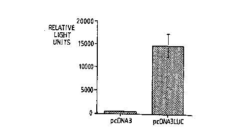
Figure 4 is a bar chart showing the results of a luciferase assay (in relative light units) for
endometrial cells s~lccec~fully transfected with a gene encoding luciferase (pcDNA3LUC)
compared to cells transfected with a control plasmid (pcDNA3).
Examples
Example 1
Mice
Nulliparous mature BALB!CJ mice were housed in a light (14h light:lOh dark; lights off
at 20.00h) and temperature (22~C) controlled Small Animal House and fed a mouse and
rat diet (Labsure; Christopher Hill Group, Poole, Dorset, UK). They were placed with
vasectomized males of the same stock ovemight and examined the next moming for the
presence of a vaginal plug. Mating was presumed to have occurred at 02.00h, time O and
day 1 was counted as the day on which the plug was first detected. Mated females were
housed individually prior to experimentation.
Laparotomy was performed using aseptic procedures under Metafane anaesthesia
(methoxyfluorane, C-Vet Ltd., Bury St. Edmunds). The uterine homs were exposed by
either mid-ventral or bilateral incisions. Injections were made either into the tip of the
horn at the tubo-uterine junction or at the base of the hom at the utero-cervical junction.
Repeated studies showed that the latter technique provided the best method of
~imini~tration but for some purposes the fommer was preferable when it was necessary to
minimi~e disturbance of the reproductive tract.
Injections of liposome/DNA (pcDNA3 construct, +/- p~-galactosidase reporter gene),
naked DNA or control solutions (25-100,ul) were performed by insertion of the tip of a flat
Stratatip into the base of the horn. Solutions were previously drawn up into the tip by
CA 02208446 1997-06-20
W 096/20013 PCTtGB95/03008 14
means of a Travesty applicator which was also used to control the slow injection of
solutions into the horn.
After injection, the incision was closed using interrupted mattress sutures and the mice
allowed to recover in their cages and provided with food and water ad libitum.
Plasmid constructs
The plasmids described in this application (by way of example) are based on the
commercially available vector pcDNA3 (Catalogue No. V790-'70, from Invitrogen, San
Diego, California, USA). Plasmid pcDNA3, without any reporter gene, was used as a
negative control. Experimental plasmids pcDNA3-~5gal, pcDNA3-CAT, and pcDNA3-Luccontained the ,B-galactosidase, chloramphenicol acetyltransferase and luciferase reporter
genes respectively. These plasmids contained the following genetic elements: Ampicillin
resistance gene, ColE1 original of replication, CMV promoter, [reporter gene], bovine
growth hormone polyA addition site, fl origin of replication, SV40 origin of replication,
neomycin resistance gene and an SV40 polyA addition site, in operable relationship such
that the reporter gene might be expressible in eukaryotic cells upon introduction of the
plasmid. Plasmid DNA was purified from E. coli by alkaline lysis and further purified
using a Qiagen ion exchange column (according to the m~n~lf~ctllrer's instructions).
Liposome preparation
The liposome used was a 3:1 (w/w) lipid formulation of DOPSA (2,3-dioleyloxy-N-
[2(sperminecarboxamido)ethyl]-N-N-dimethyl-1-prop~n~minium trifluoroacetate) andDOPE (dioleoylphosphatidyl ethanolamine) (LipofectAMINE; Gibco BRL Paisley,
Scotland). A number of DNA: lipid ratios and different injection volumes were used as
shown in table 1. The DNA/liposomes were mixed immediately prior to each experiment.
10 ,ul of DNA solution was added to 10 ,ul of lipid solution, mixed gently and left for 15
minutes at room temperature. 80 ,ul of PBS was then added to give the final concentration
of DNA and lipid as shown in table 1. This was then injected in to the uterus of the
pseudopregnant mice. (See section above for details of mice and surgery.)
CA 02208446 l997-06-20
W 096/20013 PCT/GB95/03008
Histochemical lor~ fion of ,l~-Galactosidase
Animals were killed by carbon dioxide inhalation and uterine horns flicsecte~l out, free of
fat and mesentery. Each horn was divided into 3 sections with the top and bottom section
being snap-frozen in liquid nitrogen, and stored at -70~C prior to quantification of ~5-
galactosidase content. The middle section of each uterine horn was fixed in 1.25%
gluteraldehyde in PBS for 15 minutes, rinsed in PBS twice, and placed in X-gal staining
solution (lmg/ml X-gal, SmM K3Fe(CN)6, SmM K,,Fe(CN)6, 2mM MgCl7, 0.02% NP40,
and 0.01% sodium deoxycholate) for 24 hours at room temperature. Sections were then
rinsed in PBS/3% DMSO (2 x 5 minutes), 70% ethanol (3 x S minutes) and placed in100% ethanol. Tissues were embedded in glycol methacrylate resin and 7,um sections
were cut and counterstained with neutral red prior to microscopic ex~min~tion.
Results
Table 1 below shows the various conditions employed and the resulting staining intensity
of uterine sections after ~lminictration of DNA/liposome complexes. The results shown
in the Table demonstrate the criticality of the timing of ~lminictration of the plasmid
DNA, with ~fiminictration on day 2 giving the best levels of expression, ~-lminictration on
day 3 giving reasonable levels, but a lminictration on day 4 resulted in very little
expression of the reporter gene, presumably because the endometrial cells would not take
up the construct at this time point, for reasons that are not entirely clear.
CA 02208446 1997-06-20
WO 96/20013 PcT/Gss5/03008
16
TABLE 1
Day of DNA LipidInjection volumeHistochemical
~Amini~tration Gug/ml) (,ug/ml)(ul) St~ining intensity
(autopsy day)
2(5) 2 20 50 + +
3(5) 2 20 S0 + +
4(6) ~ 20 50
2(6) 2 20 50 + + +
2(5) 8 20 20 + +
4(6) 8 20 50
4(6) 30 '~0 50 +
4(6) 2 60 50
Staining intensity + + + strong, + + moderate, + weak, - none
Controls:
An uninjected 6-day pseudopregnant mouse gave no uterine st~ining for ,B-galactosidase
activity.
A pseudopregnant mouse (~-lmini~tration day 2; autopsy day 6) injected with 50 ,ul of
pcDNA3 minus ,~-galactosidase (2 ,~cg/ml) and lipid (20 yg/ml) gave no uterine staining
for ,B-galactosidase activity.
F~min~tion of histological sections after staining with X-gal showed that the gl~n~ r
epithelium was strongly stained and the luminal epithelium was also stained but less
strongly. The optimal st~ining was seen in ~nim~l~ transfected with ~,ug/ml DNA and
20yg/ml lipid in 50,ul ~-lmini~tered on day 2 of pseudopregnancy. Fig. la shows a section
from such an animal and Fig. lb a section from a control animal which received (under
identical conditions) a plasmid which lacked the p~-gal gene.
CA 02208446 1997-06-20
WO 96/20013 PCT/GB95/03008
17
Example 2
Transfection of primary cultures of human endometrium
The inventors have also demonstrated that human endometrial epithelial cells can be
transfected in vitro at high efficiency.
The same plasmid (pcDNA3, +/- ,B-g~l~ctQcidase reporter gene) and lipids as already
described were used. Endometrial cells were prepared by the method of Smith and Kelly
(Smith et al., 1987 Prost~ n~inc 34, 553-561). Once the culture were established the
following transfection protocol was used. DNA (2,~cg) and liposome (8,ug) were each
diluted in 100,~cl of serum free medium (Opti-MEM1 BRL), mixed and incubated at room
temperature for 15 min. Following this a further 800,ul of Opti-MEM1 was added. The
cells (in 24 well plates) were washed with PBS followed by washing with Opti-MEM1.
The DNA/liposome mixture (0.5rnl) was then added to the cells and incubated at 37~C for
3hr in a C07 incubator after which 0.5ml of culture medium containing 20% foetal calf
serum was added. Cells were fixed (0.1% gluteraldehyde), rinsed and stained with X-gal
24h after transfection.
This work shows that genes can be transferred to the endometrium in vivo and this will
find utility in many endometrial (and placental) conditions, for example improving
implantation in both ~nim~lc and man, disrupting implantation (i.e. contraception),
endometriosis and menorrhagia, hyperplasia and adenocarcinoma.
Additional data were obtained relating to the transfection in vitro of purified human uterine
epithelial cells. This complements the in vivo mouse work and shows that similar cells,
after minim~l time in culture, can be efficiently transfected with the same liposome and
DNAs used in vivo.
Example 3
Transfection of human endometrial epithelium in vitro.
Human primary epithelial cells from endometrium were isolated and cultured according
to the method of Zhang et al.? (J Cell Science, 1995; 108:323-331). The cells were plated
CA 02208446 1997-06-20
W 096/20013 PCTIGB95/03008 18
in standard 6 well tissue culture plates to achieve a density of 50Yo confluence the next
day. The cells were cultured for 5 days, then replated into 24 well plates, at a density of
60,000 cells per well. The next day the cells were transfected with DNA/liposomecomplexes (DNA/LC). These were prepared as follows:
Transfection procedure
Apparatus
LipofectAMINE (Gibco Catalogue No. 18324-012), Opti-MEM I (Gibco Catalogue No.
51985-018)
24 well culture dish
Cell culture medium was as described by Zhang et al (cited above).
This consists of DMEM/HEPES, 10% FCS, Endothelial Cell Growth Supplement (Sigma
Catalogue No. E-2759), at 30,ug/ml heparin (Sigma Catalogue No. H-3149), at 90,ug/ml,
gentamycin (Sigma Catalogue No. G-127'~), at 5 ,ug/ml, and fungizone (Gibco Catalogue
No. 15290-018) at 1 ,ug/ml. Also used were Mg++, Ca++ free PBS and a 2 ml
Eppendorf tube.
Two different plasmid constructs were used containing different reporter genes.
pcDNA3CAT was obtained from Invitrogen Corporation, and contains a reporter geneencoding the enzyme chloramphenicol acetyl transferase. The second plasmid pcDNA31uc
comprised the same vector, but the CAT gene was replaced with the gene encoding the
firefly luciferase enzyme. Large scale DNA preparations of the vectors were made using
the Qiagen midiprep system. As a negative control, pcDNA3 containing no reporter gene
was used.
Preparation of DNA/Liposome complexes
1) Solution A
Dilute 1,ug of DNA into 1OO~LC1 Opti-MEM I in an Eppendorf tube. Use DNA at
1,ug/ml final concentration in the transfection medium.
CA 02208446 1997-06-20
W 096/20013 PCT/GB95/03008 19
2) Solution B
Dilute 4,ul of LipofectAMINE into 100,ul Opti-MEM I in an Eppendorf tube. Use
lipofectAMINE at 8,~g/ml final concentration in the transfection medium.
3) Combine two solutions A and B into a new tube and mix gently.
4) Incubate at room temperature for 15 minutes.
Cell rinsin~
1) Prior to transfection, rinse the cell monolayer roughly three times in FRESH PBS
without serum.
~) Re-rinse the cells monolayer twice with Opti-MEM I.
Transfection
1) Add 800f~1 (total 1.0 ml) of Opti-MEM to each tube cont~ining the DNA-lipidmixture. Final DNA concentration 1,ug/ml, and lipofectAMINE concentration
8,ug/ml.
2) Remove the Opti-MEM I in the cell monolayer.
3) Mix DNA/LC mixture gently and overlay the diluted complex solution onto the
washed cells, 0.5 ml/24 well.
4) Incubate the cell monolayer for 3 hrs at 37~C in a C07 incubator.
Further cell culture
1) 3 hrs later, remove transfection mixture, and add 2mls of the Zhang medium to each well, and culture further.
W 096/20013 PCT/GB95/03008
Quantitative assav
24 to 48 hours after transfection, the cells were extracted, and assayed for CAT or
luciferase reporter activity, as appropriate.
1. Rinse cells three times in PBS.
2. Extract cells with 300 microlitres of Lysis buffer (Promega Catalogue No. E-
3871), scraping cells off thoroughly into Eppendorf tube.
3. Freeze extract rapidly at -70~C, and store until assay.
4. To assay, thaw extracts and centrifuge at 13,000g for 5 minutes.
5. Repeat freeze/thaw/spin cycle once more.
6. Assay luciferase reporter gene activity using Luciferase assay kit from Tropix
(Catalogue No. BClOOL). Use 40,ul of each extract per tube.
7. CAT reporter gene activity was assayed using the Quan-t-CAT kit from Amersham (Catalogue No. TRK 1012).
Results
Primary endometrial epithelial cells were transfected in 24 well plates as described above.
Transfections were performed on triplicate wells with pcDNA3 (as control), pcDNACAT,
and pcDNA3LUC. After 48 hours the cells were harvested and assayed for luciferase or
CAT activity.
The CAT enzyme catalyses the transfer of acetyl groups from acetyl coenzyme A tochloramphenicol. Use of tritiated acetyl coA results in transfer of radiolabel to
chloramphenicol. The CAT activity in a sample is directly proportional to the amount of
tritiated chloramphenicol produced. Results are therefore expressed in cpm per tube. A
standard curve can be produced using lysis buffer containing known amounts of purified
CAT.
The results are shown in Figure 3, which is a bar chart showing the mean +sem for
triplicate determinations for a typical experiment. The results in numerical forrn were as
CA 02208446 1997-06-20
WO 96/200l3 PCT/GB95/03008
21
shown below, with CAT activity in the cells treated with pcDNA3CAT about 6 fold higher
than the control samples, demonstrating succeccful transfection of the endometrial cells.
pcDNA3 pcDNA3CAT
710 4480
616 4134
662 3234
mean 662 +271 3951 +372
In the assay for luciferase, the cell extract cont~ining luciferase is mixed with its substrate
luciferin, resulting in the emission of light. The light signal intensity is proportional to
the luciferase enzyme present in the extract, and can be measured by a luminometer.
Initial results are given in relative light units.
Results are shown in Figure 4, which is a bar chart illustrating the mean +sem for
triplicate determinations for a typical experiment. The results in numerical form were as
shown below. The signal from cells treated with pcDNA3LUC was over 30 fold higher
than the background signal of the control samples, again demonstrating s~lcc~s.~ful
transfection of the endometrial cells.
Examples of possible applications of endometrial gene transfer
At least seven different types of gene construct could be transfected into the endometrium
to achieve a variety of different ei~fects. Each of these different types will be described
in turn.
1) Over-expression of cytokines and growth factors.
These are conceptually the simplest types of constructs in that they will be designed to
over-express either a cytokine or a growth factor in the uterine epithelial cells. Examples
of suitable cDNAs for such over-expression include those encoding LIF, VEGF, EGF,
CSF, TNF, Amphiregulin, and a variety of interleukins and colony stimulating factors.
These have been shown to be expressed naturally in the endometrium and are thought to
CA 02208446 1997-06-20
W 096/20013 PCTJGB95/03008
22
be important in regulating endometrial function. (Stewart et al, 1992, Nature, 359, 76-79
Charnock-Jones et al, 1994, Journal of Reproduction and Fertility, 101, 421-426
Charnock-Jones et al, 1993, Biology of Reproduction, 48, 1120-1128; Das et al, 1995,
Molecular Endocrinology, 9, 691-; Tabibzadeh (1994, Human Reproduction Update, 2,
947-967) has published an extensive review of this field). These agents each affect
different aspects of reproductive function including impl~nt~tion, blood vessel development
and leukocyte biology. Therefore possible indications for the a~mini~tration of such
constructs would be where one wished to improve fertility, particularly in livestock
species, or prevent conception of humans and their companion anim~lc, and also to treat
a variety of menstrual disorders in humans.
An example of an experiment desi~ned to improve fertilitv of livestock
It has been shown that LIF is essential for the process of implantation (Stewart et al, 1992
cited above). This factor is produced by the endometrium at the time of implantation.
It is therefore possible that in species where the rates of embryonic loss are high,
increasing the levels of LIF expression from the endometrium at the time of implantation
could reduce these rates of loss. Therefore, the transfection of a gene construct desi~ned
to direct the synthesis of LIF from the endometrium at the time of implantation could
improve fertility rates in such species. Constructs transfected into the endometrium
would need to contain applopliate regulatory sequences to ensure that the LIF protein was
produced at the appropliate time. It is likely that this could be achieved by using the
promoter from the LIF gene from the species in question.
Treatments designed to alleviate menstrual dysfunction in women can also be envisaged
using endometrium gene transfer. An example of this would be to alter the blood vessel
development within the endometrium by transfection of gene encoding angiogenic growth
factors, for example VEGF. A local increase in VEGF production might be expected to
enhance capillary growth and therefore may promote endometrial thickening. Equally,
increased levels of VEGF may facilitate the repair of capillaries after menstruation and
thus alter bleeding patterns of patients treated with this type of construct.
CA 02208446 1997-06-20
Wo 96/20013 PCT/GB95/03008
23
2) Over-expression of receptors
Increasing the number and type of receptors expressed by the uterine epithelium would be
anticipated to have significant biological consequences. The types of receptors one may
wish to over express include, but are not limited to, growth factor and cytokine receptors
for e.g. EGF, TGFa, VEGF and a variety of colony stimulating factors and interleukins.
Steroid hormone receptors are also suitable for expression in the epithelial cells. Such
transfection would be expected to be useful where one wishes to improve fertility, prevent
conception, treat menstrual disorder and also to elucidate the function of orphan receptors
(orphan receptors are receptors where the ligand is currently unidentified). Orphan
receptors represent an area of great interest to the pharm~ceutical industry, since
characterisation of the ligand may well lead to generation of new drugs.
It is becoming increasingly recognized that the development of the endometrium is a
complex process mediated by an interaction of many cytokines and their receptors and that
the stimulatory effects of ovarian steroids are frequently mediated through these cytokines.
In particular, it has been shown (Nelson et al, 1992, Endocrinology, 131, 1657-1644) that
TGFa is a potential mediator of oestrogen action in the mouse uterus. Therefore
transfection of constructs directing synthesis of this factor might be expected to promote
endometrial growth, and therefore might enhance fertility in situations where the
endometrium had not developed adequately. Similarly, this factor might be anticipated
to promote epithelial surface repair after menstruation, and therefore be useful in the
treatment of menorrhagia.
There are several members of the steroid hormone receptor superfamily for which the
ligand is currently unknown. Transfection of such cDNAs into the endometrium could
be of great benefit in elucidating the biological function of these receptors, and therefore
may find application in the search for new pharmaceutical agents which act upon these
receptors, (Evans 1988, Science 240, 889-895).
3) Transfection of constructs designed to block or prevent the action of cytokine growth
factors and other hormones.
CA 02208446 l997-06-20
WO 96/20013 PCT/GB95/03008
24
The transfection of natural antagonists to cytokine and growth factors opens the possibility
for mo~ ting endometrial function. An example of such antagonists would be the
interleukin 1 receptor antagonist (Hannum et al, 1990 Nature 343, 336). ~(lmini~tration
of this protein has been shown to block pregnancy in mice (Simon et al, 1994
Endocrinology 134, 521-528). Other natural antagonists of cytokines and growth factors
include the natural soluble receptors. Soluble receptors have been described in a variety
of growth factor/cytokine systems. For example TNF (Engelmann et al, 1990, J. Biol
Chem. 265, 14497-14504), FGF (Givol et al, 1992, FASEB Journal, 6, 3362-3369),
PDGF (Tiesman & Hart, 1993, J. Biol. Chem. 268, 96'71-9628) and IL-6 (Novick et al,
1989, J. Exp. Med. 170, 1409-1414). The common feature is that the extracellular
ligand binding domain of the receptor is released from the cell as a freely soluble factor.
This is achieved either by proteolysis or by alternative splicing which generates a
truncated protein molecule lacking the transmembrane and intracellular domains. Kendall
and Thomas (1993, Proc. Natl. Acad. Sci. USA, 90, 10705-10709) described a soluble
variant of the VEGF receptor flt. This protein was able to block the action of VEGF in
vitro. We have isolated three further cDNAs encoding additional soluble variants (see
PCT/GB95/01213). The use of these natural agents has several advantages over other
antagonists (for example anti-VEGF antibodies). Since they occur naturally in the body
one would anticipate that they would not elicit an immune response and should be well
tolerated. Also, since they are derived from the membrane bound receptor, the binding
characteristics will be very similar and thus will compete very effectively for the ligand.
It is possible that other soluble receptors exist naturally or that they could be engineered
in vitro. It is also likely that if the ligand binding domain from a member of the steroid
hormone receptor family was expressed, it could act as a dominant negative receptor in
that it would compete for the ligand if expressed within the cell at high enough levels.
Alternatively, a non-activating but DNA binding "receptor" could be used to block gene
trancription. This application would be useful for antagonizing the action on natural
steroids including those which are the as yet unidentified ligands for orphan receptors
(Pemrick et al, 1994 Leukemia 8, 1797-1806). sign~lling deficient receptors from the
seven transmembrane domain receptor family could also be engineered and transfected.
The soluble interleukin-1 receptor antagonist (Eisenberg et al, 1990 Nature 343, 341) has
CA 02208446 1997-06-20
wo 96/200l3 PCT/GB95/03008
been shown to antagonize the actions of IL-1 in vivo (Simon et al, 1994 Endocrinology
134, 521-528). Thus transfection of the endometrium with a cDNA construct designed
to direct the synthesis of antagonist would be expected to block pregnancy in mice.
Other factors likely to antagonise growth factor or cytokine action include soluble variants
of natural receptors, for example the soluble variant of the VEGF receptor for flt has been
described by Kendall & Thomas (1993, cited above) and also by Boocock et al (1995 J.
Natl. Cancer Ins. 87, 506-516). Local production of such factors would be expected to
antagonise the actions of VEGF and may lead to useful therapeutic use in situations where
there is hyperproliferation of endothelial cells, for example in a variety of menstrual
disorders where it is desirable to reduce the capillary density in the endometrium. This
would include malignant disease.
4) Use of Antisense methods to prevent local production of a specific protein (or enzyme)
An alternative approach to block the action of cytokines, growth factors and hormones
would be to use antisense or ribozyme technology to block either the production of the
ligands or the production of the receptors in the appropriate cells (James, 1991 Antiviral
Chemistry and Chemotherapy, 2 191-214; Albert & Morris, 1994 Trends in
Pharmacological Sciences 15, 250-254).
Antisense technology relies on the binding of a so-called antisense oligonucleotide or
polynucleotide to a cellular mRNA. This binding prevents the translation of this mRNA,
and therefore reduces the amount of the appropriate protein produced by the cell.
Synthetic oligonucleotides or polyribonucleotides have both been used successfully for this
approach. Liposome mediated transfection of oligonucleotides or liposome mediated
transfection of DNA constructs which direct the synthesis of longer antisense
polyribonucleotides would be expected specifically and selectively to reduce protein
reduction by the transfected cells. Ribozymes also prevent protein production byselectively cleaving the RNA that encodes the specific protein in question. These too can
be transfected in as polyribonucleotides or as DNA constructs which direct the synthesis
of such polynucleotides (for reviews see James 1991, and Albert & Morris 1994, both
CA 02208446 l997-06-20
WO96/20013 PCT/GB95/03008
26
cited above). An example of such a use of an antisense ribozyme to prevent fertility
would be as follows. It has already been shown that LIF is essential for the process of
implantation in the m~rnm~ n pregnancy (Stewart et al, 1992, cited previously).
Therefore the transfection of either oligonucleotides or DNA constructs directing the
synthesis of antisense polyribonucleotides, or Abozymes directed against the LIF mRNA,
would be expected to prevent the synthesis of this factor. The lack of this factor should
then lead to a failure of implantation and therefore conception would be blocked.
A similar approach could be used to block the production of angiogenic growth factors,
for example VEGF, which would prevent the proliferation of endothelial cells required for
tumour growth. Therefore this type of therapy might be particularly advantageous where
m~lign~nt disease is being treated.
5) Local production of Immunoglobulins and fragments thereof
It is possible using modern recombinant DNA technology to generate single chain
antibodies which have nearly identical binding characteristics to the entire monoclonal
antibody from which they were derived. Such single chain antibodies have successfully
been expressed in bacteria (He et al, 1995 Immunology 84, 662-668). It is possible in
principle to engineer a construct which will direct the expression of a single chain antibody
and express this in epithelial cells. If this was carried out in the endometrium in vivo one
would anticipate that single chain antibodies directed against a steroid hormone would bind
to the steroid and prevent its action in the epithelial cells. An example of such an
antibody would be the single chain antibody derived from the antiprogesterone monoclonal
antibody DB3 (He e~ al, 1995, cited above). If this antibody were secreted into the
uterine lumen it would also bind progesterone and may have actions elsewhere in the
uterine compartment. Antibody directed against growth factors and cytokines which are
known to be active in the endometrium could similarly block their function if produced
locally in this manner.
An additional application for locally produced single chain antibodies would be in
preventir.g or treating sexually transmitted diseases. In this situation antibodies directed
CA 02208446 1997-06-20
WO 96/20013 PCT/GB95/03008
27
against the agent in question (for example papilloma virus, HlV, chlamydia) would be
secreted into the uterine lumen and prevent infection by the agent in question. Antibodies
directed against sperm or oocyte antigens could be envisaged to play a role in
contraception.
6) Active immunization for achieving mucosal immunity
An additional method which could be used to achieve local immunity would be to design
constructs which would direct the secretion of an antigen into the lumen. This would elicit
a local immune response and thus site specific mucosal immunity would be achieved. It
has been known for many years (Howe, 1967 Journal of Reproduction and Fertility 13,
563-566) that the uterine lumen contains many leukocytes. It is possible that antigen
produced by transfected endometrial cells taken up by these leukocytes and subsequently
presented to elicit a mucosal immune response. Delivery of antigens to the intestinal
lumen has resulted in such immunity and in some instances has been shown to be very
effective (for example vaccination against poliomyelitis).
7) Blocking pathogen attachment sites.
Attachment of pathogens to mucosal surfaces is frequently an essential prerequisite to the
establishment of infection. Blocking attachment of the pathogens to these sites may thus
present a method of protecting humans or animals against disease, particularly sexually
transmitted diseases. This applies not only to bacterial pathogens (such as certain
pathogenic strains of E. coli, and N. gonorrhoea), but also to viral pathogens. Many
viruses when infecting a cell attach by a cell surface "receptor". The local production of
soluble receptors may be expected to compete with the cell surface molecules and thus
prevent viral infection. Equally the saturation of the cell surface receptors with viral
mimics (which act like the viral "ligand") might also block infection, as might local
production of specific immunoglobulins or effective binding portions thereof.
. " ~