Note: Descriptions are shown in the official language in which they were submitted.
CA 02210090 1997-07-09
STENT DELIVERY SYSTEM
BACKGROUND OF THE INVENTION
Stents are well-known for use in various
medical procedures including procedures for widening
blood vessels on a permanent basis. Basically, a stent
is a typically tubular structure which is passed into a
patient, typically on a catheter, in a collapsed,
minimum-diameter form. When the stent has been properly
placed in a blood vessel, for example,.ar any other body
duct or organ, a catheter balloon within the stenc is
expanded, forcing the stent into a typically permanently
expanded configuration, where it serves rather as a
scaffold to support the interior of a blood vessel or the
like in a permanently expanded configuration.
Many different types of stents are known.
Typically, a stent is inserted into a blood vessel
through the interior of a guiding catheter which must be
preplaced in the patient, to guide the stent and the
catheter which carries it to a desired location, where
the stent will be expanded by the balloon of the catheter
that carries it. Typically, the guiding catheter is
emplaced by following a preplaced guidewire to the
CA 02210090 1997-07-09
desired position. Thus, to emplace a stent, it is
generally necessary to have at least a catheter
introducer sheath, a guidewire, a guiding catheter, a
balloon-carrying stent catheter, a-nd the stent itself.
By this invention, a stent may be implanted
without the need for a pre-emplaced guiding catheter
while body tissues through which the stent is advanced
are still protected from scraping by the irregularities
that are typically found in a stent. This is
accomplished with little increase in the diameter of the
overall system, which means that the system may be used
to place a stent deeply into small-sized peripheral
arteries or other small body lumens. Also, the stent
remains protected against bending by calcified lesions or
the like across which it must pass, prior to expansion.
Improved radiographic assessment of the
positioning of the stent can be provided in accordance
with this invention. Also, the stent is reliably held in
position on the catheter balloon which it surrounds as
the stent application system of this invention is
advanced into a patient.
DESCRIPTION OF THE INVENTION
By this invention, an implantable medical
device such as a stent may be deployed in a patient by
-
CA 02210090 1997-07-09
the steps which comprise: advancing a balloon catheter,
a stent, and a catheter sheath together into the patient,
where the catheter carries the stent in a position
surrounding a catheter balloon, and the catheter also
carries an outer, semi-flexible sheath surrounding at
least part of the catheter and the stent.
The catheter, stent, and semi-flexible sheath
are advanced together into the patient to position the
stent at a desired location while the stent is enclosed
in the sheath and surrounding the balloon.
One then retracts the sheath to expose the
stent. One can then inflate the balloon to radially
expand the stent. Then, the catheter and sheath may be
withdrawn from the patient.
By this invention, it is also preferred to,
before and/or after the inflation of the balloon and
radial expansion of the stent, to pass x-ray contrast
fluid to the vicinity of the stent between the catheter
and the sheath, to x-ray visualize the position of the
stent. Improvements in x-ray visualization are achieved
because a larger flow of x-ray contrast fluid can pass in
the tubular path between the catheter and the sheath,
when compared with the flow of x-ray contrast fluid in
the manner of the prior art, where the x-ray contrast
fluid passes through the guidewire lumen of a prior art
stent catheter after removal of the guidewire. Thus, by
- 3 -
CA 02210090 1997-07-09
this invention, the guidewire used herein does not have
to be removed, and increased fluid flow can be provided
because of the increased cross-sectional area of flow
~ between the exterior of the catheter and the sheath, when
compared with the cross sectional flow area of a lumen
within the catheter. Even before retraction of the
sheath it can be possible to provide a good flow of x-ray
contrast medium to a vessel in which the stent resides,
to obtain good x-ray visualization.
As a further advantage of this invention, the
catheter, stent, and sheath may be advanced into the
patient without the use of a guiding catheter. The outer
sheath, which travels with the catheter and stent, may
serve the function of the guiding..cath~eter, while also
serving the function of a radiographic catheter,
providing abundant quantities of x-ray contrast medium to
the position of the stent. Also, walls of blood vessels
or other body lumens may be protected by the sheath from
injury by the advancing stent, while the stent also may
be protected from bending or damage if it is forced past
a calcified area.
Preferably, the sheath used in this invention
defines side holes positioned adjacent to the stent,
typically in a distal end portion of the sheath, to
facilitate the flow of x-ray contrast fluid out of the
sheath. X-ray contrast fluid may thus be applied to the
- 4 -
CA 02210090 1997-07-09
desired stent area whether the sheath has been retracted
from the stent or not.
Preferably, the sheath has a wall thickness of
no more than about 0.015 inch, so that the increase in
maximum diameter of the stent catheter system of this
invention can be very small, when compared with
corresponding prior art catheters where the stent is open
and exposed to the exterior. Also, it is preferred for
the sheath used in this invention to be of sufficient
stiffness to avoid wrinkling and axial collapsing as the
sheath is advanced with the catheter and stent into the
patient. To accomplish this, the sheath may be made of
a material which would be quite stiff in the customary
form of a thicker tube but retains adequate flexibility
at such a small wall thickness, for example, polyimide
plastic.
Preferably, the catheter and sheath define
proximal ends, and are connected together at those ends,
each through a separate hub. One of the hubs, preferably
the hub connecting the sheath, may be slidably movable
along the catheter, permitting movement of the sheath
between an advanced position which surrounds the stent
and a retracted position where the sheath is
longitudinally spaced from the stent. Specifically, the
catheter may define a proximal portion of non-circular
cross section, for example a teardrop-shaped cross
_ 5
CA 02210090 1997-07-09
section. The hub which is connected to the sheath is
slidable on this proximal portion between the advanced
and retracted positions described above in a manner which
prevents rotation about the longitudinal axis of the
catheter. This can be accomplished by providing to the
sheath hub a passageway through which the catheter
proximal portion slides, which passageway may also be of
substantially non-circular cross section, and typically
the same non-circular cross section, thus preventing
rotation of the sheath hub about the catheter
longitudinal axis. This non-rotational sliding
relationship between the two hubs and the other parts of
the system prevents twisting of the stent as it is
advanced and manipulated within the patient.
It is also preferred for the hub connected to
the sheath to have a lock to secure the sheath hub in the
advanced position so that the sheath remains in its
position surrounding the stent, until it is specifically
desired to retract the sheath. In that circumstance, the
lock is released to permit~the sheath to be retracted.
It is also preferred for the sheath to define
a distal tip. The sheath distal end is releasably
retained by the catheter distal tip until the sheath is
retracted. This may be accomplished by forming the
catheter distal tip to have at least portions which have
an outer diameter slightly larger than the outer diameter
- 6 -
CA 02210090 1997-07-09
of the sheath at the distal end thereof. The distal tip
may also define some rings or the like which are
typically perpendicular to the catheter longitudinal
axis, so that the sheath may advance into a
circumferential pressure seal relation about the rings.
Typically, the catheter distal tip is made of an
elastomeric material, so that a good pressure seal can be
achieved with the sheath. Thus the sheath can be
advanced without engaging in an undesirable "fish mouth"
configuration, in which a portion of the sheath extends
outwardly, forming an opening to its bore or lumen.
This, in turn, can cause coring of tissue from the body
lumen in which the catheter is advancing, which of course
is very undesirable, and can be substantially eliminated
by the use of a distal tip as described above..
DESCRIPTION OF DRAWINGS
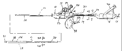
Fig. 1 is a perspective view of the stent
deploying catheter of this invention, in its position in
which the catheter and stent are to be advanced into a
patient;
Fig. 2 is a sectional view taken along line 2-2
of Fig. l:
CA 02210090 1997-07-09
Fig. 2A is a sectional view taken along line
2A-2A of Fig. 1;
~ Fig. 3 is a perspective view of the stent
deploying catheter in its position in which the stent is
being expanded by expansion of the catheter balloon;
Fig. 4 is an enlarged, longitudinal sectional
view of the distal tip of the stent deploying catheter of
Fig. 1, with the stent attached;
Fig. 5 is a longitudinal sectional view of the
two hubs of the stent deploying catheter of the previous
drawings, with the hub for the sheath~being in locked,
advanced configuration; and
Fig. 6 is a longitudinal sectional view similar
to Fig. 5, but with the hub of the sheath shown in
unlocked configuration to permit sliding of the sheath.
DESCRIPTION OF SPECIFIC EMBODIMENT
Referring to the drawings, a catheter 10 is
provided comprising a tubular catheter body 12 (Fig. 3)
which is conventionally connected to a first hub 16. A
proximal, tubular, stainless steel section or "hypotube"
_ g _
CA 02210090 1997-07-09
14 surrounds a proximal portion of catheter body 12, and
is also connected to hub 16. Tubular catheter body 12 is
enclosed in Fig. l, but visible in Figs. 2, 2A and 3.
Inner, catheter tube 13 is also cdnventionally provided
to define a guidewire lumen 15 and a balloon inflation
lumen 17 within tubular body 12.
Catheter body 12 and proximal catheter section
14 are thus of the multiple lumen type, which lumens 15,
17 respectively communicate with a pair of ports 18, 20
in hub 16. Lumen 15 connects with port 18 and comprises
a guidewire lumen. Lumen 17 connects with port 20, and
also communicates with a catheter balloon 26 carried on
catheter body 12 (Fig. 3) in cor~ventional manner. A
stent 28, which may be of a conventional crossing wire or
apertured tube design, is carried over and around balloon
26 in conventional manner.
Catheter 10 also carries an outer, semi-
flexible sheath 30, surrounding most of catheter body 12
and connected to a second hub 32. Second hub 32 defines
a side port 34, which communicates with the tubular space
36 (Fig. 2A) between catheter body 12 and sheath 30.
Second hub 32 is slidable along hypotube 14
between the two positions of Fig. 1 and Fig. 3, taking
sheath 30 along with it as it slides. In the forward
sliding position of Fig. 1, distal end portion 38 of
sheath 30 occupies a position overlying balloon 26 and
- g
CA 02210090 1997-07-09
stent 28, so that the stent is enclosed and out of
contact with the exterior. The distal end 52 of sheath
30 can engage and, if desired enter into, sealing
relation with a catheter distal tip 40, which is carried
at the distal end of catheter body 12. As shown in Fig.
4, some of distal end 38 of sheath 30 can overlie a
proximal portion 42 of tip 40, which proximal portion
carries sealing rings 43 to provide a seal between the
sheath 30 and the tip until the sheath is withdrawn.
Distal portion 38 of sheath 30 may be somewhat
radially enlarged as shown relative to other portions of
sheath 30, to provide room to receive stent 28 in its
collapsed configuration. A portioq of sheath 30 proximal
to end portion 38 may also define a series of side holes
46, for flow of x-ray contrast solution outwardly from
the interior of the catheter of the catheter to areas of
tissue surrounding stent 28.
Second hub 32 defines an annular seal 33 to
prevent fluid leakage from annular space 33, and a lock
50 for selectively preventing and permitting the sliding
motion of hub 32 and sheath 30 along the rest of the
catheter, to provide or to prevent the desired movement
between the positions of Figs. 1 and 3. A hub lock 50
comprises a rotary handle 48, which is shown in the
locked position in Fig. l, with the handle being rotated
90 degrees in Fig. 3 so that it is no longer visible, and
- 10 -
CA 02210090 1997-07-09
occupies the unlocked position. As handle 48 occupies
the position of Fig. l, rotatable lock 50 engages in a
locking relation with a recess 37 (Fig. 3) carried on the
exterior of hypotube 14, to prevent proximal motion of
second hub 32 and sheath 30. However, when handle 48 is
rotated to the position of Fig. 3, rotatable lock 50,
attached to handle 48, moves out of engagement with
recess 37 of hypotube 14, thus permitting second hub and
sheath 30 to be proximally withdrawn toward first hub 16.
With this motion, distal end portion 38 of sheath 30 is
correspondingly withdrawn, exposing balloon 26 and stent
28 as shown in Fig. 3. A similar locking recess 39 is
shown for locking hub 32 in the position of Fig. 3. .
Then, balloon 26 may be inflated through lumen
17, correspondingly causing the expansion of stent 28, to
provide its permanent emplacement in a blood vessel or
the like. Following this, balloon 26 may be deflated
again, by control of fluids through lumen 17, so that the
catheter may be withdrawn from the patient, leaving
stent 28 in its desired position.
At any time during these proceedings it can be
seen that x-ray contrast fluid can be applied to the area
of and surrounding stent 28. For example, in the
configuration of Fig. 1, x-ray contrast fluid may flow
through port 34 of second hub 32 into the annular space
36 between catheter body 12 and sheath 30. This space
- il -
CA 02210090 1997-07-09
serves as a flow channel for the x-ray contrast fluid, to
flow distally for about the entire length of the catheter
until it encounters the holes 46 near the distal sheath
end portion 38. The x-ray contrast fluid can flow out of
the catheter at that point in an abundant flow, provided
by the relatively large flow cross section of tubular
space 36, to provide a clear indication to. the surgeon as
to the location of stent 28.
Then, after emplacement of the stent as in Fig.
3, it still is possible for x-ray contrast fluid to flow
through tubular space 36 and out of the side portions 46
and the distal end 52 of sheath 30, to provide good x-ray
visibility of the location of stein 28, particularly if
distal end portion 38 of the sheath is upstream in terms
of blood flow from the stent. It should also be noted
that the application of x-ray contrast medium can be
applied without removal of a guidewire 56 (Fig. 4) , which
may extend through the catheter from guidewire port 18
along its entire length, and out of distal aperture 54 of
distal tip 40. Accordingly, the guidewire may remain in
its position through the entire operation and use of this
catheter, including a step or steps of the use of
contrast medium to provide x-ray visualization of the
position of stent 28.
Following the implantation of stent 28, the
catheter and sheath are easily withdrawn, while the
- 12 -
CA 02210090 1997-07-09
guidewire 56 may remain in position if desired.
Port 34 may have its flow controlled by a
stopcock 35, for selective application of x-ray contrast
medium.
Sheath 30 may comprise a tube having a wall
thickness of no more than about 0.015 inch, to be of
sufficient stiffness to avoid wrinkling and axial
collapsing as the sheath is advanced with the catheter
and stent into the patient. Thus, it is generally
preferred for the sheath to be made of a strong, semi-
flexible plastic, for example, polyimide plastic or other
material having similar properties of good stiffness plus
a desired measure of flexibility.
Hypotube 14 can be seen..in Fig. 2 to have an
outer periphery of non-circular cross section,
specifically of generally teardrop configuration,
although other shapes may also be used, for example
rectangular or the like. Hub 32 may define a central
aperture 37 that slides along and surrounds 14 as hub 32
moves between the two positions shown in Figs. 1 and 3.
Bore 37 may also be of similar, close-fitting, non-
circular cross section, specifically a teardrop-shape of
similar size and shape to the outer surface of hypotube
14, so that hub 32 is non-rotatable relative, to hypotube
14. This avoids accidental rotation, which can damage .
stent 28.
- 13 -
CA 02210090 1997-07-09
Typically, sheath 30 is of circular cross
section, as is catheter body 12.
Thus, a catheter is provided which can be
advanced into a patient without the need for a guiding
catheter. Then, the stent or other medical device of
implantation can be located by means of flow of x-ray
contrast media passing out of the distal end portion of
the catheter, without removal of the guidewire along
which the catheter may be advanced, with improved
contrast media flow rates. The sheath that surrounds the
implantable medical device such as a stent can be
retracted when the device is properly positioned, and the
device may be emplaced with great-,reliability and ease.
The above has been offered for illustrative
purposes only, and is not intended to limit the scope of
the invention of this application, which is as defined in
the claims below.
- 14 -