Note: Descriptions are shown in the official language in which they were submitted.
CA 02210523 1997-07-24
WO 96/25103 PCT/US96/01920
SURG~C~I INSTI7.UMENT
This invention relates to surgical instrumentsl and in particular
to powered arthroscopic surgical instruments.
Powerl3d arth,~s _~ic surgical instrurnents typically include a
rigid, slc-lion~ry outer tube within which a rigid inner tube is rOtdt-J
by a motor. A cutting implement, such as a blade or abrading burr,
is disposet on the distal end of the inner tube. Tissue or bone is
10 ~Yrc~s~ to the cutting implement through an Gpenirl~ in the distal
end of the outer tube, and tissue or bone r,ay",~, IL:a cut by the
rotating blade or burr are drawn thro~h the interior of the inner tube
along with i"i~ g fluid by the use of suction applied at the
pr~,~i" ,al end of the instrurnent. Examples of such sur~ical
15 instruments are described in ~J.S. Patent Nos 4,2031444, 4,274,414,
4~834,729, and 4,842,578, all of which are assigned to the present
assignee
Some arthrosco~ic surgical instruments are linear, that is,
ZO straight between their proximal and distal ends. Others are curved
to rdcilita~ positioning the cutting i",ple".t:"l against tissue to be cut
without requiring that the instrument be rernoved from the body and
reinserted through an ~dr1itional puncture In a curved instrument, a
region of the inner tube is flexible to enable the inner tube to accept
25 the curvature imposed by the outer tube while lldnsrl,iLli, ,~ the
torque ~pplied by the motor to the blade.
The cutting implernent ~ ros~ at the distal end of
instruments of the prior art have tended to lack ~ alilit~ in their
30 control with the e~cision of tissue being ~all or nothing" event. In
certain circumstances, the cutting of tissue may necessit~te careful
and delicate use of the cutting i.I~p~e,~ t. In other circumstances, a
CA 022l0523 l997-07-24
W O96/25103 PCTrUS96/01920
more aggressive approach may be needed. Instruments of the prior
art tend to la~k this versatility and thus the ~ucoP~-s of an operation
may lie more in the skill of the surgeon to control the cutting
implement than might otherwise be desired.
It is an object of the pr~ser,l invention to overcome, or at least
alleviate, this proble,...
Accordingly, there is provided a surgical instrument
10 comprising a base, a suppor~ member extending distally frorn said
base and carrying at a distal region a window defining an openirlg,
a surgical tool a~ least partially disposed in said distal region
and movable to cut tissue eAb,.Jing through said opening
15 characterised by the fact that said support r..ei ,.ber is provided with
a shield at least partially ~icposed in said distal region and movable
with respect to said window to at least partially cover said opening.
One general aspect of the invention is a surgical instrument in
20 which a movable shield can be positioned to at least partially cover
the distal opening in a support, I ,e, . ~L ~r Another general aspect of
the invention is a method for using the surgical instrument. In still
another general ~spe~t the support ,.,~"l~e:r has a pair of openings
in its distal end. either of which can be seleetively covered by the
25 movable shield.
Among other advd"~as~es, the invention allows the user to
partially or completely cover the Gpe~ l9 by moving the shield,
thereby preventing at least some tissue frorn entering into the
30 instrument through the opening and being cut by the surgical tool.
.
CA 02210523 1997-07-24
WO 96t25103 PCT/US96/01920
As a result, the cutting action of the surgical tool can be reduced or
s~'~ by appro~,.i..Le positioning of the shield.
Moreover, v~lhere the surgical instrument has a pair of
openings at its distal end, the invention allows the user to partially or
completely cover one of the oper.i- I95 by moving the shield, thereby
preventing at least some tissue from entering into the instrument
through that op~r,i"~ and being cut by the surgical tool. As a result,
by a~l,,upriate positioning of the shield, the user can select be~vccn
10 windows that have, for example, different cutting configurations and
different rot..liGnal ol ier.l; Itions.
For il.ald,-ce, one window can be configured for more
dyy,essive cuKing than the other. The preferred degree of cutting
15 can thus be Lhose~ l by moving the shield to cover the opening of the
~i.ld~,v: having the ur,Jasired cutting ~,lldr~.;~istics. Moreover, the
windows may be locAt~l at different ~utdlio"al .Jrit:~lldtiuns around
the distal region of the support rllel"ber. l hus, even if their cutting
char~Leri~tics are idenlicdl, the w;nclo~rs ~.an be selectively covered
20 and uncovered to ~Jldrl9e the di,~clion of eutting of the instrument.
Preferred embodirnents include the following features.
In a particularly useful embodiment, an act~ting member
25 (e.g., a tube Go~Yi~lly disposed ol~tside the support rllelllbar)
e~ ls distally from the base, and transmits a lot.~iG~ ~al force
applied at a proximal end to move the shield, which is attached to a
distal end of the actuating member. The ~ru~i,,Idl end of the
~ctll~ti-lg ~ ."~cr is rigidly secured to a knob ~ dtably mounted to a
30 stationary portion of the base The knob may be selectively rotated
to a plurality of discrete posiliGns with r~s~e~l to the base. allowing
CA 02210523 1997-07-24
W O 96/25103 PCTrUS96/01920
the shield to be positioned to a corresponding plurality of discrete
rotational o.ientdlions. 8ec~ ce the ~ t~ tirl9 member is rotatably
coupled to the base, the openings may be selectively covered and
uncovered while the instrurnent remains in situ within the patient.
A drive member (e.g., a tube dispose~l coaxially within the
support member) c~ nl~s distally from the base, and transmits a
rotational force applied at a ,uro~ al end to move at least a portion
of the surgical tool, a cutting implement attached to a distal end of
10 the drive member. As the drive mernber rotates, the edges of the
cutting implement move toward and closely past the edges of the
windows. A hollow p:~c~ge in the tubular drive member is adapted
to receive suction at its proximal end, transporting body material cut
by the cutting imp!e,..,~nL away from a surgical site while the
15 instrument remains in situ for further cutting.
The support member (e.g., a tube) couples to the base in a
manner that allows it to slide axially with ,~:spe~ to the ba-~e. During
assembly, the support tube is inserted into the ~otl~tor tube, and
20 the A~tu~tor tube is atta~hed to the base. When the drive tube is
then inserted into the support tube, the outer surface of the distal tip
of the drive tube bears against the inner surface of the distal tip of
the support tube Because the support tube can slide axially with
respect to the base, this forces the supporl tube distally until the
25 outer surface of the distal tip of the support tube bears agai"st the
inner surface of the distal tip of the aoh~tnr tube. Thus, when
ass~i"bl~d, there is little or no gap ~ JJccn the distal tips of the
various tubes This reduces the amount of severed tissue, fluid, and
other ",atl::,ial that would othe~ise pass into the annular regions
30 separating the three tubes.
CA 02210523 1997-07-24
WO 96125103 PCT/US96/01920
In all~:nlate embodiments of the present invention, the
support tube is bent, and an actuating .,lel.,ber extending distally
from the base is relatively flexible at least in the bend region,
allowing the actuating member to transmit force through the bend
S region to move the shield For ioatance, the ~ ting member (e.g.,
a tube r~ispos~-i outside the support r~mLt r) and the drive tube
may both be relieved with a series of axially sp~ed slots in the
region of the bend. This arrangement provides the actuating and
drive tubes with the requisite transverse flexibility to accommodate
10 the bend, and the n~cess~ry torsional ~tirr"ess to rotate the shield
and the cutting implement, r~ ctively RF!r~llsf~ it is bent, the
instrument may be used to operate on surgical areas that would
otherwise be difficultto reach with a sllaiyl,t-shafted instrurrent.
In other embodiments, a relatively rigid sheath is dispo~e~l
co:~Yi-"y with, and is axially s~ with respec~ to, the bent support
"-~n,~:r, which is relatively d~ru.-"a~le at least in the bend region.
By sliding the rigid sheath fore and aft along the support m t:."L:r to
selectiYely cover and uncover the bend region, a surgeon may
20 ~ ,ye the angle of offset provided by the bend region, all while the
instrument f~",ains in sity within the patient.
Other features and advantages of the invention will ~e~o",e
apparent from the r~ L ~J;n9 de'~ s~ iun, and from the
daims
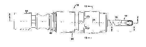
~ig. 1 is a top view of a surgical instrument.
Fig. 2 is a sectional side view of the surgical instrument, taken
along line Z-2 of Fig. 1
Fig. 3 is a side view of the surgical instrument, taken along
30 line 3-3 of Fig. 1
CA 02210523 1997-07-24
WO 96/25103 PCT/US96/01920
Fig. 4 is a cross-s~l;tiGnal side vjew of a distal region of the
surgica1 instrument.
Fig. 5 is a perspective view of a distal region of the
i"ler",ediate tube of the surgical instrument.
Figs. 6 and 7 are top and side views, respectively, of the
distal region of the inter",~id~ tube of the surgical instrument.
Fig. 8 is a sectional view of the distal region of the
intermediate tube of the surgical instrument, taken along line 8-8 of
Fig. 7.
Fig. 9 is an end view of the distal region of the intermediate
tube of the surgical instrument, taken alon~ line 9-9 of Fig. 7.
Figs. 10, 11, and 12 show inner, i"k,r",~:diate, and outer
tubes, respectively, o~ the surgical instrument.
Fig. 13 is a sectional view of a ratchet mechanism of the
1~ surgical instrument, taken along line 13-13 of Fig. 1.
Fig. 14 sho~vs the surgical instrument in use.
Fig. 15 shows another embodiment of a surgical instrument.
Fig. 16 shows another e",boJi~nent of a surgical instrument.
As shown in Figs. 1, 2, and 3, a surs3iGal instrument 10
suitable for pe,rur-,-ing, e.g., closed, arthroscopic surSery on the
knee with a surgical tool 12, includes an inter,l,ediate tube 14 within
which a rotating innertube 16 is c~Yi~~y di~po5ed. In turn,
intermediate tube 14 is a~Yi~ly disposed within a rotatable outer
25 tube 18. Tubes 14, 16, and 18 extend distallyfrom a base 20.
Referring also to Fig. 4, a distal region of outer tube 1~ is
partially cut away to form an aperture 22, which extends to the
longitudinal axis 24 of instrument 10. The remaining, solid portion of
30 the distal region of outer tube 18 co" ",, ises a shield 26. As outer
tube 18 is rotated relative to i"l~,r"~diate tube 14, shield 26
CA 02210523 1997-07-24
Wo 96/25103 PCT/US96/01920
altemately covers and uncovers an incisor window 28 and a
synovator ~indow 30 locatPd on oF,posil~: sides o~ a window
dSS~ y 31 carried at the distal end of i"l~r" lediate tube 14.
As shown in Figs ~-9, the edges 32 of incisor window 28 are
sha, ~ e. Iell and serrated, and the edges 34 of synovator windo~v 30
are sharpened and smooth. Referring to Fig. 4, a window 35 fc~(".ed
by the sharpened, smooth edges 37 of a cutting impiement 36
carried at the distai end of inner tube 16 is periodically ex~,osed
10 through incisor window 28 and synovator ~r~ o~l/ 30 as inner tube
16 rotates. Thus, tissue entering through either incisor window 28 or
synovator v~indow 30 (dq,en~Jing on the ,~Jtdtional oric' ,Ldtion of
shield 26) can extend into the interior of inner tube 16. As inner
tube 16 rotabs, edges 37 o~ cutting implement 36 move toward and
15 closely past edges 32, 34 of W;l~vrs 28, 30 in ~J;.Id~N asse,.~61y
31, severing the tissue projecting It.orell,r~ugh. Together, cuffing
implernent 36 and window ass~",bly 31 cu"-prise sursical tool 12.
Inner tube 16 is made from metal or other rigid material, such
20 as stainless steel As shown in Fig. 10, the distal end 40 of inner
tube 16 supports cutting il "plel l .ent 36 (made frorn, for example,
stainless steel and attached to tube 16 by uI~hli, ,~ or brazing).
Cutting implement 36 is sized to provide a close-running fit with the
inner portion of w;, ,du~ assembly 31 for t:r~ jF .l cutting. The
25 o~ui"g defined by window 35 in i-l,ple~ nt 36 is an extension of a
central pz~5s~3~e 46 in inner tube 16 that runs the entire length of
tube 16.
rlu~i",al region 48 of innertube 16 is rigidly mounted to a
30 drive shaft 50 that rotates within base 20, sho~vn also in Fig. 2.
Centra1 p~ss ~e 46 te,l"i,lates in a vacuum source opening 5Z In
=
CA 022l0523 l997-07-24
W O96/25103 PCTrUS96/01920
drive shaft 50. The pr~Ai",dl end 54 of drive shaft 50 fits into a
handpiece 110 (~ig. 14) which includes a motor 112 for lvl~Ling
drive shaft 50 and inner tube 16 with respect to both interrnediate
tube 14 and outertube 18. One l::AdlJ~ e of such a handpiece is
~escribed in U.S. Patent No. 4 705 038 e"ti~l~ "Surgical System
for Powered Instruments", and assigned to the present assignee
which is incorporated by reference. Opening 52 i~ coupled to a
vacuum source 114 (Fig 14) during operation to remove severed
tissue and irrigating fluid from the surgical site via p~c5~9~ 46 in a
10 manner des. ~ ed in debil below.
.
Fig. 11 shows illlellrl~.liaLt: t~be 14 which is also made ~rom
a rigid material such as stainless steel or other metal. Distal end 56
of in~e""adiate tube 14 supports wi.ld~JJv assembly 31 (made from~
15 for C~dn~ E, stainless steel and attached to tube 14 by welding or
brazing). The inner and outer~ ",e:le,~ o~window assembly 31 are
substantially equal to the inner and outer di~rll~L~ of tube 14.
Intermediate tube 14 is hollow along its entire length to
20 provide a pass~ge 60 that receives inner h~be 16 and outting
i" ~lo .,enl 36 which exten-l~ to the partially closed distal end 62 of
window assembly 31 The openings defined by windows 28 30 in
window assel"bly 31 are u ~t~nsions of ~ J~ 60. The inner
diameter of i,.te""ediate tube 14 is only slightly larger than the outer
25 diameter of inner tube 16 ~e.g. by appru~i--,ately 0.002 inches or
0.051 mm) This allows inner tube 16 to ratate freely but helps
minimize wobbling of tube 16 to keep sharp cutting edges 37 of
cutting implement 36 and edges 32, 34 of v~lindo~vs 28 30 closely
aligned.
CA 02210523 1997-07-24
W O96/2S103 PCTnUS96/01920
The proximal end 63 of intermediate tube 16 is rigidly
mounted to a coupling 64 loc~ted within a cavity 66 of a hub 68 of
base 20, shown also in Fig. 2. Cavity 66 includes an axially
extending keyway 70 sized and lc~c.lled to recei~e a key 74 on
5 coupling 64. Thus, although coupling 64 can move axially with
respect to hub 68, key 74 prevents coupling 64 from rotating.
Cavity 66 in hub 68 communicates with p~ss~ge 60, and is
configured to receive drive shaft 50. During as~c:,,,l,ly,after outer
10 tube 18 has been attached to hub 68 in the manner desc, iLe~
below, innertube 16 is in~ d through hub 68 into p~s~ 60 of
inter",adiate tube 14. When the distal tip 78 of cutting implement 36
(Fig. 10) contacts the inner surface of the distal tip 62 of window
assembly 31, coupling 64 and intermediate tube 14 are forced
15 distally, until the outer surface of distal tip 62 contacts the inner
surface of the partially closed distal tip 80 of oubr tube 18. Thus~
because inter",~Jiale~ tube 14 can slide axially with respect to hub
68, the gap between the distal tips of inl~rlll~cliate tube 14 and outer
tube 18, as well as the gap between the distal tips of inner tube 16
20 and i"~r,~ Jidte tube 14, are essentially zero. This reduces the
amount of severed tissue, fluid, and other Illate~ that would
othe~ise pass into the annular regions separating tubes 14, 16, 18.
With inner tube 16 i"~l "e ', a pliable fitting 82 retains drive shaft ~0
within hub 68. Fitting 82 provides a fluid-ti3ht seal when base 20 is
25 il,s~,led into handpiece 110.
- Outer tube 18, shown in Fig 12, is also made from a rigid
material such as stainless steel or other metal. Aperture 22 is
- .Jefi"~d by SlllU~tjl, unsharpened edges 84 of tube 18. Edges 84
30 extend, parallel to axis 24, from a point p~uxi, llal of distal tip 80 to
CA 02210523 1997-07-24
WO 96/25103 PCT/US96/01920
distal tip 80. Aperture ~2 is an extension of a central r~cs~~ 88 in
outer tube 18 that runs the entire length of tùbe 18.
Pruxi",al region 90 of outer tube 18 is rigidly mounted to a
5 knob 92 that rotatably couples to hub 68 of base 20. As shown in
Fig. 11, a pair of fingers 9C extends distally from base 68, parallel to
axis 24, and a raised shoulder region 96 encircles base 68
immediately proximal of the point where flngers 94 attach to base
68. When i,-~",l~diate tube 14 is inserted into r~ss~ge 88 of outer
10 tube 18 and knob 92 and base 20 are forced toge~h~r, a mating
shoulder 98 on the inner surface of the proximal end of knob 92
engage~ shoulder 96, as shown in Fig. 2, preventing knob 92 and
base 68 from separating longitudinally.
As shown in Fig 13, fingers 94 are quasi-pentagonal in cross-
15 section. VVith knob 92 installed, the radial outermost point 100 of
each finge~ 94 rests in an a mating apex 102 on the inner surface of
knob 92. Apexes 102 are ~r,~,ed by the intersection of adjacent
arcuate sur~aces 104 of a wall 106 of knob 92. Fingers 94 and
arcuate surFaces 104 eoaGt to allow the relative l~tdliCil ,al orientation
20 be~ween knob 92 and hub 68 to be changed~ in a ratchet-like
r~sl,ion, in LliSCIel~, 180~ steps. In particular, outertube 18, knob
92, and fingers 94 are oriented so that incisor window 28 is fully
Govered by shield 26 vvhen knob 92 is rotated to one step, and
synovator window 30 is fully covered by shield 26 when knob 92 is
25 rotated to the other step.
As knob 92 is rotated with l~s,.,. _L to hub 68, outerrnost points
100 move across arcuate surfaces 104, initially forcing fingers 94
radially inward. When ou~erl,lost points 100 move past the
30 recpective midpoints of surFaces 104, the elastic energy stored in
the ~ rl-c~l flexible fingers 94 forces the fingers radially ou~Nard
CA 02210523 1997-07-24
W O96/25103 PCTrUS96/01920
until the relative rotational Orierlt~liGn beh~ n knob 92 and hub 68
has changed by 180~, and fingers 98 rest in the c",posite apex 102.
Thus, fingers 94 positively urge oul~lr.,Ost points 100 into each
~ssor;~t~d apex as it is encountered, thereby giving the surgeon
S kinesthetic feerlh~rk as to the amount by which outer tube 1~ has
been rotated, and also helping to avoid z~ccidPntal rotation o~ outer
tube 18 with ~~spo_L to w;, Idu)~S 28, 30. Nloreover, a pair o~
dia~ ,ally opposed bulges 108 on the c: uter sur~ace of knob 92
are oriented adjacent to, and at the same circl~mferential loc~lio" as,
1 o ar~xes 102. Bulges 108 thus make knob 92 easier to grasp, and
further indicate to the surgeon when knob 92 has been rotated a
sumcient degree. Together, fingers 94 and knob 92 conlprise a
ratchet asse,"Lly.
1~ Ref~. ,i"~ also to Fig 14, in operation, surgical instrument 10
is inserted into the distal end of a hanrlrieGe 110. Outer tube 18 is
then introduced as shown through a punc~ure wound 120 into the
knee joint 122, below the ,uat~ Light is prl ,e-~cl into the joint via
a second puncture 124 using a fiber optic light source 126, and a
20 visual image of the s~rgical site is retumed through a separate
optical path to a television camera 128. The image is d~li.r~red by
camera 128 onto a television screen 130 ~or viewing by the surgeon.
(Alternatively, the surgeon can view the image using an e~ or
the image can be r~or-Jed.)
The surgeon ~ t s surgical tool 12 by activating motor
112, which receives Gp~ tiny F,ult,l,~ial and current from power
supply 116. Motor 112 engages and rotates drive shaft 50, thereby
applying rotdlional force to inner tube 16 and lutali~l~ tube 16 with
30 ,t:~pe~t to tubes 14, 18. The surgeon controls ruldliulldl speed and
direction (either unidi, t:~lional or oscillatory) using foot swi~ches
CA 02210523 1997-07-24
Wo 96/25103 PCT/US96/01920
116a, 116b, which control the magnitu~e and polarity of operating
potential and current provided by power supply 116 to motor 112.
Motor 1 12 is capable of rot~li"~ inner tube 16 over a wide range of
speeds, e g., between about 100 rpm and 5000 rpm, and can deliver
5 a torque of up to 25 oz. inches (0 177 Nm).
Dir~trtnt types of surgical instruments such as instrurnent 10
have rotational and torsional limits. To prevent the surgeon from
inadvertently operdli"g instrument 10 at dangerously high speeds
10 and torques, instrument 10 identifies to sensors (not shown) in
handpiece 110 what type of instrument it is, and the speed of and
torsion applied by motor 112 is co"lr_~lc-~ so that these limits are not
.qY~eede~. (This control technique is described in the
. . ,antioned U.S. Patent No. 4,705,038.)
During the surgical proced-lre, the body joint is dist~n~d vvith
fluid intro~ ed through a third puncture wound 132 frorn a fluid
source 134. The fluid i"i~les the site and renders tissue 136
(which is, e.g., synovial tissue) mobile so that it floats and can be
20 ~lispl~ ed (similar to the movement of scawccd in water).
The surgeon progressively cuts away synovial tissue 136 by
movlng surgical instrument 10 frorn side to side and in the axial
direction using handpiece 110 (while viewing television screen 130).
25 For instance, if incisor window Z8 is exposed to the joint tissue (that
is, if synovator window 30 is f~llly covered by shield 26), instrument
10 will cut tissue ayyr~ssively, h~ç~nce of the conhguration o~
serrated edges 32. I~ during the procedure the sur~3eon desires
instea~ to cut tissue less aggressively, the ,~r~:s~r.l invention allows
30 him to do so simply by holding knob 92 fixed, and lul~lilly
handpi~ 110 (and thus hub 68) until incisor window 28 is fully
CA 02210523 1997-07-24
Wo 96/25103 PCT/US96/01920
vered by shield 26. Thls e~oses the l~ss-aggresshre. smooth-
ed~ed synovator window 30. The ratchet mechanism provides the
surgeon with Icinesth~lic feedback, indicating when th~ handpiccc
110 I)as been rotated the requisite 1~Qn. (Alternatively, handpiece
110 can be held fixed and knob 92 rotated. Because incisorwindow
28 is located on the opposite side of window as~embly 31 T'rom
synoYator window 3~, in order to resume cu/;ting the same tissuc as
before, the ~urgeon would then rot3te instrumcnt 10 180~ 3bout 8Xi6
24.)
The 3urgeon cun change the ,utcllional orienlation of 6hield
26 with respect to windows 28, 30 at any time. For example, innQr
tube 16 c~n be driven by motor 112 or may b6 stationary while the
eurgeon rotates shield 26. T~e surgQon can resume more
aggressivo tissue-cu';ting at any time simply by rotatlng knot~ 92 nr
1~ handpiece 110 in eitherdi,~c~;on.
Tissue fra~men~s and other ~ody material cut by surgical tool
12 are wlthdrawn from the surgical site along with irrigatiûll fluid via
central passage 46 of inner tube 1~ (Fiys. 2, 10) in response to
Z0 suction applied by vacuum source 114.
Other embodiments ~re within the scope of the following
claims.
For example, although surgical inr~trument 10 is straight
between it~ proximal and distal and~, a surgical instrurnent 210
embodying the teachings d;s~l~ced herein could instead include a
bend region 212, as shown in Fi~. 15. ~end re~lon 212. whlch is
di~po.c~d sli~htly proximal ot the dlstal end 214 of outer tube 21~,
an~ularly o~tsets surgical tool 218 frorTI a generally straight axis 220
of surgioal instrurnent 210. Bend region 212 ena~les surgical
CA 02210523 1997-07-24
WO 96/25103 PCT/US96/01920
in~trument 210 to opsrate on sur~ical areas that are difficult to reach
with a straight-shafted instrument.
In order to rota~e a shield 222 at a dlstal region of outer tube
5 216 to selectlvely coYer and uncover an incisor window 224 and a
synovator wlndow (not shown) located Dn ~Jpo5;~e sides of an
assembly carried at the distai end of a bent, rigid intermediate tube
22~, wter tube 21~ is flexible at least in bGnd region 212. The inner
tube (not shown) i5 likc,/~isc flexible at lea~t in bend region 212,
10 allowing it to transmit torque through bend region 212 to operate
surgic~l tool 218. ~Itern~tively, the inter"led;~l~ and inncr tube6
may be flexible, and the outer tub~ may be rigid. In thi6 latter
embodiment, interrnediate tube 226 i6 rota~ed to 6electively cover
~nd uncover the inci60r and 6ynovator window~, and outcr tube 216
16 (and thu6 al60 shiald 222) remains ~t~tionary. Similar flexible tube
arrang~m0nts are disclosed in copending app' ~ on serial no.
08/2û0,662, filed on Febr iary 23, 1994, which is a continuation-ln-
part of ~pp'ic ~ ~ on serial no. 08/011,3~4, filed on January 29. 1993,
which are both assi~ned to the present as~iignee and i,,~.un~o,d~ed
20 hereln by re~erence In their entirety.
Various tube configurations exhibit the requi3ite flexibility ot
least in the bend reQion to be employed in surgiccl instrument 210.
For instance, a region of an otherwise rigid tubc or tube6 may be
Z5 r~l,ev~ with a series of sxisliy spaccd, circumferentially extending
3iot9 228 (only slots 228 in o~ter tube 216 shown in Fig. 15).
Slotting a rotatable tube for flexibility and torque t.dns",ission is
de6cribed in IJ,S~ P~hnt No. 5,152,744, assigned to the present
assignee and incorporated herein by reference in its entlrety~ To
30 prevent tissue fra~ments or other body materlal frorn catching on Dr
passin~ throu~h the slots In the inner tube. the slots can be covered
CA 02210523 1997-07-24
Wo 96/25103 PCT/US96101920
- with a pliable nlaterial ~uch as silicone R~V or a heat-shrinkable
polymeric sheath (not shown).
The flexible regicn or regions can instead be comprised of a
5 series of discrete. interengaging segrnents as di~Glosed in
copendinQ application serial no. 03/228 083 filet on April 15 1994
whioh is as~igned to the pre~nt ~3s;gnee snd incorporated herein
by reference in its entirety. Alternatively, the tube~ can be
con~prised, at lea~t in the bend region of a flexible or elar,~v"~r;c
10 m~tcrial, such as rubber, pla6tic, or other polymer.
Inter",ediiat~ tube 226 is comprised of a material (e.g.
~tainlo~s ~teel or other metal ceramiG or pla~tic) sufficiently ri~id to
retain the sllape and orient;~"ion of bend re~ion 212 durln~ normal
1~ surgical use of instnlment ~1û. Althou~h bend reglon 212 Is often
pr~fo,.,led durin~ manufacture ~e.g. by moldlng in~ "~diate tube
226 to the deslre~ shape or by bending it around a mandrel). if
Illter"l~ e tube 22~ is c-,"".,ised at least in the bend region of a
plasticly deformable material, bend region 212 can alternatively or
20 additionally be p~ ,haped or reshaped by the surgeon prior to or
during the procedure to best rnatch the contour3 ~nd chor~cteristics
of the ~urgical site.
Anothcr cmbodiment of the precent inv~ntion ~urgical
26 instrument 310, i~ ~hown in Fig. 16. A bend region 312 in sur~ical
in~trument 310 is provided by a curved intermedi~te tube 314 and
an inner tube 316 and an outer tube 318 of the instrument are
flexible at least in bend re~ion 312. (Alternatlvely. similar to the
alternative embodlment descrlbed above in conneclion with Fig. ~5
30 outertu~e 318 could be curved and intemlediate tube 314 could be
flexible at least in bend region 312.) A window ~ssembly 320
CA 02210523 1997-07-24
W O96/25103 PCTrUS96/01920
16
camed at th~ dictal end of Inter"l~dlate tube 314 has opposit~ly
disposed inclsor and synovator v.~ d-J.~ 322. 324, witilin which a
cuttlng Implement (not shown) disj~osed at the distal end of
Innertube 31~ rotates A shield 326 carried at the distal end of outer
5 tube 3t8 can be rotated to selectively cover either of windows.322,
324.
Similar to the instrurnent dis~losed in U.S. Patent No.
5,282,821, incorporated herein by raf~rence in its entirety,
int~r-"cdiate tube 314 of ~urgicai inctrumant 310 is resilient, and a
10 straight, rigid cheath 328 i~ dispo~cd co~xially outside outer tube
318. Sheath 328 i~ axially slidable with respect to outer tube 318
and in its rest position (shown in Fig. 16) the distal end ~f sheath
328 ter",i"ales at a point just pr~ f bend re~ion 312. 511dlng
sheath 328 distally (i.e., in the dil~LLion indlcated by arrow 330) ~ver
15 bend region 312 causes intermedlate tube 314 to straighten out.
decreasin~ the an~le ot offset provided by bend region 312. Slidin~
sheath 328 pru~l",ally 13ack to its rest posilion allows bend region
312 to recover its prerormed curvature. Thus, the angle of offset of
tile ciistal region 332 of instrument 310 with respect to the proximQI
20 region 334 of the instrument can be selectivcly changed while the
instrument ~",~;"c ~ihl within the pstient.
Other types of sur~ic31 toolc, CUch a~ abr~ders, may be
ernployed with any of curgical instruments 10. 210, 310. Moreover,
25 the surgical tool may be removably connected to the distal end of
the inner tube, allowing just the ~ur~ical tool to be replaced should It
b~come worn or dama~ed, as disclosed In U.S. Patent No.
~,32û,~3~, assigned to the present assignee and incorporated
herein ~y re~erence In its entirety. Als~, the sur~ic~l tool and the
30 inner tube need nc~t be comprised of the same material. Rather, the
~urgical tool will cften i~e comprised of a hard material well-suited to
-
CA 02210523 1997-07-24
W O96/25103 PCTrUS96/01920
- cutting, v,/h_reas the inner tube m~y be COI ~ l~ri5~ lead of a more
compliant mat~rial (at least in the bend region) for tral~s."illi"y
torque throu~h the bend region.
In addition, the inner, int~r",~Ji~te, and outer l-,el.,Le,~ need
not bê tubes, but Gould instead be, e.~., solid ~le~lber~ or cables.
Moreover, the ~,ro~ ,al end of the outer member can be coupled to
a source of ..lt~tior,al power, such as a motor (not shown), ~
the shield to be either power-rotated or manually actuated. Fùrther,
10 the interrllcJi~l~ tube of the instrument may be provided with more
or fewer windows, and the ui"dow confisu,alio"s may be tc~ilolLd to
dirLr~, .l cutting sr.~ i.,nO. For in~tance, in some applications it
may be u~eful to provide a sin~le large window, and to ~/ary thê size
of ths Y,~;.l '~w opening by partially covering it with the shield. In
15 such an arpliG~tion, it might be desirable to remove the ratchet
mechanism to allow knob 92 to rotate ~.lloothly with ~ l to hub
68, or to modify the ratchet mechanism to rotate in smaller
..,ental ~têp~i.
In ~ iti~", the shield need not be attached to a member thst
tA~ends from the base. Rather, the shield may be a cap mounted
on the distal end of the inter~l,Gdiate tube. In such a construction,
the instrument would have to be .~it~.dr -.v., from the joint space in
order to select a Ji~,_r.. n ~vindow ~or cuttin3. Also, the shield need
25 not rotate, but could be configured il.~ I to slide along the
intermediate tube to selec~ively cover and uncover eith~r or both of
- the v~i~ Ido~/,'S, The inner "~e,~ ~ber could likewise translate axially to
operate the distal tip surgical tool.
While the invention has been dcscriL~d in terms of surgical
30 instruments for s,li-ruscut ~, the invention may also be used with
CA 02210523 1997-07-24
wo 96/25103 PCT/US96/o1s20
18
other types of instruments, for example, in3truments con~i~ured for
other kind~ of endoscopic procedures and ~or biopsy epplicdlions.