Note: Descriptions are shown in the official language in which they were submitted.
CA 02210894 1997-07-18
W096/220S4 PCT/AU96/00024
THOD OF ~SSUEREPAIR
l~nNlCAL FIELD
The present invention relates to methods for joining
living tissues, including veins, arteries, microvessels,
tubes, nerves, organ tissues and biological surfaces,
such as peritoneum, omentum, fascia, shin, artificial
tissues, and to ph~rm~ceutical products useful in joining
these tissues.
P~CR~OVND ART
Joining tissues such as veins, arteries,
microvessels, tubes, nerves, tissues and biological
surfaces such as the peritoneum and skin has mainly been
carried out clinically to date by suturing and
microsuturing.
Microsuturing requires considerable skill and is a
time consuming procedure. Frequently, tissues which have
been joined by microsuturing form considerable scar
tissue. Some of the difficulties encountered with
microsuturing can be better understood by considering the
example of rejoining damaged peripheral nerve tissue.
Peripheral Nerves
The electrical signals that control the body's
organs and transmit information back and forth to the
central nervous system (CNS) travel along peripheral
nerves. The structure of these peripheral nerves is
analogous to telepho~e cables. In a telephone cable
there is a strong protective outer coating that protects
all the inner compon~nts. The copper wires are often
grouped in separate insulating tubes that lead to
different systems. Each of the inner copper wires is a
single line that can transmit electricity in either
direction and has an insulating coating around it so that
it does not interfere with the lines next to it.
A peripheral nerve (Figure 1) has an outer membrane
consisting of connective tissue such as collagen. This
CA 02210894 1997-07-18
W 096/22054 PCT/AU96/00024
membrane (epineurium) protects and holds the separate
nerve bundles together. The nerve bundles which lie
inside this membrane are called fascicles. These
fascicles also have a collagen based surrounding membrane
and their task is to group together llerve axons supplying
a similar area of the body. Inside the fascicle membrane
the axons are surrounded by loose colmective tissue. The
axons are a long extension from a cell body which is
contained within the CNS in the spine or the brain.
Sensory axons transmit to the CNS and motor axons
transmit from the CNS. Nerve metabo]ism is sust~lnp~ by
the vascular system from both outside the nerve and along
the centre of the nerve.
Peripheral nerves can have very small diameters.
For instance, the mature median nerve at the wrist is
approximately 1 cm in diameter and contains an average of
forty fascicles, each of which can contain up to 4500
axons. When a peripheral nerve is cut all axons distal
to the wound change their properties as axon flow is cut
off from the cell body. Even when the nerve is
reconnected, these axons continue to degenerate distally.
The Schwann cells which norm~lly wrap themselves around
the axons as insulation guide regenerating axons.
Joining nerves as accurately as possible by lining up
corresponding fascicles enables the axons to more
efficiently regenerate.
Operating upon nerves has been facilitated by using
magnification and special microsurgical equipment.
Accurate repairs need to be effected at the fascicular
level ensuring that regeneration is along the correct
bundle leading to the original area those axons supplied.
The current technique of peripheral nerve repair uses
microsuturing (Figure 2). This technique requires a
dedicated, trained surgeon as microsuturing of just one
of the many fascicles with three or more microsutures
(using say a 70 micron diameter needle and 30 micron
thread) can take very long operating times.
CA 02210894 1997-07-18
W096122054 PCT/AU96100024
Microsuturing is at present clinically used where
the skills are available. Unfortunately, there are
relatively few surgeons who have the necessary
manipulative skills for operating at high magnification.
Even a reasonable microsuturing technique results in long
operating times with added damage to the inner axons due
to sutures penetrating the thin insulating perineurial
sheath. The use of sutures results in some scarring of
the repair due to foreign body reaction. There is also
evidence which indicates that in the long term scar
tissue formation and scar maturation can lead to
impairment of the joined nerve.
Work has been performed on the use of lasers alone
in effecting nerve joins. One of the problems of laser
welding has been the fact that the intact gel-like nerve
tissue of the axons is actually under pressure within the
fascicle. When the fascicle is cut this material
extrudes. This can lead to the direct laser weld being
formed on nerve tissue rather than the surrounding
membrane of the fascicle, causing nerve damage. To date
the welds have typically been made using infrared lasers
such as CO2 lasers which rely on water absorption for
energy transfer. Tissue preparation before welding
relies on overlapping the nerve membranes. This is
difficult due to the extruding gel-like axons and so can
lead to denaturation of the nerve axon material. The
affected tissue tends to scar and the fibrous tissue that
proliferates as a result is a poorer electrical conductor
than nerve tissue. The bonds formed to date as described
in the prior art using laser welding have typically
lacked strength. These laser joins alone tend to fail so
microsuturing has been used in addition to welding to
strengthen these joins.
To deal with at least some of the deficiencies of
laser welding, various glues have been used in forming
the welds. These low protein concentration, fluid glues
tend to run between the ends of the nerve that are being
joined which may result in damage to the axoplasm of the
CA 02210894 1997-07-18
W096/22054 PCT/AU96/00024
-- 4
nerve fascicle and also hinder regeneration. They are
also applied around the join which is then
circumferentially welded. These joins later show thick
scarring which causes stricture of ~he nerve. Moreover,
the joins tend to be weak.
The welding techniques so far available also tend to
lack precision. Factors that influence the precision of
this approach adversely include differences in: the
consistency of the glue used; the aperture of the needle
or other device used to apply the g]ue; and the pressure
exerted in applying the glue.
DESCRIPTION OF T~E lNvh~llON
The present invention provides a method for joining
tissue comprising:
aligning and abutting edges of the tissue to be
joined;
applying a solder, across the aligned and abutted
edges; and
exposing the solder to an energy source under
conditions which provide a transfer of energy from the
source to the solder to cause the solder to bond to the
tissue surface adjacent the edges thus providing a weld
holding the edges together.
In addition to causing the solder to bond to the
protein of the underlying tissue, the energy transfer can
affect the structure of the solder itself leading to
bonding within the solder and an enhancement of the
strength of the solder and hence the join.
Drops of solder are typically used where the solder
is a fluid solder, and are "painted" across the edges.
The solder can also be provided as a preformed solid
strip.
The energy source is typically a laser.
A variety of tissue types can be joined using this
method. The method is applicable to anastomoses of
biological tubes including veins, arteries, lymphatics,
nerves, vasa efferentia, fallopian tubes, bile ducts,
CA 02210894 1997-07-18
W096/22054 PCT/AU96/00024
tubes of the alimentary canal, the ureter, the urethra,
tear ducts, bronchi and any other such bodily tubes as
well as to repairs of incisions or tears of biological
organs such as kidneys, liver or spleen, or of biological
surfaces such as the peritoneum and skin. It will
therefore be understood that the method can be used in a
variety of join situations including the joining of
cylindrical anastomoses and the closure of linear defects
such as incisions.
Where the tissue repair is with respect to nerve
tissue or other tissue tubes where the tube contents need
to be protected from damage, it is especially important
that the weld should not be concentrated on the edges
being joined as this can damage extruded tissue. Rather,
the weld should be distributed across the planar or
tubular surface in which the discontinuity lies.
Where the tissue to be repaired is an essentially
hollow body tube such as a blood vessel, the repair can
additionally comprise the insertion of a thin-walled
hollow cylinder of solder inside the tube under repair so
that the cylinder spans the severed portions of the tube.
Typically, while the severed tube and cylinder assembly
is held together, energy from the energy source is
directed through the tube wall to bond the cylinder to
the tube ends. The cylinder may incorporate a dye, as
hereinafter described, to attract energy to the cylinder
for more efficient welding. The repair is completed by
the application of at least one strip or drop of solder
across the edges on the outer surface and treating the
applied solder as described above.
Where the repair is with respect to tissue surfaces
such as peritoneum, it will be understood that it is less
important to avoid concentration of welding on the edges.
The method can also be modified for the repair of
other discontinuities in tissue surfaces such as holes,
resulting from accident or surgery. In this form of the
invention the solder may be spread or pre-cut to conform
to the shape of the repair site, and the edges of the
CA 02210894 1997-07-18
W096/22054 PCT/AU96/00024
repair site may not need to be aligned or abutted for the
repair to be effected.
A typical nerve repair using the method of the
invention is one in which the edges are ends of a cut
peripheral nerve fascicle that are to be joined together
or an end of a nerve fascicle and the fascicle of
substitute nerve graft material. This latter situation
is particularly applicable where nerve repair is required
but a section of the nerve under repair has been severely
damaged or is unavailable, so that the available ends of
the fascicle are too remote from each other to be
directly joined. The actual nature of the damage
sust~nP~ by the nerve and whether the repair is a
primary or secondary repair are factors affecting
recovery but in any case the edges of nerve fascicles to
be joined are cleanly cut at right angles prior to
joining.
Application of the solder as a strip or strips, with
space between for natural co-aptation of the surfaces
themselves permits the nerve under repair to
revascularise. Circumferential welding, by comparison,
can inhibit the body's natural healing process and so
slow down blood capillary access needed for the area of
repair. Laser soldering and suturing techniques
ultimately rely on the body regenerating connective
tissue to hold the nerve together after either solder or
suture connections break down and are replaced by the
healing process. The present inventors have shown in in
vivo experiments that successful regeneration can be
achieved by the methods of the present invention without
restriction on surrounding tissue movement after the
operation. In the case of nerve repair operation on
human patients it is routine to initially restrict the
movements of the joints of the operated limbs to assist
in reducing tension across the repair site.
Typical biodegradable, biological solders useful in
the method of the invention include protein solders.
CA 02210894 1997-07-18
W 096/22054 PCT/AUg6/00024
It is envisaged that other naturally occurring
biomolecules could be used as alternatives. Further
analogues of biological, biodegradable polypeptides could
be used. Analogues of biological, biodegradable
polypeptides useful in the invention include synthetic
polypeptides and other molecules capable of forming a
viscous "glue~ that does not react adversely within the
tissue undergoing repair.
The protein solder may be a solid or a fluid solder
composition.
Fluid protein solder compositions useful in strip
welding typically comprise between 100 and 120 mass ~ of
protein relative to water. Preferably, fluid protein
solders comprise between 100 and 110 mass ~ protein
relative to water.
The fluid solder strip is typically 50 to 200~m in
thickness. Its length is selected to suit the join to be
formed but typically is of the order of 2 to 3 mm in
length. It is typically painted across the join.
Solid protein solder compositions useful in strip
welding typically comprise between 120 and 230 mass
protein relative to water. Preferably the strip
comprises 170 to 230 mass ~ protein and more preferably
about 210 mass ~.
It will be understood that different proteins will
have different degrees of solubility in water or
appropriate solutions which in turn will affect the
optimum concentration of protein in the composition for
different protein solders. Appropriate ranges for
particular proteins in both solid and fluid solders can
be determ~ned based on the known properties of the
proteins.
Typically, the solid protein solder composition is
provided as a preformed strip. Solid solder strips are
easier to manipulate than fluid solders. Under the moist
conditions inherent in surgery fluid solders may run
mzking it difficult to laser denature the solder before
it has spread. The solid solder strips can have a paste
CA 02210894 1997-07-18
W096/22054 PCT/AU96/00024
like or more rigid consistency. They are typically
placed across the join with microforceps. In one form of
the invention, it is envisaged that the solder strips
will be substantially rectangular in shape. However,
different shape strips may be required in different
repair situations. It may also be desirable to provide a
plurality of strips joined together for efficient repair
of a large or a substantial nu-mber of repair sites.
The protein solder m~y comprise a single protein of
which albumin is a typical example or alternatively the
solder may comprise more than one protein.
Albumin has desirable qualities for solid solder
strip formation since it has a high proportion of B sheet
structure which gives rigidity to the strips. Fibrin is
another example of a protein with significant B sheet
structure. Incorporation of ~ helical protein in the
solder can assist in making the strips more malleable and
thus retain a flatter profile which is particularly well
suited for joining nerve ends. An example of a suitable
proportion of ~ helical protein is between 1 and 10~ by
weight of the protein used. About 5~ is a preferred
amount. Collagen, tropoelastin and elastin are examples
of suitable ~ helical proteins.
Protein used in the solder is selected to ml n; m; se
the risk of adverse host reactions and should therefore
preferably be an autologous protein for the host or a
foreign protein of low antigenicity.
The proteins may be obt~; n~ from any suitable
source. Recombinantly or synthetically produced proteins
as well as purified naturally occurring proteins may be
used.
Preferably, when the solder is to be used with a
laser which produces energy at a suitable wavelength the
composition includes a substance, such as a dye, which
absorbs energy at the wavelength produced by the laser
with which the solder is to be used. It is preferable to
choose the combination such that the dye or other
substance absorbs the energy transmitted by the laser
CA 02210894 1997-07-18
W O 96/22054 PCT/AU96/00024
efficiently but the underlying tissue to be joined
absorbs the transmitted energy poorly. The dye or other
substance assists in making the welding specific to the
solder used which in turn assists in m; n; m; sing
accidental tissue heating damage to the underlying
tissue.
The process of bonding, where protein solders are
used, relies on protein molecules being available for
cross-linking. This occurs when the protein molecules
are unfolded. Upon laser irradiation of, for instance,
an albumin and indocyanine green cont~; n; ng solder at a
nerve tissue join, albumin molecules are heated through
energy transfer from the indocyanine green molecules,
allowing them to unfold and bond between themselves and
to neighbouring tissue surface such as the fascicle
membrane.
Dyes which contrast with the tissues being repaired
can also be useful in making the solder easier to see.
An example of a dye with this property is indocyanine
green.
When the laser used is a C02 laser, a dye will not
assist the energy transfer, as the energy transfer is by
water absorption.
The energy provided by the energy source should be
sufficient to bond the solder to form the weld while
m;n;m; sing damage to the underlying tissue. The
temperature required to denature a protein solder is
typically at least 50~C and may exceed 100~C. A
preferred range is 50~ to 90~C. A particularly preferred
range is 80~ to 90~C.
The time of treatment for each join to be effected
can vary depending on such factors as ambient conditions,
altitude, and of course the nature of the tissue to be
joined. The duration of treatment is typically short. A
30 second passage for laser treatment of a 0.4 mg strip
is an example of the time involved although it will be
understood that shorter or longer treatment times could
CA 02210894 1997-07-18
W096/22054 PCT/AU96/00024
- 10 -
be required. It will be understood that solid solder
takes longer to denature than fluid solder.
In a second aspect the present invention provides a
protein solder composition comprising protein and a
suitable solvent for the protein. Water is typically
used as the solvent for water soluble proteins.
In a third aspect the present invention provides a
kit for use in joining tissues comprising, in a
preferably sterile pack, a plurality of protein solder
strips and/or shapes of the second aspect of the
invention. Preferably a plurality of strip lengths
and/or shape sizes are included in the pack.
The kit preferably includes means for sterile
manipulation of the strips. The kit also preferably
includes means for measuring the strips.
The kit may also comprise an energy source such as a
fibre coupled laser system.
BRIEF DESCRIPTION OF T~E DRAWINGS
Figure 1 shows the structure of a peripheral nerve
in schematic form.
Figure 2 shows the joining of a peripheral nerve by
prior art microsuturing techniques.
Figures 3 a) and b) shows in schematic form joining
of a nerve fascicle with a) fluid solder and b) solid
strips.
Figure 4 shows the repair site of a 0.3 mm diameter
tibial nerve immediately after: a) diode laser strip
welding, and b) microsuturing.
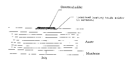
Figure 5a shows a rat tibial nerve welded by the
laser solder methods of the present invention. The
solder and the membrane are denatured but no significant
change to the axons has occurred (x100 Giemsa).
Figure 5b shows a rat sciatic nerve joined by
microsuturing using 10-0 nylon. Localised perineural and
~nn~ 1 damage occurs.
CA 02210894 1997-07-18
W096/220S4 PCT/AU96/00024
Figure 6 shows in schematic form joining of a blood
vessel using internal biodegradable solid solder cylinder
and external solid solder strips.
Figure 7 shows in schematic form a cross-section of
a repaired nerve fascicle.
Figure 8 shows the method used for measuring tensile
strength of repaired nerves.
Figure 9 shows a solid solder strip positioned upon
a severed rat tibial nerve just prior to laser welding.
Figure lOa shows regeneration of myelinated axons in
a laser nerve repair that has regenerated for 3 months.
Figure lOb shows fibrous tissue around a suture in a
sutured nerve that has regenerated for 3 months.
Figure 11 shows muscle action potential results for
repaired nerves.
BEST ~ ~O~ OF CA~URYING O ~ T~E INV~:N11ON
Tissue repair is performed using a laser to activate
a protein solder applied across the ~issue edges to be
joined. This solder denatures upon laser irradiation and
bonds with itself and the neighbouring membrane to form
the join. The procedure is shown schematically in
Figures 3 and 7 for a repair to a nerve fascicle. The
solder is applied in longitn~;n~l strips across the join.
Nerve Repair
Repair to severed nerve tissues is effected by the
placement of solder across the severed edges and exposure
of the solder to laser as described above. In order to
repair nerve tissue without damage to the contents of the
nerve it is desirable to avoid concentrating the weld on
the edges as extruded nerve contents may be damaged.
Rather the weld should be distributed across the planar
or tubular surface in which the discontinuity lies.
CA 02210894 1997-07-18
W O 96/22054 PCT/AU96/00024
HQ11OW Body Tube Repair
When repairing hollow body tubes it is preferable to
insert an internal cylinder of solder into the tube so
that it lies between the discontinuity. The severed ends
of the tube are placed over opposite ends of the solder
cylinder. The arrangement is shown in Figure 6.
Lasering can then be effected to cause bonding of the
cylinder to the tube being joined while the arrangement
is held in place. If there is a good fit between the
tube and the cylinder this laser step may not be
required. The join is completed by the addition of
external solder as for nerve repairs.
Tissue Surface Repairs
Surfaces such as peritoneum are planes of tissue in
which joins without sutures can be achieved by the
application of solder across the discontinuities to be
joined and welding as described above. In this case it
is less important to avoid concentration of welding on
the edges.
~aser and Solder System Suited to Nerve Fasicular Repair
To denature the protein solder, a GaAs/GaAlAs laser
diode with a nn~;n~l power of 250 mW (Spectra Diode ~abs,
San Jose, California) is used. The laser light is
coupled into a 100 ~m diameter core optical fibre which
is hand held in a fibre chuck. The diode is operated in
continuous mode at 75 mW during the laser soldering.
Because this laser is Class 3b, and is not eye safe,
protective glasses must be worn at all times when using
this laser.
A suitable protein solder is a mixture of water,
albumin and indocyanine green (ICG) dye (Becton
Dickinson, Missouri). Indocyanine green has a m~;mllm
absorption coefficient at a wavelength of 805 nm of 2 x
105 M~1 cm~l. The percentages of albumin and dye compared
to the water were 110~ and 0.6~ respectively for fluid
solder. 210~ albumen was used in preparing solder
CA 02210894 1997-07-18
W096/22054 PCT/AU96/00024
- 13 -
strips. It is notable that ICG dye appears to
preferentially bind with the albumin ensuring that heat
is efficiently transferred to denature the protein
solder.
Laser Solderinq Technique
When conducting the surgery an operating microscope
or some form of magnification is preferable. For a laser
solder repair of a tubular join a section of thin gauze
material is placed under the join to assist in a rotation
technique. The tissue edges are prepared in accordance
with st~n~rd techniques for the tissue type and geometry
of the repair.
Using micro forceps the edges are aligned and butted
together. A 2 mm long stripe of ~luid solder is
"painted" longitll~;n~lly across the junction of the edges
using a 30 gauge needle freshly coated in the solder.
Alternatively a strip solder is laid across the join
using microforceps. The solid strip repair method is
simpler. A solid strip is held in special microforceps
and placed across the junction parallel to the length of
the structures to be joined. The laser output is then
directed at the solid strip and the .solid solder changes
colour signalling denaturation which causes it to adhere
to the underlying tissue membrane. The process is
repeated with further strips to ensure a strong union of
surface.
The diode laser output from the 100 ~m optical fibre
is then used in a 30 second continuous pass to denature
solid solder into a strip weld. At a diode output power
of 75 mW, the solid solder strip turns brown on the
surface and opaque underneath from the single pass,
signalling denaturation. When using fluid solder
denaturation occurs more quickly. A two second laser
pass can be sufficient to denature the fluid solder.
Generally a second layer of fluid solder is applied to
the strip in order to increase the strength of the weld
and the two second laser pass is repeated. The gauze
CA 02210894 1997-07-18
W096/220S4 PCT/AU96/00024
- 14 -
under the join is then used with the micro-forceps to
rotate the join so that other strips can be applied.
Preparation of Fluid Protein Solder
Composition: - Albumin (fraction V powder from
Sigma, St. Louis, Missouri) at least 100
to 110~ by weight compared to water.
- Indocyanine Green (Becton Dickinson,
Missouri) approximately 0.6~ by weight
compared to water.
~ Water (injection grade)
Procedure: A solution of ICG in water was prepared in
a minitube. The albumin was added to the tube. The
albumin and solution were mixed using a vortex mixer.
This causes the protein structure to change leading to
linkage of protein molecules to each other rather than to
water molecules.
Preparation of Solid Protein Solder
Composition: - Albumin (fraction V powder from
Sigma, St. Louis, Missouri) 210~ by weight
compared to water
- Indocyanine Green (Becton Dickinson,
Missouri) approximately 0.6~ by weight
compared to water
- Water (injection grade)
Procedure: The ICG was dissolved in the water and the
albumin was added to this solution in a minitube. This
combination was mixed using a vortex mixer and a needle.
The combination was mixed (for approximately 3 minutes)
until it became a homogenous, malleable, green paste.
The phase of the mixture changed und$r this mixing
technique to provide an almost solid composition with
mainly protein to protein linkages rather than protein to
water linkages. The system is no longer a solution at
this stage. The protein paste was malleable and could be
cut into strips for up to about 30 minutes after m;~;ng.
CA 02210894 1997-07-18
W096/22054 PCT/AU96100024
- 15 -
After this time the paste hardened due to dehydration and
became too hard to cut.
The resulting strips were between 50 and 100 ~m in
thickness, about 0.6 mm wide and 1.5 to 3.5 mm long. It
will be understood that where the strips are used in
m~n~;ng nerve fascicles that the desired width and length
are dictated by fascicle ~;m~n~ions. The width,
thickness and length mentioned here are those suitable
for use with a rat tibial nerve which has a diameter of
0.2 to 0.8 mm. The ratio of strip width to nerve
circumference is typically:
Width ~ 1/5 circumference
EXAMP~E 1
A 100 ~m core optical fibre-coupled 75 mW diode
laser operating at a wavelength of 800 nm has been used
in conjunction with a protein solder to stripe weld
severed rat tibial nerves, reducing the long operating
time required for microsurgical nerve repair. Welding is
produced by selective laser denaturation of the protein
based solder which contains the dye indocyanine green.
Operating time for laser soldering was 10 +/- 5 min.
(n=24) compared to 23 +/- 9 min. (n=13) for
microsuturing. The laser solder technique resulted in
patent welds with a tensile strength of 15 +/- 5 g,
while microsutured nerves had a tensile strength of 40
+/- 10 g. Histopathology of the laser soldered nerves,
conducted tmm~ tely after surgery, displayed solder
adhesion to the outer membrane with m;n;m~l damage to the
inner axons of the nerves. An in vi~o study, with a
total of fifty-seven adult male wistar rats, compared
laser solder repaired tibial nerves to conventional
microsuture repair. Twenty-four laser soldered nerves and
thirteen sutured nerves were characterised at three
months and showed successful regeneration with average
Compound Muscle Action Potentials (CMAP) of 2.4 +\- 0.7
mV and 2.7 +/- 0.8 mV respectively. Histopathology of the
in vivo study, confirmed the comparable regeneration of
CA 022l0894 l997-07-l8
W 096/22054 PCT/AU96/00024
- 16 -
axons in laser and suture operated nerves. A faster,
less da-maging and long lasting laser based anastomotic
technique is presented.
Material~ and Method~
1. Animals
A total of fifty-seven young adult male Wistar
rats weighing between 400 and 550 g at the outset
were used in this study. Thirty-four rats received
laser solder repair and the r~m~;n~ng twenty-three
received st~n~rd microsuture repair as detailed
below. Five rats of each repair method were used
for tensile strength measurements and light
microscopy immediately after surgery and the
r~m~;n;ng thirty-seven rats were subjected to a
study of functional recovery using electrophysiology
and histopathology.
2. T~ Rer solder sYstem
To denature the protein solder, a GaAs/G~ R
laser diode with a n~m~n~l power of 250 mW (Spectra
Diode Labs, San Jose, California) was used. The
laser light was coupled into a 100 ~m diameter core
optical fibre which was hand held in a fibre chuck.
The diode laser was mounted on a heat sink, and the
diode current and temperature were controlled by a
SDL-800 diode driver. The diode was operated in
continuous mode at 75 mW during the laser soldering,
corresponding to a m~;mnm power density of 955 W/cm
at the tissue. The laser output power was measured
with a Scientech (Boulder, Colorado) power meter.
~ecause this laser is Class 3b, and is not eye safe,
protective glasses were worn at all times when using
this laser.
The solder used in this study was an albumin
based protein mixture, also cont~; n; ng indocyanine
green (ICG) dye (Becton Dickinson, Missouri).
Indocyanine green has a m~; mnm absorption
-
CA 02210894 1997-07-18
WO96/22Q54 PCT/AU96100024
coefficient at a wavelength of 805 nm of 2 x 10⁵ M~
cm~l. It i5 notable that this dye appears to
preferentially bind with the proteins ensuring that
heat is efficiently transferred to denature the
protein solder.
3. SurqerY
Anaesthesia was maint~;nP~ during surgery using
a mixture cont~; n; ng Fluothane (4~ during induction,
2~ thereafter) in ~2 (lL/min~. Using a OPMI 7
operating microscope (Zeiss, West Germany) the
sciatic nerve of the left leg was exposed at the
sciatic notch so that the nerve branches could be
distingll;s~e~. The tibial branch, just below the
sciatic notch, was exposed from the surrounding
subcutaneous tissue for a length of 1 cm. For a
laser solder repair, a section of thin gauze
material was placed under the tibial nerve to assist
in rotation of the nerve, and for the suture repair,
a section of plastic was placed under the nerve to
allow easier suturing. The tibial nerve was then
severed with serrated micro-scissors and left for 3
minutes for the normal extrusion of axoplasm to
occur. This was then trimmed with the serrated
micro-scissors as required, after which the nerve
was repaired with either four laser solder strips or
four 10-0 perineurial sutures.
The laser solder method involved aligning both
stumps of the severed nerve with micro-forceps then
a 2 mm long strip of solder was "painted"
longitn~;n~lly across the junction of the severed
ends using a 30 gauge needle freshly coated in the
solder (Figure 3a). The diode laser output from the
100 ~m optical fibre was then used in a continuous
two second pass to denature the solder into a strip
weld. At a diode output power of 75 mW, the solder
was observed to turn brown on the surface and opaque
~n~rne~th from the single pass, signalling
CA 02210894 1997-07-18
W096/22054 PCT/AU96/00024
- 18 -
denaturation. A second layer of solder was applied
to the strip and the two second laser pass was
repeated. The gauze under the nerve was then used
with the micro-forceps to rotate the nerve so that
three other two layered stripes could be applied,
each approximately 90~ apart.
Seven rats were operated with a more advanced
version of the organic solder, which is still an
albumin based protein mixture but it has the
advantage to be dehydratated and cut into solid
rectangular strips (Figure 9). The average surface
area of the solder strips was 1.5 +\- 0.5 mm2 and the
thickness was 0.15 +\- 0.01 mm. Four strips were
positioned along the tibial anastomized nerve and
then radiated with the same procedure adopted for
the fluid solder. The solid strip was fused with
the perineurium of the tibial nerve by the laser
radiation, joining the extremites of the sectioned
nerve.
For all operation the time of anastomosis was
recorded and a photographic record was taken for
later reference. The ~n;m~l S were placed in their
cages with no restriction of movement for 3 months.
4. Immediate Measurement of Tensile Stren~th and
Histo~athology
In ten of the operated rats, the 1 cm long
section of the laser and suture repaired nerves was
harvested ; mm~ tely for tensile strength
measurements. Fine silk was tied to each end of the
tibial nerve. One end was then attached to a
calibrated force transducer (FT30C, Grass
Instruments, Quincy, Mass) and the other to a screw
driven translator (Figure 8) As the screw was
turned the translator would stretch the nerve in a
slow and steady m~nner. The applied tension was
observed on an oscilloscope connected to the output
of the force transducer. Tension was applied until
CA 02210894 1997-07-18
W096/22054 PCT/AU96/00024
- 19
the nerve separated, and the breaking force was
recorded. The nerves were kept moist, as upon
drying, the tensile strength can be increased.
For light microscopy the anastomosis site of the
tibial nerves were fixed in 5~ formalin, alcohol
dehydrated, imbedded in paraffin, longit~ n~l ly
sectioned and stained with either Masson's trichrome
or Giemsa.
5. Functional Assessment : Histopatholoqy and
Electrophysioloqy
Three months post operatively the rats were
reanaesthetised using the method described in
section 3. The site was exposed and the anastomosis
of the tibial nerve observed. The two other
branches of the sciatic nerve, the peroneal and
sural nerves were then severed so that only the
tibial nerve branch of the sciatic nerve could
conduct electrical stimulation of the sciatic nerve
to the muscles of the hind foot. Two days later the
rats were positioned on their side and insulated
from the table by a folded surgical drape. An
infrared lamp was used to maintain their rectal
temperature above 36~C.
A clinical electromyograph (Cadwell Sierra
EMG/EP) was used for stimulation and recording. Two
25 gauge stimulating electrodes were placed lOmm
apart on each side of the sciatic nerve above the
sciatic notch, near the hip. The nerve was
activated using rectangular pul~es (0.1 to 0.3 ms; 0
to 30 mA; 1 Hz). Compound muscle action potentials
(CMAPs) were recorded from the plantar muscles of
the foot in response to supr~m~;m~l stimulation of
the sciatic nerve. A set of three recording
electrodes were used. A 25 gauge ground electrode
was inserted subcutaneously between the stimulating
and recording electrodes 1, 2. A 30 gauge reference
electrode was inserted into the heel pad and a 30
CA 02210894 1997-07-18
W096/22054 PCTIAU96/0002
- 20 -
gauge recording electrode was inserted into the
plantar muscles of the foot . The CMAPs were
recorded and processed to determine their negative
wave peak value.
Histopathology of the sutured and laser soldered
nerves, was conducted after the Electrophysiology
test with the same procedure as adopted in section
4.
Results
At the completion of surgery all anastomoses were
successful. The operating procedure was found to be
easier for laser soldering than for microsuturing. This
resulted in the shorter operating times for laser solder
repairs {10 ~ 5 min (n=24)} than {23 + 9 min (n=13)} for
microsuture repairs. The tensile strength of five laser
solder repaired nerves immediately after the operation
was 15 ~ 5 g and the tensile strength of the microsutured
nerves, 40 i 10 g.
Histopathological ~Am; nA tion of the anastomosis
sites ;mm~; Ately after surgery ~mm~trated that the
albumin and ICG dye based laser solder does bond well
with the outer membrane of the nerve, the perineurium,
while the inner axons remain unheated. In Figure 10a, a
tibial nerve fascicle weld produced by the diode laser
and albumin/ICG dye solder is shown in section. Both the
protein solder and the perineurium have denatured forming
the bond. On the lower side of the bond, the axoplasm
has its normAl wavy structure. Note that since heating
is concentrated at the dye, only denaturation of the~0 solder and adjacent perineurium occurs.
One of the promising aspects of laser anastomosis is
the potential for reduced damage to the axoplasm by
removing the need for sutures. A section showing the
effect of microsuturing nerve fascicles using 10/0-nylon
is shown in Figure 5b. This section stained with Giemsa,
displays axon extrusion at the join, as well as localised
perineurial and A~on~l damage due to the suture.
CA 02210894 1997-07-18
W096/22054 PCT/AU96/00024
Histopathology at 3 months shows regeneration of
myelinated axons in laser nerve repairs (Figure lOa),
with no discontinuity of either the fibers and their
sheaths, or the fibrous perineurium. No evidence is seen
of inflammation or myelin phagocytosis. Full
restoration, as assessed by light microscopy, of the
histologic integrity of the tibial nerve has been
achieved by the laser weld.
The sutured nerves also show successfull anastomosis
with myelinated axon regeneration, however, it is still
evident that the nylon thread is surrounded by fibrous
tissue, which creates an obstacle to the directionality
of the regenerated axons (Figure lOb).
The electrophysiological measurements of the in vivo
study were performed on twenty-four laser solder repaired
rats and thirteen microsuture repaired rats having three
months recovery. Of this group all twenty-four laser
solder anastomoses were patent as were the thirteen
microsuture anastomoses. The average amplitude of the
muscle action potentials, resulting from suprAmA~;mAl
stimulation of the nerve above the repair site was 2.4
+/- 0.7 mV for the twenty-four laser soldered tibial
nerves and 2.7 +/- 0.8 mV for the thirteen microsutured
nerves. The normal muscle action potential produced by
stimulating the tibial nerve suprAm~;mAlly was recorded
at 8.7 _ 3 mV from ten rats (Figure 11).
Di~c--~R~ ~n
Clinically, when a major peripheral nerve is
severed, forty or more fascicles may need to be
individually rejoined. With three or four microsutures
per fascicle, suturing tends to be prolonged, as it must
be meticulous. In a nerve graft, where two anastomoses
are needed, the suturing time is doubled. We have sought
a suitable method of nerve anastomosis that could at
least duplicate the end result but was significantly
faster than the present hand sewing microsuture
technique. A bonus of the described laser soldering
CA 02210894 1997-07-18
W096/22054 PCT/AU96/00024
method was the ~Pm~n.~trated lack of change to the A~on~1
components beneath the denatured perineurial layer seen
; mm~ tely after surgery. Three months later comparable
regeneration was demonstrated by electrophysiological
nerve conduction studies.
lNV~l~IAL APPLICABILITY
The present invention has application in the field
of surgery where it is of application in joining together
tissue edges, in end to end, side to end and side to side
applications.
-
CA 02210894 1997-07-18
W096/22054 PCTIAU96/00024
S
1) R. Malik, S. Ho and D.B. Church: A new method for
recording and analysing evoked motor potential from
dogs. Journal of Small Anim~7 Practice (1989) 30,
13-19.
2) R. Malik, S. Ho: Motor nerve conduction parameters
in the cat. ~ournal of Small Anim~7 Practice (1989)
30, 396-400.
~0 3) Laser activated protein bands for peripheral nerve
repair. A hauto, R Trickett, R Malik, J Dawes, E
Owen. European Biomedical optics Week - BIOS Europe
195 12-16 September 1995 (Proceeding in Press)