Note: Descriptions are shown in the official language in which they were submitted.
P-2581 PCT
CA 02211595 1997-07-28
1
DUAL CHAMBER PACING SXSTEM WITH
OPTIMIZED ADJUSTMENT OF THE AV ESCAPE INTERVAL FOR
CARDIOMYOPATHY
BACKGROUND OF THE INVENTION
This invention relates to cardiac pacing systems and methods generally and,
in particular, to dual chamber cardiac pacing systems and methods for
delivering
ventricular pacing pulses synchronized to atrial signals so as to benefit
patients with
cardiomyopathy and forms of congestive heart failure (CHF), and in particular
Hypertrophic Obstructive Cardiomyopathy.
Hypertrophic Obstructive Cardiomyopathy (HOCM) is characterized by a
narrowed left ventricular outflow tract (LVOT), which causes a significant
increase in
the left ventricular end systolic pressure. The narrowed LVOT is caused by an
increased thickness of the interventricular septum which obstructs blood flow
during
systole, the time of cardiac ejection.
Symptomatic improvement of patients with HOCM can be obtained in some
cases with the use of standard pharmacotherapy. However, drugs in use for this
therapy have disadvantages which have been cited in the literature. Likewise,
surgical
intervention, e.g., septal myectomy or mitral valve replacement, is another
optimal
treatment. However, such surgical treatments carry a significant operative
mortality
and have not been shown to alter the natural history of the disease. See,
"Permanent
Pacing As Treatment For Hypertrophic Cardiomyopathy, " by Kenneth M. McDonald
et al., American Journal of Cardiology, Vol. 68, pp. 108-110, July 1991.
The value of dual chamber cardiac pacing and treatment of patients
suffering from HOCM has been recognized in the literature. Modern multiple-
mode,
dual-chamber cardiac pacemakers are designed to maintain AV synchrony for
damaged
or diseased hearts that are unable to do so on their own. For example, a DDD
pacemaker has electrical connections to both the atrium and the ventricle,
senses
electrical signals in both chambers of the patient's heart, and delivers
atrial pacing
stimuli in the absence of signals indicative of natural atrial activation, and
ventricular
pacing stimuli in the absence of signals indicative of natural ventricular
activation.
Such a dual chamber pacemaker maintains the AV synchrony of the heart by
delivering
ventricular pace pulses at a controlled AV interval following each atrial
event.
AMENDED SHEET
CA 02211595 2000-10-10
X6742-624
2
Studies have indicated that patients suffering from
HOCM may benefit from a specific mode of dual chamber pacing
wherein a ventricular pacing pulse is delivered in timed
synchrony with the sensed or paced atrial depolarization.
Pacing the right ventricular apex before spontaneous atrio-
ventricular conduction activates the ventricles is understood
to alter the ventricular septal activation pattern. Since the
right ventricle is caused to contract first, it pulls the
septum toward the right ventricle thereby reducing the LVOT
obstruction.
The literature uniformly acknowledges the potential
advantages of synchronized A-V pacing for HOCM patients,
stressing the importance of achieving ventricular capture.
Causing "complete ventricular capture" is important to obtain
the above-described septal movement, while selecting the
longest AV delay that results in complete ventricular capture
is important in order to maximize the atrial contribution to
ventricular filling. The delivered pacing pulse should provide
"pre-excitation," i.e., depolarization of the ventricular apex
before the septum. This altered pattern of septal contraction,
as well as optimal left ventricular filling, is generally
recognized as being important to this mode of pacemaker
treatment. Further, it appears to be established that such
synchronized AV pacing provides HOCM patients a longterm
benefit, i.e., the benefit remains even after cessation of
pacing, since such AV pacing causes a reduction in the
obstruction of the LVOT which persists in sinus rhythm after
cessation of pacing. However, the duration of the benefit is
not certain.
CA 02211595 2000-10-10
X6742-624
2a
The literature suggests that the AV escape interval
should be set at the longest duration that maintains
ventricular capture at different exercise levels. See the
above-cited McDonald article. It has been suggested that the
AV escape interval which allows for maximal pre-excitation of
the ventricle by the pacing pulse can be selected by
determining the AV escape interval that produces the widest
paced QRS complex duration, as seen on a surface
electrocardiogram. See "Impact of Dual Chamber Permanent
Pacing in Patients With Obstructive Hypertrophic Cardiomyopathy
With Symptoms Refractory to Verapamil and ~-Adrenergic Blocker
Therapy," by Fananapazir et al., Circulation, Vol. 8, No. 6,
June 1992, pp. 2149-2161.
CA 02211595 2000-10-10
X6742-624
3
In the U.S. Patent N0. 5,507,782 assigned to
Medtronic, Inc. and issued April 16, 1996, the pacemaker
periodically checks to determine a value of intrinsic AV
conduction time (AVC) and subtracts therefrom a ventricular
sense offset interval (VSO) to get the AV escape interval.
After a waveform of the ventricular depolarization resulting
from complete capture is noted and recorded for comparison, the
AV escape interval is set to a lengthened value, resulting in
one or more ventricular sense events. The value of AVC is
determined as the time difference between the atrial event and
the sensed R-wave. Following this, the pacemaker AV escape
interval is reduced further until the pacemaker finds an R wave
with a waveform that indicates good capture. The difference
between AVC and the capture value of A-V is VSO, and the
pacemaker thereafter sets AV = AVC - VSO.
The prior art techniques for AV synchronous pacing of
HOCM patients recognize the necessity to periodically evaluate
the AV delay, or AV escape interval. The patient's spontaneous
atrio-ventricular conduction time generally will change with
heart rate, i.e., from rest to exercise. Moreover,
simultaneous drug treatment such as beta blockers may also
modify AV conduction time and require renewed evaluation of the
AV delay. The importance of periodically making an accurate
determination of the optimized AV interval thus takes on
significance. If the AV delay is adjusted to a value which is
too short, in order to ensure complete ventricular capture, the
atrial contribution to ventricular filling may be compromised.
However, if the AV escape interval is adjusted to too great a
value, ventricular capture is compromised, and there may be
episodes of no ventricular pacing or the ventricular pace may
CA 02211595 2000-10-10
66742-624
3a
not contribute the best possible reduction of the LVOT
obstruction. Accordingly, it is important in this therapy to
be able to continuously or periodically adjust the AV escape
interval to optimize it for HOCM therapy.
CA 02211595 1997-07-28
WO 96/25975 . PCT/US96/00951
SUMMARY OF THE INVENTION
This invention provides an apparatus and method for adjustment of the AV
delay for dual chamber pacing therapy in patients with HOCM. The apparatus and
method are based upon an improved method for determining the optimum AV escape
interval, including both the means of detecting data from which the optimum
interval
can be derived, and the operating algorithm for fording an optimized operating
value of
AV delay. The terms AV delay and AV escape interval (AVes~) are used
interchangeably.
In a first preferred embodiment, the pacemaker and method of this invention
locate the far field R-wave sense (FFRS) and utilize data from the FFRS
signals for
determining the optimum AV interval. As is known, the FFRS is a
representation, or
measure of the QRS, but sensed in the atrium. More specifically, one
embodiment is
based upon our observation that patients with HOCM and like conditions are
likely to
produce an FFRS which is late relative to the delivered ventricular pacing
pulse.
Accordingly, a method of the invention is to adjust the AV interval through a
series of
respective values, and measure the time between each ventricular pacing pulse
and the
following FFRS or QRS, i.e., the VP-FFRS or VP-QRS time. The pacemaker
determines the AVesc corresponding to the longest VP-FFRS time, which longest
time
corresponds to the latest septal activation and accordingly represents an
optimized value
of AV escape interval. The AVesc is then reset in accord with the determined
optional
AV value. More specifically, the pacemaker incorporates an algorithm for
determining
the knee of the VP-FFRS or VP-QRS curve, and sets the AV interval to a value
just
slightly less than the knee. Likewise, the FFRS duration, or QRS duration or
"width"
reaches a maximum value as the AV interval is shortened to about the longest
value
consistent with good capture. A second method thus involves similarly
adjusting the
AV escape interval, e.g., scanning from a relatively high AV value resulting
in natural
ventricular depolarizations, toward shorter values which result in capture and
evoked
R-waves, and measuring corresponding values of FFRS or QRS duration. After the
duration data is obtained from the scan, an algorithm analyzes the data and
determines
the AVes~ value corresponding to the breakpoint where QRS or FFRS duration
reaches
a high value plateau.
'~ 4
CA 02211595 2000-10-10
66742-624
The invention can be practiced either by adjusting AV
escape interval when the patient presents for programming, or
when the patient is ambulatory. In the case of a patient whose
pacemaker is in communication with a programmer, the algorithm-
s driving data may be obtained from the ECG as recorded from skin
electrodes which are connected to the programmer; from sub-Q
electrodes as used in a syncope monitor; or from the far field
electrogram as recorded from the atrial channel of the
pacemaker and communicated to the programmer. The programmer
collects and displays the appropriate data so that the
physician can inspect it and pick the desired AV setting or,
alternatively, the pacemaker system can automatically select
the optimum setting and present it to the physician as a
recommended value. In the case of an implanted pacemaker, the
pacemaker can continuously or periodically, e.g., once a day or
more frequently, determine a new adjusted AV escape interval
and override the previously programmed value.
The invention may be summarized as a dual chamber
pacemaker system, having atrial sense means for sensing signals
from a patient's atrium, ventricular sense means for sensing
ventricular signals from a patient, ventricular pace means for
generating and delivering ventricular pacing pulses to said
patient's right ventricle, AVesc means for setting and timing
an AV escape interval from the occurrence of a sensed atrial
signal, sync control means for controlling delivery of
ventricular pacing pulses at the time out of said AV escape
interval in the absence of a sensed ventricular signal, and
FFRS, QRS or ventricular septal pre-excitation sense means for
detecting FFRSs, QRSs or ventricular septal pre-excitation
following delivered ventricular pacing pulses, further
CA 02211595 2000-10-10
X6742-624
5a
comprising analyzing means for analyzing said detected FFRS,
QRSs or ventricular septal pre-excitation and determining from
variations in said detected FFRS, QRSs or ventricular septal
pre-excitation therefrom an indication for adjustment of said
AV escape interval, said AVesc means having adjusting means for
adjusting said AV escape interval in accordance with said
indication.
BRIEF DESCRIPTION OF DRAWINGS
Figure 1 is a perspective representation of the
pacemaker system of this invention showing an implantable
pacemaker connected to a patient's heart.
Figure 2 is a block diagram of the pacemaker system
of this invention, showing a pacemaker inter-connected with an
external programmer and with ECG leads.
Figure 3 is a block diagram of the primary functional
components of a pacemaker used in the system and method of this
invention.
Figure 4A is a generalized flow diagram illustrating
steps taken in synchronous pacing in accordance with this
invention, including adjusting AV escape interval for
optimizing HOCM therapy; Figure 4B is a flow diagram
illustrating the primary steps of a pacemaker routine which
includes searching to determine a HOCM-optimized AV escape
interval.
Figure 5A is a representative data plot of QRS or
FFRS duration as a function of pacemaker AV escape interval;
Figure 5B is a representative plot of VP-FFRS or VP-QRS time
interval as a function of pacemaker escape interval.
CA 02211595 2000-10-10
66742-624
5b
Figure 6A is a flow diagram illustrating steps taken
by the pacemaker system of this invention in acquiring data for
a determination of AV interval adjustment; Figure 6B is a flow
diagram of a routine for determining optimized AV escape
interval
CA 02211595 1997-07-28
WO 96/25975 PCT/US96100951
from data representative of FFRS or QRS duration; Figure 6C is a flow diagram
of a
routine for determining optimized AV escape interval from data representative
of the
time interval between ventricular pace pulses and evoked QRS or FFRS signals.
nFTAILED D SCRIPTION OF THE PREFERRED EMBODTMFNTS
Figure 1 illustrates the external configuration of a dual chamber pacemaker
6, which is provided with a hermetically sealed enclosure 8, typically
fabricated of
biocompatible metal such as titanium. Mounted to the top of the enclosure 8 is
a
connector block assembly 12, which receives electrical connectors located on
the
proximal ends of leads 14 and 16. bead 16 is an atrial pacing lead, carrying
two
electrodes 20 and 21. Electrodes 20 and 21 are used both to sense atrial
depolarizations
and to deliver atrial pacing pulses. Atrial pacing pulses may be delivered
between
electrode 20 and electrode 21 or between electrode 21 and the housing 8 of the
pacemaker 6. Sensing of atrial depolarizations may occur between electrode 20
and
electrode 21 or between either of electrode 20 and 21 and the housing 8 of the
pacemaker 6. Also, alternately, FFRS signals may be detected by electrodes
placed at
other positions, e.g., at locations 24, 25.
Similarly, lead 14 represents a ventricular bipolar pacing lead, carrying two
electrodes 28 and 29. As discussed above in conjunction with atrial lead 16,
electrodes
28 and 29 are used to sense and pace the ventricle. Ventricular pacing may be
accomplished between electrodes 29 and 28 or between electrode 29 and the
conductive
housing 8 of pacemaker 6. Sensing of ventricular signals, including
depolarizations
(QRS-waves) and repolarizations (T-waves) may be accomplished between
electrodes
29 and 28 or between either of electrodes 29 and 28 and the housing 8 of the
pacemaker
6.
As discussed in the present application, the preferred embodiments of the
pacemaker 6 operate in a DDD or DDDR pacing mode, wherein pacing pulses are
delivered to both atrium and ventricle and wherein atrial and ventricular
depolarizations
are both effective to inhibit delivery of the next scheduled pacing pulse in
the chamber
in which they are detected. While the present invention is believed optimally
practiced
in a pacemaker operating in DDD pacing mode, in some patients there may also
be a
benefit to operating the device in VDD or DVI mode, which provides ventricular
pacing pulses synchronized only to sensed atrial depolarizations or only
delivered to
6
CA 02211595 1997-07-28
WO 96/25975 PCT/US96100951
atrial pacing pulses, respectively, depending upon the specific underlying
heart
condition of the patient. However, DID mode~is expected to be the mode most
widely
used to practice the present invention.
Figure 2 illustrates the pacemaker 6 in block diagram form, coupled to a
human heart 10, in conjunction with an external programmer/display apparatus
corresponding to those typically employed to program modern, multi-
programmable
implantable pacemakers. Within the housing of the pacemaker are located the
pacing
circuitry 320, which includes circuitry performing all of the basic timing,
stimulation
and sensing functions of a cardiac pacemaker and a microprocessor circuit 302,
which
controls the timing intervals provided by the pacing circuitry 320. Pacing
circuitry 320
also includes a bidirectional telemetry circuit coupled to an antenna 334,
allowing
transmission of information from external programmer 4 into the pacemaker 6 to
modify its parameters and allowing transmission of information from the
pacemaker 6
to the external programmer 4, again generally corresponding to telemetry and
programming systems presently existing in commercially marketed mufti-
programmable
in implantable pacemakers.
The programmer 4 also includes a corresponding antenna 100 coupled to a
telemetry/antenna driver circuit 102 which serves to demodulate telemetry
signals
received from antenna 334 of the pacemaker, and to apply them in parallel or
serial
digital format to input output (I/O) unit 108, where they in turn may be
applied to a
video monitor 112 via graphic interface 110, and/or provided to central
processing unit
114 and/or printer 118. Microprocessor 114 controls the operation of the
prog_r_a_Itl_m__e_r,/d_i_cpl_ay apparatu_C~ and 1C rPCpllnClVe t(1 phys,'_rian
enter rl ~~m~,~, anrle vio
eu ~aan~cauu.7 - ~ 1u
keyboard 116, for controlling programming signals sent to the pacemaker, as
well as
for controlling operation of the video display 112 and printer 118. Also
illustrated is
an ECG interface 104, coupled to three ECG electrodes 106 which can be placed
upon
the patient's body. ECG interface 104 provides sensed electrograms to
input/output
device 108, where they in turn may be provided to the video display 112, the
central
processing unit 114 or the printer 118. The ECG capability is used for
treatment
according to the method of this invention for a patient who is available for
initial or
subsequent programming.
7
CA 02211595 1997-07-28
WO 96/25975 PCT/US96/00951
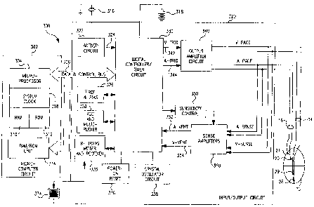
Figure 3 is a block functional diagram of the pacemaker illustrated in Figure
l, as connected to a human heart 10. The circuitry illustrated is all located
within the
conductive housing or can 8 of the pacemaker, as illustrated in Figure 1, and
the
bipolar leads 14 and 16 are illustrated schematically as coupled directly to
the circuit.
However, of course, in the actual device they would be coupled by means of
removable
electrical connectors inserted in the connector block 12, as illustrated in
Figure 1.
The pacemaker is divided generally into a microcomputer circuit 302 and a
pacing circuit 320. A pulse generator circuit 340 includes a ventricular pulse
generator
circuit coupled to the heart 10 by means of electrodes 29 and 28 on lead 14,
as well as
an atrial pulse generator circuit coupled to the heart 10 by means of atrial
electrodes 20
and 21, located on lead 16. Similarly, pacing circuit 320 includes atrial and
ventricular
sense amplifiers in sense amplifier circuit 360, coupled to the atrium and
ventricle by
means of leads 14 and 16 as well. The ventricular sense amplifier provides for
separate
detection and identification of QRS-wave signals, in a known manner; it may
also
provide for detection and identification of T-wave signals. The atrial sense
amplifier
provides for respective identification of P-waves and FFRS signals. The output
circuit
340 and sense amplifier circuit 360 may contain pulse generators and sense
amplifiers
corresponding to any of those presently employed in commercially marketed
cardiac
pacemakers. Control of timing and other functions within the pacemaker circuit
is
provided by digital controller/timer circuit 300, which includes a set of
timers and
associated logic. Digital controller/timer circuit 330 defines the basic
pacing interval of
the device, which may take the form of an A-A escape interval initiated on
atrial
sensing or pacing and triggering atrial pacing at the expiration thereof, or
may take the
form of a V-V escape interval, initiated on ventricular sensing or pacing and
triggering
ventricular pulse pacing at the expiration thereof. Digital controller/timer
circuit 330
similarly defines the A-V escape interval, AVesc, discussed in detail below.
The
specific values of the intervals defined are controlled by the microcomputer
circuit 302
by means of data and control bus 306. Sensed atrial depolarizations and FFRSs
are
communicated to the digital controller/timer circuit 330 on A event line 352;
and
ventricular depolarizations (QRS-waves) are communicated to the digital
controller/timer circuit 330 on V event line 354. In order to trigger
generation of a
ventricular pacing pulse, digital controller/timer circuit 330 generates a
trigger signal
8
CA 02211595 1997-07-28
P-2581 PCT
9
On V trig line 342. Similarly, in order to trigger an atrial pacing pulse,
digital controller/timer circuit 330 generates a trigger pulse on a trig line
344.
Digital controller/timer circuit 330 also defines time intervals for
controlling operation of the sense amplifiers in sense amplifier circuit 360.
Typically,
digital controller/timer circuit 330 will define an atrial blanking interval
following
delivery of an atrial pacing pulse, during which atrial sensing is disabled,
as well as
ventricular blanking intervals following atrial and ventricular pacing pulse
delivery,
during which ventricular sensing is disabled. Digital controller/timer circuit
330 will
also define an atrial refractory period during which atrial sensing is
disabled, this
refractory period extending from the beginning of the A-V escape interval
following
either a sensed or paced atrial depolarization, and extending until a
predetermined time
following sensing of a ventricular depolarization or delivery of a ventricular
pacing
pulse. Digital controller/timer circuit 330 similarly defines a ventricular
refractory
period following ventricular sensing or delivery of a ventricular pacing
pulse, which is
typically shorter than the portion of the atrial refractory period following
ventricular
sensing or pacing. Digital controller/timer circuit 330 also controls
sensitivity settings
of the sense amplifiers 360 by means of sensitivity control 350. In the
embodiment
illustrated in Figure 3, the pacemaker is provided with a piezo electric
sensor 316
which is intended to monitor patient activity, in order to allow provision of
rate
responsive pacing, such that the defined pacing rate (A-A escape interval or V-
V
escape interval) increases with increased demand for oxygenated blood. Sensor
316
generates electrical signals in response to sensed physical activity which are
processed
by activity circuit 322 and provided to digital controller/timer circuit 330.
Activity
circuit 332 and associated sensor 316 may correspond to the circuitry of the
type
disclosed in U.S. Patent No. 5,052,388, issued to Betzold et al., and U.S.
Patent No.
4,428,378, issued to Anderson et al. cited here to show what is known and may
be
used for this invention. Similarly, the present invention may be practiced in
conjunction with alternate types of sensors such as oxygenation sensors,
pressure
sensors, pH sensors and respiration sensors, all well known for use in
providing rate
responsive pacing capabilities. Alternately, QT time may be used as the rate
AMEr~IDED SHEET
CA 02211595 1997-07-28
P-2581 PCT
9-A
indicating parameter, in which case no extra sensor is required. Similarly,
the present
invention may also be practiced in non-rate responsive pacemakers.
AME;',~n-~; ~~EEi
CA 02211595 1997-07-28
WO 96/25975 PCT/US96100951
Transmission to and from the external programmer 4 illustrated in Figure 2
is accomplished by means of antenna 334 and associated RF transmitter and
receiver
322, which serves both to demodulate received downlink telemetry and to
transmit
uplink telemetry. Crystal oscillator circuit 338 provides the basic timing
clock for the
circuit, while battery 318 provides power. Power on reset circuit 336 responds
to
initial connection of the circuit to the battery for defining an initial
operating condition
and similarly, resets the operative state of the device in response to
detection of a low
battery condition. Reference mode circuit 326 generates stable voltage
reference and
currents for the analog circuits within the pacing circuit 320, while analog
to digital
converter ADC and multiplexor circuit 328 digitizes analog signals and voltage
to
provide real time telemetry of cardiac signals from sense amplifiers 360, for
uplink
transmission via RF transmitter and receiver circuit 332. Voltage reference
and bias
circuit 326, ADC and multiplexor 328, power on reset circuit 336 and crystal
oscillator
circuit 338 may correspond to any of those presently used in current marketed
implantable cardiac pacemakers.
Microcomputer circuit 302 controls the operational functions of digital
controller/timer 330, specifying which timing intervals are employed, and
controlling
the duration of the various timing intervals, via data and control bus 306.
Microcomputer circuit 302 contains a microprocessor 304 and associated system
clock
308 and on processor RAM circuits 310 and 312, respectively. In addition,
microcomputer circuit 302 includes a separate RAM/ROM chip 314. Microprocessor
304 is interrupt driven, operating in a reduced power consumption mode
normally, and
awakened in response to defined interrupt events, which may include delivery
of atrial
and ventricular pacing pulses as well as sensed atrial and ventricular
depolarizations.
In addition, if the device operates as a rate responsive pacemaker, a timed
interrupt,
e.g., every cycle or every two seconds, may be provided in order to allow the
microprocessor to analyze the sensor data and update the basic rate interval
(A-A or V-
V) of the device. In addition, in a preferred embodiment of the invention, the
microprocessor 304 may also serve to define variable A-V escape intervals and
atrial
and ventricular refractory periods which may also decrease in duration along
with
decreases in duration of the basic rate interval. Specifically, the
microprocessor is used
to carry out the routines illustrated in Figures 4A, 4B and 6A-6C.
P-2581 PCT
CA 02211595 1997-07-28
11
The illustrated circuitry of Figure 3 is merely exemplary, and corresponds
to the general functional organization of most microprocessor controlled
cardiac
pacemakers presently commercially available. It is believed that the present
invention
is most readily practiced in the context of such a device, and that the
present invention
can therefore readily be practiced using the basic hardware of existing
microprocessor
controlled dual chamber pacemakers, as presently available, with the invention
implemented primarily by means of modifications to the software stored in the
ROM
312 of the microprocessor circuit 302. However, the present invention many
also be
usefully practiced by means of a full custom integrated circuit, or any
combination of
hardware and software.
Referring now to Figure 4A, there is shown a generalized flow diagram of
steps taken by a pacemaker system in accordance with this invention in
performing
synchronous pacing, with adjustment of AVes~ for optimal HOCM therapy. The
steps
of this flow diagram are suitably carried out by microcomputer circuit 302.
This is a
simplified flow diagram setting forth only steps pertinent to controlling
AVes~, and
does not include many other steps and responses that occur during each cycle
of a
typical dual chamber pacemaker. The illustrated logic of Figure 4A recognizes
that
the intrinsic AV conduction time following an atrial pace pulse is greater
than
following a sensed atrial depolarization, by an amount described as "atrial
sense
offset", or ASO. The AVesc following an atrial pace is defined as PAV; the
AVesc
following an atrial pace is defined as PAV; the AVes~ following an atrial
sense is
defined as SAV; and PAV = SAV + ASO.
At block 401, the routine of Fig. 4A is waiting for what is expected to be
an atrial event. When an event occurs, the routine goes to block 402 and
determines
whether there has been timeout of the atrial escape interval, Aesc. If yes,
this indicates
that an atrial pace (AP) should be delivered, and this is done at block 404.
Following
this, the routine sets AVesc to PAV, and initiates timeout of AVes~. Returning
to 402,
if there has been no timeout of Aes~, the pacemaker proceeds to 408, and
determines
whether there has been an early ventricular sense (VS). If yes, the routine
branches to
block 409 and resets the timing appropriately, whereafter it returns to block
401.
However, as would normally be the case, if at 408 the event is not a VS,
meaning that
it has been an atrial sense (AS), the routine proceeds to block 410 and sets
AVes~ to the
At.~,cNDED SH~E~
CA 02211595 1997-07-28
WO 96/25975 PCT/US96/00951
current value of SAV. Following this, the routine goes to 412 and initiates
timeout of
the atrial escape interval (Aesc), and timeout of the AV escape interval,
AVesc (either
SAV or PAV). Then, at 414, the pacer waits for the next event, normally a
ventricular
event.
At 415, the pacemaker responds to an event by first determining whether the
event was a timeout of AVesc. If no, meaning that there was a ventricular
sense, the
pacemaker proceeds to block 417 and resets PAV and SAV to a shorter value
which
ensures capture by the next ventricular pace pulse. For example, each of these
values
can be decremented by 20 or 50 ms, to ensure that succeeding timeouts of AVes~
occur
early enough for complete capture. It is to be noted, however, that the
algorithms
discussed below are designed to avoid an occurrence of VS, such that the
pacemaker
should rarely take this path.
If at 415 there has been a timeout of Ves~, then the pacemaker proceeds to
block 418 and delivers a V pace. Then, at block 419, the pacemaker determines
whether it is programmed to go into the AV adjust routine. If no, the routine
is done
and it exists back to 401. If yes, the pacemaker goes to the adjust AV routine
at block
420. Here, the pacemaker analyzes collected data, e.g., VP-FFRS time; FFRS
duration; or FFRS or QRS amplitude. With this data in hand, the pacemaker
system
can adjust the values of PAV and SAV, in accordance with a predetermined
algorithm
for changing AVes~ so as to optimize resultant pre-excitation. Following this,
the
routine returns to block 401 and waits for the next atrial event.
Note that the pacemaker can be programmed for automatically monitoring
AV data and adjusting AVes~ each pacemaker cycle, or these steps can be taken
on some
other periodic or user-programmed basis, within the scope of the invention.
For an
implanted pacemaker which is set to automatically adjust AV, the pacemaker
goes
directly to 420. Similarly, for a pacemaker system in accordance with this
invention
which adapted to be programmed specifically by a physician, the routine exits
unless
the programming sequence has been activated.
Figure 4B is a simple flow diagram of the primary steps of an adjust AV
routine that includes a "search", or scan, whereby AVes~ is varied in accord
with a
predetermined program. At block 426, the pacemaker system monitors the data
from
which an indication of AV optimization is derived, e.g. FFRS duration or VP-
FFRS
12
CA 02211595 2000-10-10
66742-624
13
time. Following this, at 427, the monitored data is analyzed
and a decision is made as to whether the AV delay requires
adjustment based upon the monitored data. Specific embodiments
of this determination are set forth in Figs. 6A-6C. The
routine then branches to 428 and adjusts the value or values of
AV delay. However, if no adjustment is indicated, the routine
proceeds to 429 and determines whether AV search is to be
undertaken. If no, the routine exists, but if yes the routine
goes to block 430 and carries out a search whereby typically
the AV escape interval is incremented cyclically or every n
cycles toward a value corresponding to the patient's intrinsic
conduction. For example, AVesc can be incremented 5 ms every
cycle, or every n cycles, until either fusion is detected, or
there is a ventricular sense. Figure 6A gives a specific
example of a search.
Referring now to Figure 5A, there is shown a plot of
data representative of QRS or FFRS duration (ms) as a function
of pacemaker AV escape interval (ms). It is to be noted that a
particularly reliable measure of QRS duration can be obtained
from the FFRS signal in and around the "fusion" range between
full capture by the pacing pulse, and ventricular sense. As is
seen in Figure 5A, the QRS duration is relatively low at higher
AV intervals which are greater than the patient's intrinsic PR
conduction time, i.e., where a VS occurs before timeout of
AVesc~ However, as AVesc is shortened, it comes into a fusion
area where QRS increases up to a knee value (illustrated at
about 150 ms)~ at shorter intervals, where a VP results in full
capture, QRS duration is substantially constant. The portion
between full capture and failure to capture is termed the
fusion area, or range, and the ability to detect duration
10-31-00 09:21 1D=613 232 8440 P.02
66742-624
13a
changes in this area, as seen from FFRS signals, provides the
basis for one embodiment of this invention. Although Figure 5A
illustrates QRS data, the FFRS data corresponds directliy, and ,
~.n paxticular is characterized by the same knee, or breakpoint,
between the fl~sion range and the lower full capture ral~ge. The
knee is seen t4 be at the onset of fusion.
Referring now to Figure 5S, there is shown aiplot of
the time between a delivered ventricular pacing pulse ;(VP) and v
the sensed fFR6, i_e., ~t = VP - FFRS. The VP-FFRS duration is
measured from the time of delivery of the ventricular lacing
pulse to the time when the leading edge of the FFRS is~detected
to rise to a predetermined threshold amplitude. The variation
of VP-QRS follows the same form, .
ia5 31/10/2000 a 9:16 X613 232 8440 i0receiveri
CA 02211595 2000-10-31
CA 02211595 1997-07-28
WO 96/25975 PCTIUS96I00951
i.e., the duration is longest corresponding to short AV intervals when the
delivered
pacing pulse captures the heart, and drops during the fusion range. What is
important
is that the Ot/AV curve exhibits the same knee characteristic as seen in the
QRS/AV
curve of Fig. SA. As used herein, the phrase "VP-FFRS knee" refers to the
point on
the VP-FFRS vs. AV interval curve where VP-FFRS starts to drop from its
maximum
value toward lower values at higher AV intervals.
Referring now to Fig. 6A, there is shown a flow diagram of more detailed
steps for carrying out a search routine to obtain data from which an adjusted
SAV is
determined. At 515, the pacemaker system determines whether the ventricular
event
has been a V sense. If no, meaning that a ventricular pace pulse was
delivered, the
routine goes to block 535 and determines whether a search flag has been set.
If no,
meaning that no search is currently in operation, the routine goes to block
536 and
determines whether to initiate a search. A search may be triggered either by
an
external program signal, or by a signal generated automatically by the
pacemaker, e.g.
after a predetermined number of cycles or a predetermined amount of time. If
no
search is indicated, the routine exits. However, if a search is indicated, at
529 the
pacemaker first decrements the AV delay by a small increment 02, to provide
that the
search starts at an AV delay which is safely short of the fusion area.
Following this, at
537 the search flag is set.
Returning to 535, if it is found that the search flag is set, the routine goes
to
block 540 takes initial steps for obtaining data. For an embodiment which uses
VP-
FFRS time, the pacer starts a clock to time out the time from the delivered
ventricular
pace pulse to the detected FFRS. The pacemaker also generates a sense window
connected through control 350 for a predetermined duration adjusted to exclude
the T-
wave, e.g. up to 300 ms. The sense window acts on the atrial sense amplifier,
and the
FFRS is channeled through line 352 to circuit 330, where it is detected as
shown at
flow block 542. Following this, at block 544, the pacemaker system gets and
stores the
value of the applicable parameter, e.g., VP-FFRS time (TNX). Thus, the time is
obtained from the clock which had been set at 540, and the variable TNx is
stored. In
the embodiment where the width of the FFRS signal is utilized, this width is
obtained
from the FFRS signal and stored. In the embodiment where the amplitude of the
FFRS
is utilized, the amplitude is obtained and the variable ANx is stored.
i4
CA 02211595 1997-07-28
WO 96/25975 PCT/US96/00951
The steps 542, 544 of sensing and processing the FFRS signal are
accomplished by standard hardware, preferably also using digital processing
techniques. For getting the time of VP-FFRS, a standard edge detector may be
utilized
in circuit 330 to sense when the leading edge of the FFRS signal has reached a
predetermined level, or has increased by a predetermined percentage. For
determining
width, or duration, the signal is processed to determined when it first rises
to a
predetermined level, and when it falls back below such level. And amplitude is
measured by either a simple peak detector, or other standard amplitude
detection
circuitry. These standard circuits may be supplemented or replaced by known
digital
processing techniques, carried out with the aid of microprocessor system 302.
Following the operations at 544, the routine goes to 545 and determines
whether the variable X has reached a maximum. This variable corresponds to the
number of cycles that data has been taken at the same AV value. If X has not
yet
reached XmaX, e.g. 5, the routine increments X at 546. If X does equal X~X,
the
routine sets X equal to 0 at block 548, and at block 560 increments the value
of AVN,
setting AVN = AVN_1 + D3, where O3 is a predetermined increment, e.g. 2 or 5
ms.
At 561, N is incremented by 1, for purposes of accurate storage at block 544.
In this
manner, X measurements can be taken at N representative search values of
AVes~.
Returning to step 515, if a V sense is detected, AV is immediately
decremented at 528, e.g., by D2, = 20 ms, to prevent further cycles without
pacing
capture. At 552, the system determines whether the search flag has been set.
If no,
this means that there has been a V sense without a search, and the routine
exits. If yes,
this means that AV has been lengthened to the point where capture is lost. The
search
flag is reset at 554, and the variable N is set equal to 0 at 555. Then, at
560, the
system initiates the Find SAV routine, as described more fully in the
embodiments of
Figures 6B and 6C. Initiation of the Find SAV routine may be done
automatically
within the pacemaker, or the data can be downloaded to the programmer for
analysis
and determination of an optimum value of SAV.
Referring now to Figure 6B, there is shown a first embodiment of the Find
SAV routine 560 for obtaining an adjusted SAV as a function of FFRS width
(duration). At 601, the average width value (WN) corresponding to the X values
of
each AVN during the search is determined. This may be done by any suitable
CA 02211595 1997-07-28
WO 96/25975 PCT/US96/00951
processing technique, preferably obtaining a sample rolling average. Following
this
step for each value of N, resulting in M values of average width, the variable
N is set
equal to 1 at 602. At 604 the pacemaker compares the difference of WN-Wrr+i
against
a predetermined increment 0. This step thus determines whether the QRS width
(WN+1), as represented by the measured FFRS width, is significantly shorter
than the
value at the next shorter AV interval (WN). To allow for fitter and timing
vagaries, the
algorithm preferably is set to determine a substantial change in width as
being only
greater than D, e.g. 15 ms. If such a differential is not found, the routine
goes to 605
and determines whether N has stepped through the maximum number of values for
which data is available, i.e., N = M. If no, at 607N is incremented by 1 and
the
routine returns to step 604. At the point where the differential between
adjacent AV
values exceeds D, the routine branches to block 608 and determines a new SAV
to be
equal to the just prior value of AV, i.e. SAV (N - 1). Following this, the
determined
value of SAV is displayed at 610. Alternately, for an implanted pacemaker, the
new
value of SAV can be automatically adopted.
Referring back to the illustrative plot of Figure SA, for this data the
algorithm of Fig. 6B proceeds to the point where it determines that the AV
interval of
about 160 ms is the first to have an averaged width which falls outside the
allowed
range, i.e., the differential of AVN+i to AVN is greater than 0. The algorithm
then
assumes that the AV interval of about 150 ms, AV(N), is an optimum point, and
subtracts one AV interval increment to obtain AV (N-1), at approximately 140
ms. By
this means, an AV value at or just less than the knee is determined.
Referring now to Fig. 6C, there is shown a flow diagram that corresponds to
Figure 6B, but which determines the optimum value of SAV in terms of TN, the
time
between the ventricular pace pulse and the evoked response as detected through
the
FFRS (VP-FFRS). At block 620, the average of TN is obtained from the X
measured
values corresponding to each value of N. This produces an array of values of
TN
corresponding to the M different values of AVN utilized during the search
routine.
Following this, at 622 the variable N is set equal to 1. At 624, TN is
compared to
TN+1, to see if the difference is greater than a predetermined increment ~.
Note that as
AV interval increases, the system is looking for the knee corresponding to a
decrease in
TN. When this decrease first exceeds the predetermined increment; this
indicates the
16
CA 02211595 1997-07-28
P-2581 PCT
17
Onset of fusion, and the routine branches to block 628 and sets SAV equal
to SAV(N-1). The premise in this subroutine is the same as for Figure 6B,
i.e., the
first AV interval which corresponds to a substantial decrease in time is just
down the
slope from the knee. Accordingly, selecting the just prior value of AV,
corresponding
to TN+i, represents a factor of safety. It is seen that if no interval
difference as
computed at 624 exceeds the predetermined increment, the routine loops
continuously
until N = M, at which time the information is displayed. The physician can
inspect
this data and choose from it an optimum value of SAV.
There has thus been disclosed a pacemaker system and method for dual
chamber synchronous pacing optimized for cardiomyopathy therapy, and
particularly
for HOCM therapy. In a preferred embodiment of this invention, the pacemaker
system detects the FFRS and processes the signal to determine at least one
characteristic thereof. The system collects data representative of a selected
FFRS
characteristic or several characteristics, over a range of values of AV escape
interval,
which values include the fusion range or zone. The FFRS characteristic is
suitably
VP-FFRS time; FFRS duration; or FFRS amplitude; or any combination of these
variables. Thus, the parameter for deternlining AV may be X, where X = fn
(amplitude) + fn (duration) + fn (timing). In another embodiment of the system
and
method of this invention, the R-wave may be monitored directly and a
characteristic
derived from it, e.g. amplitude or VP-QRS time, in which case the system
utilizes
these characteristics in the same manner to determine the optimum adjustment
of AV
escape interval.
The novel technique of using the FFRS to determine optimal AVeS~ has been
illustrated with the preferred embodiment of scanning, or searching to
determine the
"knee" also sometimes called the bending point of the VP-FFRS curve, from
which a
new value of AVes~ is determined. However, the pacemaker of this invention
further
includes monitoring an FFRS characteristic, e.g., VP-FFRS time, or VP-QRS
time, to
determine when operation may be in the fusion range. Thus, referring back to
Fig.
4B, the monitored data can simply be inspected each cycle to see whether there
has
been a decrease in the interval, i.e., whether a shortening of the VP-FFRS
duration
indicates the onset of fusion. In this case, even though no search as such has
been
conducted in order to determine the knee, the pacemaker of this invention
senses the
AMENDED SHEET
CA 02211595 2001-04-12
66742-624
onset of fusion and adjusts AVesc bY shortening it. The scope
of the invention thus embraces ongoing cyclical monitoring of
an FFRS characteristic:, as well as searching to acquire batch
data from which an ac<~urate determination of the knee is
.'~ obtained.
It is further_ noted that the system and method as
claimed can utilize a number of different configurations.
Thus, an implantable pacemaker used in this invention can
contain hardware and/~:~r_ software for control of AVesc upon
1~ command from an external. programmer; upon command from a
"patient activator"; automatically, based on internal logic,
e.g., elapsed time or number of pacemaker cycles; or based on
some other parameter or criteria being met, e.g., change in one
or more sensor levels. Also, the practice of the invention
15 embraces the use of an external pacemaker and the like, and
available technology for transmitting data to and from the
patient location.
17a