Note: Descriptions are shown in the official language in which they were submitted.
CA 0221~600 1997-09-16
-1-
PROCESS AND DEVICE FOR DETERMINING THREE-DIMENSIONAL
STRUCTURES IN THE SUBMICRON RANGE
The invention relates to a method as stated in the preamble of Patent Claim 1, and a
device as specified in Patent Claim 8. Structures in the submicrometer range are generally
measured, for example, using sc~nning microscopes, wherein the object to be measured is moved
through a servo device and the surface structure of the object being measured is scanned using a
precision probe tip. The precision probes used in such methods are frequently damaged,
resulting in undesirable int~llul~Lions in operation. In addition, the forces exerted by the
precision probe on the surface of the object range from 0.1 to 1.10-9 newtons. Even these slight
forces can result in a shift in positions on the object being measured.
The object of the invention is to devise a method and a device that will not require a
mechanical element to scan the surface of the object being measured.
Rather than using geometrical optics in the invention, in contrast to optic~l microscopes,
the spatial and temporal complex amplitudes (intensity and phase angle distribution) proceeding
from an object to be measured are identified and processed.
The concept of different beam positions for the two partial beams is understood to mean
positions which generate different radiation fields created by ~ùpelilllposing the two partial
beams at the locus of the detectors on the detector field. The phase angles of these various
radiation fields differ at one and the same detector locus by different fractions of a complete
wave oscillation. Thus, from preferably at least 3 measurements taken at one and the same locus,
the amplitude and phase of the su~ illlposed field can be positively ~letPrmin~l Since one of
CA 0221~600 1997-09-16
the partial beams comes from the object being measured, the measured, superimposed radiation
field contains the information on the structure of the object.
As described in detail below, different beam positions can now be generated, for
example, within one beat cycle of a beat frequency state for the two partial beams which exhibit
slightly different radiation frequencies. Of course, one partial beam may also be slowed down
with regard to the other by wavelength fragments, and its beam configuration may be modified.
Such a deceleration can, for example, be effected using elecko-optical, acoustic-optical,
magneto-optical components, mechanical phase shifting elements, etc.
At least three measurements are preferably taken per locus for phase determination. It is
possible, however, to proceed with fewer measurements if the measurements from adjacent
detectors are compared with one another and used as ratios.
One practical application that uses spatial intensity and phase angle distribution is
known-in-the-art from holography. With the measuring method specified in the invention for
determining the structures of m~gnified objects, rather than viewing an interference structure
generated by a reference beam, the complex amplitude is determined point by point. This
complex amplitude is then used to calculate the associated phase-angle values from the measured
values. The phase values are superelevated using a multiplication factor that determines the
m~gnification. From these elevated phase values and the real components of the measured,
original complex amplitude, a second, complex amplitude is calculated, from which, using the
spatial coordinates of the detectors, a m~gnified structure of the measured object (as, for
example, a hologram) can be created which can then (following further calculations revisions) be
displayed via a plotter or some other image-generating device.
CA 0221~600 1997-09-16
-3-
Preferably, in determining the phase-angle values of the complex amplitude, using the
radiation that is reflected back from the structure of the object being measured, superimposed
beams exhibiting a low beat frequency are used. The beat frequency that is used is based upon,
among other things, the speed of the recording cycle for the individual detector values read from
the detectors of the detector field.
Below, examples of the process specified in the invention, along with the device used to
implement this process, are described in greater detail using the attached diagrams. Further
advantages of the invention are discussed in the following descriptive text. The diagrams show:
Fig. 1: a block diagram of the device;
Fig. 2: a system of coordinates used to calculate the superimposition of plane waves;
Fig. 3: an illustration of the calculation of phase relationships of plane and spherical waves,
based upon points in the environment of the focusing point of a lens; and
Fig. 4: an illustration of the computation of errors in calculating transversal resolution.
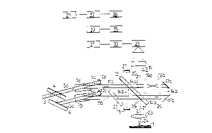
The device illustrated in Figure 1 for defining three-dimensional structures in the
submicrometer range of an object 1 uses coherent radiation, preferably coherent laser radiation.
The coherent beam 3 is split into two partial beams 5a and 5b using a beam splitter 4. A
deflecting reflector 6 is used to orient the partial beam 5b parallel to the partial beam 5a. Each of
the partial beams 5a and 5b then passes through an element 7a and 7b that is designed to shift its
radiation frequency. The frequency shift is effected via an acoustic-optical modulator 7a or 7b
for each beam. The acoustic modulation frequencies of the two modulators 7a and 7b differ here,
for example, by one hundred hertz. In other words, the radiation frequencies of the two beams fa
and fb are shifted toward one another by one hundred hertz. The transmitted beam 9a or 9b in the
CA 0221~600 1997-09-16
acoustic-optical modulator 7a or 7b that is not deflected by the density wave of the modulation
frequency is absorbed by an absorber 10a or 10b. Only the deflected beams 1 la and 1 lb having
the frequencies fa and fb are further processed. Preferably, however, radiation occurs below the
Bragg angle, so that nearly all of the irradiated energy falls within the first deflected level.
The beam 1 1 a now reaches a second beam splitter 13 . A first beam section 1 4a of the
beam 1 1 a is transmitted by this beam splitter and the second, other part 1 4b is reflected and
collected in an absorber 15. The partial beam 14a passes through two confocally oriented
identical lenses 1 6a and 1 6b, and, following a displaced re-reflection via the two mirrors 1 7a and
1 7b, is sent back to the beam splitter 13, from which it is reflected onto the detector field 19 of a
CCD [charge-coupled device] camera.
The beam 1 lb passes through a plane-parallel plate 20 that is inclined toward the axis of
the beam and is oriented parallel to the beam splitter 13, and impinges upon the rear side of the
beam splitter 13, striking it at the same location as the beam that has been reflected back by the
two mirrors 1 7a and 1 7b. At this point, a first partial beam 21 from the beam 1 lb is reflected and
is focused via a lens 23 onto the object 1 at the locus 24. The focusing diameter, depending upon
the laser beam that is used, is slightly less than one micrometer. As described below, the beam
25 is now reflected back from the locus 24, is converted to a greater or lesser extent by the
focusing lens 23 into a plane wave, is transmitted to the beam splitter 13, and becomes
superimposed with the beam 14a on the detector field 19.
One partial beam from the beam 1 lb which is not further illustrated here is transmitted as
a beam 26 to the beam splitter 13, is reversed by the two mirrors 1 7b and 1 7a, is transmitted to
CA 0221~600 1997-09-16
-5-
the two lenses 16b and 16a, is reflected back to the beam splitter, and is then absorbed by the
absorber 27.
On the detector field 19, the partial beam 14a (5a - 1 la) having the beam frequency fa,
and the beam 25 (5b - 1 lb - 21), which has been reflected back from the locus 24 on the object 1,
having the beam frequency fb, which differs from the frequency fa by one hundred hertz
(differential annular frequency Q = 2~ I fa - fbl ), now become superimposed.
In order to obtain a perfect superimposition, care must be taken to ensure that the optical
pathway I of the partial beam 5a - 11 a - 14a and the optical pathway II of the partial beam 5b -
1 lb - 21 - 25 lie within the coherence length of the beam 3. In order to prevent a dispersion of
the group velocities on the two pathways I and II, the path lengths are selected, through materials
other than air - for example the material of the beam splitters 4 and 13, and of the lenses 16a,
16b, and 23 - such that they are equal in length.
Pathway I thus has one (glass) tr~n~mi~ion thickness in the beam splitter 4 and three
(glass) tr~nsmi~ion thicknesses in the beam splitter 13, before reaching the detector field 19.
Pathway II has two (glass) tr~n~mi~ion thicknesses in the beam splitter 4, one (glass)
tr~n~mi.~ion thickness in the plane-parallel plate 20, and one (glass) transmission thickness in
the beam splitter 13, before reaching the detector field 19. Because of the identical optical
design of the two beam splitters 4 and 13, along with the plate 20, the two pathways I and II have
an equal number of (plane glass) "tr~n~mi~ion thicknesses."
On Pathway I the beam is transmitted once to the two lenses 16a and 16b. On Pathway II
the beam is transmitted twice to the lens 23. With the identical optical design of the lenses 16a,
16b, and 23, the two pathways I and II have the same number of (spherical) "tr~n.~mi~ion
CA 0221~600 1997-09-16
thicknesses." The optical material used for the plane-parallel plate 20, the beam splitters 4, 13,
and the lenses 23, 17a, and 17b (for example glass) should possess nearly identical optical
properties for the (laser) radiation used. This will prevent a dispersion of the group velocities.
The detector field is comprised, for example, of 1024 x 1024 CCD elements, which are
positioned at a distance of approximately 6.8 ,um from one another. These detectors are
connected to an evaluation unit 29, which automatically switches on the detectors three times
within one period of the 100 Hz beat frequency, reads out the measured values, and places these
values, per detector, in first memory units 30, with a corresponding number of detectors and
sc~nning cycles in at least 300'000 individual memory units.
Because now three measured values can be evaluated per beat frequency period and
detector (locus), the complex amplitude (intensity and phase angle) at each of the detectors can
be determined by a first calculating unit. The complex amplitude is a superimposition of the
radiation reflected off of the object being measured (beam 25) and the unaffected radiation (beam
14a). The structural information about the object l is contained in the complex amplitude that is
calculated.
The phase angle values now calculated for each detector are stored in second memory
units 33 (also with at least 300'000 individual memory units) along with the corresponding
amplitude values (intensity values), which require at least another 300'000 individual memory
units. The phase angle values stored in the second memory units 33 are multiplied, via a
multiplier unit 35, by a value that determines the m~gnification of the structure of the locus 24,
and are stored in the third memory units 36. With the m~nified phase angle values now stored
in the third memory units 36, and the associated intensity values from the second memory units
CA 0221~600 1997-09-16
33, using the mathematical algorithm, stored in a second multiplier unit 37, for a two-
dimensional Fourier transformation, such as is described in the publication by Ulf Schnars, et al.
"Digital Holography - a New Method of Laser Measuring Technology", Laser und
Optoelektronik [Lasers and Optoelectronics], 26(5), 1994, pp. 40 - 45; and in U. Schnars, "Direct
Phase Determination in Hologram Interferometry with Use of Digital Recorded Holograms", J.
Opt. Soc. Am. A, 11, (7), 2011 - 2015, July 1994, an image is calculated point by point, which
can be displayed via an output unit 39. The output unit 39 may be a screen or a plotter, for
example. The image generated here represents a m~gnification of the structure found at the locus
24. In contrast with traditional light-optical microscopic images, this image is no longer limited
by diffraction. It can illustrate spatial structures.
Below, some mathematical approaches are given to clarify the above submicroscopic
m~gnification process. To make it clearer, two plane wave fronts A and R, which are
superimposed at a single point at the distance z, are used. They can be plotted using the
following equations (1) and (2):
A = Ao ~ cos(wt - kz + (I)d) (l)
R = Ro cos[(w + Q)t - kz] (2)
Ao and Ro represent the given amplitude values for the radiation; w represents the lower annular
frequency of the radiation fa (w = 2~fa); (w + Q) represents the higher annular frequency of the
radiation fb, wherein Q represents the beat frequency. K is the wave vector.
The phase angle q) of a wave at a distance d from a reference point thus becomes shifted
in relation to the phase angle at this point by
(~)d= k d. (3)
CA 022l~600 1997-09-l6
A now represents the wave that proceeds from a point on the structure at the distance d
from a reference point, and R represents the reference wave. For a superimposition at the point
of the detector field 19, not accounting for the lens 23, the following now applies for the intensity
I measured by the detectors:
I=(A+R)2
I = (Ao cos(wt - kz + q)d)+ Ro cos[(w + Q)t kZ])2
The detectors now cannot follow the optical frequency fa or fb and thus form the average
value <I>, from the intensity that is received on them:
<I> = I/2Ao2 + I/2Ro2 + 2AoRo <cos[wt-kz+q)d] cos[(w+Q)t - kz]>
<I> = I/2Ao2 + I/2Ro2 + AoRo cos[Qt - q)d] (4)
Thus a beat signal AoRo cos[Qt - q)d]iS obtained, from which phase angle values having
a precision of 10-3 can be determined by experiment. In other words, an interval d can be
determined in accordance with the equation (3) d = 10-3/1~, using a laser wavelength of 500 nm,
with
10-3
d= 500 [nm] = 8 10-2 [nm].
2~
As described above, the phase angle is now determined in that the values of the detectors
are read out at equal temporal intervals, three times per beat frequency Q.
If the two waves A and R are plane waves that are inclined toward one another by an
angle o, then the following results at a distance s from a reference axis 41 that passes through the
reference position, similar to the above performance for the wave:
A = Ao cos [wt - k ~ s + (l)d] (5)
. CA 0221~600 1997-09-16
g
The reference wave R does not change.
I = (A + R)
I = (Ao cos(wt - k ~ s + ~d)+ Ro cos[(w+Q)t kz])2
The following results from the averaging process from the detectors:
<I> = I/2Ao2 + I/2Ro2+AoRo <cos[(2w+Q)t-k-~ s+q)d] cos[Qt+k-~ s-q~d]~
<I> = l/2Ao2 + l/2Ro2 + AoRo cos[Qt + k ~ s - q)d] (6)
The change in the phase angle q)tr at a position that is at the distance s from the reference
axis 41 (in the detector plane 19), thus yields
~ tr= k ~ s - ~d (7)
If q)d= Ois inserted, then for the angle-dependent phase shift only this follows:
~tr
~= (7a)
k s
Thus, the above assumptions, with a laser wavelength of 500 nm and a distance of 1 mm,
which corresponds approximately to the boundary detectors of the 1024 x 1024 CCD detector
field, yield a resolution of
10-3 500 [nm]
X = . = 8 . 10-8 (8)
2~ 1 [mm]
In order now to convert this angular resolution into a spatial resolution, the spherical lens
23 having a focal length f is used, for example. Waves origin~ting from the points Pl and P2 of
the object 1, as illustrated in Figure 3, are thus transformed into plane waves. The wave
CA 0221~600 1997-09-16
-10-
proceeding from the point Pl and having the coordinates x = O, y = O and z = O can be described,
analogous to the above equation (1), with
A=Ao cos(wt- kz) (9)
The wave proceeding from the point P2 and having the coordinates x = -h, y = O, and z =
O can be described, analogous to the above equation (5), with
A = Ao - cos (wt - k ~ h) (10)
The wave proceeding *om the point P3 and having the coordinates x = O, y = O, and z = g,
since P3 is no longer in the focus of the lens 23, is now no longer a plane wave, but is a spherical
wave at a distance z~, *om a virtual center. This distance Zv can be determined using the lens
equation, wherein f23 is the focal length of the lens 23:
1 + g/f23 g
~ - = (1 1)
Zv + f23 f23 f23 ~ g f23 f23 f 23
or
_f223 _f223
Zv = - f23 ~ ( 1 2)
g g
For g - O, in other words P3 slides into the focal plane, Zv opposes c~ and a plane wave is again
obtained. For a locus O in the detector plane 19 at distance d, a phase shift is yielded:
~ = k (~ Zv2 + U2 - Zv) (13)
- CA 0221~600 1997-09-16
Assuming that u <~ z~, the following is true:
k (~ 2+ U2 ~)= k (~1 + (u/~) - ~) =
k z, k.u2
= k (z~, (1 + I/2(ulz~)2) -Z~) = (ulz~,)2 =
2 2z~,
If equation (12) is inserted into this equation, then for the phase shift ~q with the wavelength lw
of the radiation
k.u2 k.u2.g -~ u2 g
= = = (14)
2z~, 2.~23 lw.f223
If the example values for u = lmm used above, which corresponds approximately to the
boundary detectors of the 1024 x 1024 CCD detector field, a wavelength l of 500 nm, and a focal
length for the lens 23 of f = 2 mm are inserted, then the following is true for the phase shift:
g
l~gl=
4 lw
Because phase shifts with a magnitude of 10-3 can now be determined using measurement
techniques, a resolution of
4 . lw
g = q)g = 0.64 nm
is obtained.
CA 0221~600 1997-09-16
-12-
A plane wave proceeds, after the focusing lens 23, at the angle ~ from point P2, which is
displaced transversally in relation to point Pl, and has the coordinates x = -h, y = 0, and z = 0.
The following thus applies:
h
=
f23
With equation (7a), it then follows that
~ tr lw ~tr' f23
h= f=
k-s 2~-s
If the sample values already used above of 1 = 500 nm, q)tr= 10-3, f23 = 2 mm, and s (or u) = 1
mm, are inserted into this equation, the result is a transversal resolution of 0.16 nm. This
resolution is greater than the elevated resolution g. This product is derived from the calculation
only by approximation.
In accordance with Figure 4, the following estimate can be employed with a focal length
of 2 mm and a distance of 1 mm between a (boundary) detector and the reference axis:
m=~f223+S2-f23=~22+ 12-2=0.23
This distance m can now be compared with the maximum distance f23 = 2 mm:
m 0.23
~ O. 1
f23 2
The above theoretical resolution is ~1imini~hed by this value 0.1.
CA 0221~600 1997-09-16
-13-
From these performances it can be seen that resolutions can be produced using the
method and the device specified in the invention that are considerably better than those produced
using the optical microscope, which is limited by the effects of optical diffraction.
Based upon the optical condition of the object 1, the above-described inventive
measuring method or the measuring device specified in the invention can now be used to
measure the surface structure or to determine the inner spatial radiation that has penetrated the
object 1.
In contrast to holographic measuring methods, the measuring method specified in the
invention is no longer assigned to detectable or recordable interferences between the measuring
beam and the reference beam. Using the calculated elevation of the phase values "the measuring
process creates the interferences necessary for the desired resolution itself."
Rather than generating the frequency shift of the two partial beams 5a and Sb toward one
another through one or two acoustic-optical modulators 7a and 7b, the positions of the two
mirrors 1 7a and 1 7b may be periodically changed. In addition, rotating gratings and electric-
optical modulators may be used, with the phase shift being dependent upon the applied voltage.
The beat frequency ¦ fa ~ fb ¦ should be selected to be as great as possible, so that thermal
or other path length changes between the two partial beams 5a - 1 la, 14a and 5b - 1 lb - 21 - 25
produce no measured value distortions. This is, however, limited by the reading speed and
storage speed of the measured values from the detectors, as well as their sensitivity.
Use of the two-dimensional Fourier transformation can be omitted. In that case, rather
than a directly viewable image, a holographic image is generated, which then can be viewed with
the corresponding coherent radiation.