Note: Descriptions are shown in the official language in which they were submitted.
CA 02215650 1999-04-27
PATENT
P-3693
DNA MICROWELL DEVICE AND METHOD
Cross Reference to Related Application
Related subject matter is disclosed and claimed in co-pending, commonly-
assigned European application EP 0790861 published on August 27, 1997 and
entitled
"Device and Method for DNA Amplification and Assay".
Field of the Invention
The present invention relates generally to devices and methods for
carrying out biological or chemical processes on liquid samples, and is
particularly concerned with an integrated DNA amplification and assay device
for carrying out homogeneous DNA fluorescence polarization assays.
Background of the Invention
The processes of nucleic acid (DNA) amplification and subsequent nucleic
acid probe assay are well known and have been implemented in a variety of
formats. While these formats are highly effective, they are somewhat difficult
to
perform in the clinical laboratory. Generally, DNA amplification and assay
reactions are performed sequentially on the sample to be assayed; that is, the
CA 02215650 1999-04-27
-2-
DNA amplification reaction is first carned out to completion, and the DNA
probe assay is then performed on the fully amplified sample. This is referred
to
as an end point assay.
One problem with end point assays is that the amplified DNA
(amplicons) from the DNA amplification reaction must be physically transferred
to the subsequent DNA probe assay. Because of the transfer, the potential
exists
for contaminating the laboratory environment with the DNA amplicons. In
addition, the general risk of misidentifying a given sample or confusing it
with
other samples increases each time that, a physical transfer of the sample
takes
place.
A number of proposals have been made for self contained test units that
are capable of carrying out an integrated nucleic acid amplification and assay
on
a liquid biological sample while the sample remains confined within the test
unit. One such proposal, which employs an external roller to force sample and
detection reagents through flexible compartments and passageways in the test
unit, may be found in U.S. Patent No.5,229,297, to Paul N. Schnipelsky et al.
Another
example, in which the flow of sample and reagent liquids is controlled by
centrifugal
force rather than by a roller, is disclosed in co-pending, commonly-assigned
European
application EP 0693560 published January 24, 1996. The disadvantage of both of
these
proposals is that they require controlled fluid movements to be carried out
within the test
unit, and this renders the construction of the test unit somewhat more complex
than might
be desired.
In addition to the end point assays discussed above, homogenous methods
of nucleic acid assay also exist. Homogeneous methods do not require the
physical transfer of the amplified material to a separate assay site, but
rather
function simultaneously with the amplification reaction. Homogeneous
methods are preferred because of their simplicity and reliability. Moreover,
CA 02215650 1997-09-15
-3-
since homogeneous assays are usually performed in a closed tube, they have the
advantage that there is little risk of contaminating other samples with their
reaction products (amplicons). Examples of known homogenous assay methods
include fluorescence polarization, fluorescence energy transfer and light
absorbance.
Homogeneous nucleic acid assay methods generally employ a
polypropylene "microtube" as the reaction container. This is less than
satisfactory for several reasons. For example, a typical microtube has a
volume
of 200 p.L, while a typical liquid biological sample to be assayed has a
volume of
50 ~.L to 100 pL. This leaves a volume of air (known as "head space") above
the
liquid sample into which the reagents of the reaction can evaporate and
subsequently condense. This is an undesirable condition and requires external
heaters, applied to the top of the tube, to prevent condensation.
Another disadvantage of conventional microtubes is that nucleic acid
amplification chemistries are very sensitive to starting temperature and
require
that a certain minimum temperature be achieved before the reaction is allowed
to start. If this condition is not met, an undesired background reaction,
caused
by what is known as "mis-priming", will occur. The requirement for a certain
minimum starting temperature is known as a "hot start".
When the homogeneous assay method relies upon fluorescence
polarization, polypropylene microtubes cannot be used and glass reaction
containers must be substituted. This is due to the fact that most plastic
processing methods, such as injection molding and thermoforming, create
stresses in the material of the finished part These stresses have random
polarization effects, and interfere with the transmission of polarized light
that is
required for a fluorescence polarization assay.
In view of the foregoing, it is an object of the present invention to
provide a low-volume reaction device that has virtually no head space, does
not
CA 02215650 1997-09-15
-4-
require that external heaters be provided on top of the device, and is not
subject
to evaporation and condensation of the liquid biological sample contained
within the device.
It is another object of the present invention to provide a reaction device
and method that can achieve a "hot start" of a nucleic acid amplification
reaction, thereby avoiding invalid assay results due to mis-priming.
It is a further object of the present invention to provide a reaction device
that can be constructed largely or entirely of plastic materials, but that has
the
optical properties necessary for carrying out a fluorescence polarization
assay.
It is a further object of the present invention to provide an integrated
nucleic acid amplification and assay device in which all of the reagents
needed
for both amplification and assay are contained, in dried form, within the
device,
so that the addition of a liquid biological sample is all that is needed to
carry out
a nucleic acid assay.
It is a still further object of the present invention to provide an integrated
nucleic acid amplification and assay device that ca.n be sealed after the
introduction of a liquid biological sample, thereby preventing amplicon
contamination of the laboratory environment.
Summary of the Invention
In accordance with a preferred embodiment of the present invention, the
disadvantages and limitations of the prior art are substantially avoided by
providing an integrated nucleic acid amplification and assay device which
comprises a sample well, an optical window element that is receivable in the
sample well, and a closure device. The optical window element is held in the
sample well in a manner such that a thin capillary chamber is defined between
CA 02215650 1997-09-15
_$_
an inner surface of the window element and a confronting interior surface of
the
sample well. Dried nucleic acid amplification and assay reagents are provided
in
the capillary chamber. In use, a liquid biological sample is drawn by
capillary
force into the capillary chamber, and the closure device is then used to seal
the
capillary chamber. Within the capillary chamber, the liquid biological sample
is
spread into a thin layer that can be heated relatively quickly, thereby
avoiding
mis-priming of the amplification reaction. An optical detection step may be
carried out through the optical window element, without the need to remove
the liquid sample from the reaction device.
In one aspect, therefore, the present invention is directed to an apparatus
for carrying out a biological or chemical reaction on a liquid sample. The
apparatus includes a sample well having an interior portion defined by a
substantially flat, upwardly facing bottom wall surface and upstanding side
wall
surfaces, and an optical window element which is receivable in the sample
well.
The optical window element has a substantially flat, downwardly facing bottom
surface which is maintained in opposed, spaced-apart relationship with the
bottom wall surface of the sample well to define a capillary chamber
therebetween. An opening is provided for allowing a liquid sample to be
introduced into the capillary chamber and to be drawn into the capillary
chamber by capillary action. A closure device is provided for sealing the
opening
after a liquid sample has been introduced into the capillary chamber.
In another aspect, the present invention is directed to an apparatus for
carrying out a homogeneous nucleic acid amplification and nucleic acid assay
on
a liquid biological sample. The apparatus comprises a sample well having an
intxrior portion defined by a substantially flat, upwardly-facing bottom wall
surface and upstanding side wall surfaces, and an optical window element which
is receivable in the sample well. The optical window element has a
substantially
flat, downwardly-facing bottom surface which is maintained in opposed, spaced-
CA 02215650 1997-09-15
-6-
apart relationship with the bottom wall surface of the sample well to define a
capillary chamber therebetween. An opening is provided to allow a liquid
biological sample to be introduced into the capillary chamber and to be drawn
into the capillary chamber by capillary action. A closure device is provided
for
sealing the opening after a liquid biological sample has been introduced into
the
capillary chamber. Dried homogeneous nucleic acid amplification and assay
reagents are adhered to the interior of the capillary chamber for reacting
with
the liquid biological sample in the chamber.
In a still further aspect, the present invention is directed to a method for
carrying out an integrated nucleic acid amplification and homogeneous nucleic
acid fluorescence polarization assay on a liquid biological sample. The method
comprises the steps of providing a sample well having a substantially flat,
upwardly-facing, bottom interior surface; inserting into the sample well an
optical window element having a substantially flat, downwardly facing bottom
surface; maintaining the bottom surface of the optical window element in
opposed, spaced-apart relationship with the bottom interior surface of the
sample well to define a capillary chamber therebetween; introducing a liquid
biological sample into the capillary chamber; bringing the liquid biological
sample into contact with a dried nucleic acid amplification reagent and a
dried
homogeneous nucleic acid fluorescence polarization assay reagent in the
capillary
chamber; sealing the capillary chamber; incubating the sample well to allow
the
liquid biological sample to react with the nucleic acid amplification reagent
and
the nucleic acid fluorescence polarization assay reagent; and detecting
fluorescence polarization in the liquid biological sample through the optical
window element.
CA 02215650 1997-09-15
_7_
Brief Description of the Drawings
The various objects, advantages and novel features of the invention will
be more readily appreciated from the following detailed description when read
in conjunction with the appended drawing figures, in which:
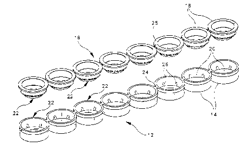
Fig. 1 is an exploded view illustrating a strip of eight connected DNA
sample wells and a strip of eight connected seals for the sample wells,
together
forming a series of sample well assemblies constructed in accordance with a
preferred embodiment of the present invention;
Figs. 2A, 2B and 2C are top, side sectional and bottom views,
respectively, of the strip of connected DNA sample wells;
Figs. 3A, 3B and 3C are top, side sectional and bottom views,
respectively, of the strip of connected seals for the DNA sample wells;
Figs. 4A and 4B are exploded and assembled views, respectively, of a
single DNA sample well assembly;
Figs. 5A and 5B are exploded and assembled sectional views, respectively,
of a single DNA sample well assembly, with the optical window element of the
assembly shown installed in the sample well in both views;
Figs. 6A, 6B and 6C are sequential sectional views illustrating the manner
in which a liquid biological sample is introduced into a partially assembled
DNA
sample well assembly by means of a pipette; and
Figs. 7A, 7B and 7C are sequential sectional views illustrating the manner
in which the DNA sample well assembly of the present invention may be used to
carry out a nucleic acid amplification and homogeneous nucleic acid
fluorescence
polarization assay using dried reagents affixed within the reaction area of
the
assembly.
'Throughout the drawings, like reference numerals will be understood to
refer to like parts and components.
CA 02215650 1997-09-15
_8_
Detailed Description of the Preferred Embodiment
A multiple-well apparatus 10 adapted for carrying out an integrated
nucleic acid amplification and assay procedure in accordance with a preferred
embodiment of the present invention is illustrated in Fig. 1. The apparatus 10
comprises a first strip 12 of eight connected sample wells 14, and a second
strip
16 of eight connected seals or caps 18. Each sample well i4, when combined
with its corresponding seal 18 and with an inserted optical window element 20,
forms a sample well assembly 22 in which an integrated nucleic acid
amplification and assay procedure can be carried out on a discrete liquid
biological sample. The individual sample wells 14 in the strip 12 are
connected
to each other in a linear fashion by means of integral tabs 24, which can be
broken by the user to subdivide the strip 12 if fewer than eight samples are
to be
assayed Similar breakable tabs 25 are used to connect the seals 18 in a linear
arrangement as shown. The strip 12 of sample wells 14 is preferably formed in
one piece by injection molding, using a suitable plastic material such as
polypropylene. The strip 16 of seals 18 may be formed in the same manner, and
is preferably made of the same material. In the preferred embodiment, the
individual sample wells 14 are generally cylindrical in shape with an outside
diameter of approximately 0.320 inch, an outside height of approximately 0.175
inch and a wall thickness of approximately 0.015 inch. The center-to-center
distance from one sample well 14 to the next (and from one seal 18 to the
next) is
approximately 0.354 inch.
As shown in Fig. 1, each sample well 14 contains an inserted optical
window element 20 in the form of a transparent circular disk supported by a
plurality of ribs 26 which are spaced circumferentially around the interior of
the
sample well. As will be described in more detail hereinafter, the lower
surface of
each optical window element 20 is spaced from the bottom wall of the
CA 02215650 1997-09-15
-9-
corresponding sample well 14 by a small distance (preferably about 0.020 inch)
to
create a capillary chamber within the bottom of the sample well. A liquid
biological sample to be assayed is introduced into the capillary chamber,
preferably through an annular gap or opening which exists between the outer
edges of the optical window element 20 and the vertical interior wall surfaces
of
the sample well 14. One of the seals 18 is then fitted to the sample well 14
to
close this opening, and the liquid biological sample is allowed to react with
dried
nucleic acid amplification and assay reagents contained within the capillary
chamber. After the reaction has progressed to a point at which detection can
begin, an optical detection step is carried out through the optical window
element 20 without the need to remove the liquid biological sample from the
sealed sample well 14. In this manner; the possibility of cross-contamination
with other liquid biological samples is minimized.
The detailed configuration of the sample wells 14 is illustrated in Figs.
2A, 2B and 2C. In the preferred embodiment, each sample well 14 is generally
cylindrical in shape, with a circular top opening 28, upstanding cylindrical
side
walls 30, and a flat circular bottom wall 32. Spaced equidistantly around the
interior circumference of the sample well 14 are six wedge-shaped ribs or
spacers
26 with notches 36 facing toward the center of the sample well. In the
preferred
embodiment, the ribs 26 are carried by the bottom wall 32 of the sample well
14,
and are formed integrally therewith during the plastic molding operation. As
will be described hereinafter, the notched ribs 26 serve to locate and retain
the
optical window element 20 at the proper location within the sample well 14.
The details of the seals or caps 18 are shown in Figs. 3A, 3B and 3C. Each
seal 18 is generally annular in shape, with an upwardly-facing circular rim or
flange 38, downwardly extending cylindrical side walls 40, and a central
circular
aperture 42. Surrounding the lower opening of the aperture 42 is a downwardly
extending, frusto-conical extension 44 which is formed integrally with the
side
CA 02215650 1997-09-15
-10-
walls 40 and tapers inwardly toward the central axis of the annular seal 18.
As
will be described shortly, the lower edges of the extension 44 is brought into
contact with the periphery of the optical window element 20 when the sample
well assembly 22 is fully assembled, in order to seal the capillary chamber
below
the optical window element 20 after a liquid biological sample has been
introduced into the capillary chamber. The capillary chamber is also sealed by
means of a circular sealing ring 46 that is formed around the outside of the
cylindrical side walls 40 of the seal 18. The sealing ring 46 frictionally
engages
the interior side wall surfaces 47 of the sample well 14 (visible in Figs. 2A
and
2B) in order to hold the seal 18 in place on the sample well.
The manner in which each of the sample well assemblies 22 is assembled
prior to carrying out an integrated nucleic acid amplification and assay
procedure is illustrated in Figs. 4A and 4B. The optical window element 20 is
inserted into the sample well 14 and is received and retained in the notches
34
formed in the radially arranged ribs 26. (This is preferably - although not
necessarily -- done during the manufacturing process, so that the sample well
14
reaches the user with the optical window element 20 already installed.) The
plastic material of which the ribs 26 and sample well 14 are made is
sufficiently
resilient to allow the ribs 26 to flex or move slightly as the optical window
element 20 is inserted into the notches 34, allowing for a positive "snap"
engagement between the optical window element 20 and the notches 34. The
upwardly facing surfaces 48 of the ribs 26 are preferably inclined downwardly
toward the center of the sample well 14 at an angle of about 45°, as
shown, in
order to provide guide surfaces for directing the edges of the optical window
element 20 into the notches 34.
After the optical window element 20 has been inserted into the sample
well 14, a liquid biological sample is introduced into the capillary chamber
located below the optical window element 20, as will be described below. The
' CA 02215650 1999-12-23
-11-
seal 18 is then placed on the sample well in order to seal the capillary
chamber.
When the seal 18 is in~ place, the sealing ring 46 is in frictional engagement
with
the interior side walls 47 of the sample well, and the lower edge of the
extension
44 is in contact with the peripheral portion of the optical window element 20.
The fully assembled condition of the sample well assembly 22 is shown in Fig.
4B.
Figs. 5A and 5B are sectional views illustrating the internal configuration
of the sample well assembly 22, with the seal 18 shown removed in Fig. 5A and
fully installed in Fig. 5B. As best seen in Fig. 5A, the optical window
element 20
is held by the notched ribs 26 in a parallel relationship with the bottom wall
32
of the sample well 14, with a uniform gap (preferably about 0.020 inch in
height)
being maintained between the upwardly-facing surface 50 of the wall 32 and the
confronting, downwardly-facing surface 52 of the optical window element 20.
This gap or space defines a cylindrical or disk-shaped capillary chamber 54
between the surfaces 50 and 52. A spot 56 containing dried homogeneous
nucleic acid amplification and fluorescence polarization assay reagents is
affixed
to the surface 50 at a central position within the capillary chamber 54. When
a
liquid biological sample is introduced into the capillary chamber 54, the
reagents
in the dried spot 56 are rehydrated by the sample to initiate the desired
amplification and assay reactions. In order to allow for the introduction of a
liquid biological sample into the capillary chamber 54, the sample well 14 and
the optical window element 20 are dimensioned such that an annular gap 58 of
approximately 0.020 inch is provided between the outer periphery of the
optical
window element 20 and the confronting interior wall surfaces 47 of the sample
well 14. In the preferred embodiment, this is accomplished by forming the
sample well 14 with an inside diameter of approximately 0.290 inch, and by
forming the optical window element 20 with an outside diameter of
approximately 0.250 inch.
CA 02215650 1997-09-15
-12-
In Fig. 5B, the sample well assembly 22 is shown fully assembled with a
liquid biological sample 60 present within the capillary chamber 54. The
liquid
biological sample 60 substantially fills the capillary chamber 54, which has a
volume of about 20 pL. By virtue of the narrow spacing between the
confronting surfaces 50 and 52 of the bottom wall 32 and optical window
element 20, respectively, the liquid biological sample is drawn into the
chamber
54 by capillary forces and is spread into a thin film or disk having a height
(thickness) of about 0.020 inch and a diameter of about 0.250 inch. In this
configuration, the liquid biological sample 60 has a large surface area
relative to
its volume and equilibrates to the temperature of the sample well 14 in a
matter
of a few seconds. In addition, by spreading the liquid biological sample into
a
thin film, a large optical target is achieved relative to the volume of the
sample.
This is desirable when an optical detection step is performed, as in the case
of a
fluorescence polarization assay.
The manner in which the seal 18 closes off the capillary chamber 54 will
be evident from Fig. 5B. Closure of the capillary chamber 54 occurs along two
separate zones, one defined by the circular line of contact between the
sealing
ring 46 and the internal vertical walls 47 of the sample well 14, and the
other
defined by the circular line of contact between the lower edge 62 of the
frusto-
conical extension 44 and the upper surface 64 of the optical window element
20.
The sealing ring 46 prevents the liquid sample 60 from passing between the
internal walls 47 of the sample well 14 and the external walls 61 of the seal
18,
and also serves to frictionally retain the seal 18 within the sample well 14.
The
lower edge 62 of the extension 44 provides a similar sealing zone between the
seal 18 and the optical window element 20, and also assists in retaining the
optical window element 20 within the notches 34 of the ribs 26. In addition,
the
aperture 42 in the seal 18 provides a free optical path to the optical window
element 20 when the sample well assembly 22 is fully assembled, thereby
CA 02215650 1997-09-15
-13-
allowing an optical detection step to be carned out on the liquid biological
sample 60 while the sample remains coned within the capillary chamber 54.
Figs. 6A, 6B and 6C illustrate the manner in which a liquid biological
sample 60 is introduced into the capillary chamber 54 of a sample well
assembly
22. Prior to introducing the liquid biological sample into the capillary
chamber,
the sample well assembly 22 has been partially assembled by inserting the
optical
window element 20 into the notched ribs 26 of the sample well 14. A pipette 66
containing the liquid biological sample 60 to be assayed (typically consisting
of a
prepared blood sample or other body fluid sample that is to be tested for a
specific pathogen) is positioned with its opening just above the gap 58
between
the periphery of the optical window element 20 and the interior vertical walls
47
of the sample well 14, as shown in Fig. 6A. The pipette 66 will typically be
of
the disposable type and may be carried either by a manual pipetting apparatus
or
by an automated {robotic) pipetting apparatus. In either case, the pipetting
apparatus is operated to dispense the liquid biological sample 60 from the
pipette
66 onto the top surface of the optical window element 20 in the region near
the
gap 58. As this occurs, capillary forces automatically draw a measured volume
of
the liquid sample 60 into the gap 58 and into the capillary chamber 54 below
the
optical window element 20, as shown in Fig. 6B. The capillary forces cause the
liquid biological sample 60 to be spread into a thin film disk which
substantially
fills the capillary chamber 54 with no head space, as illustrated in Fig. 6C.
As
noted above, this configuration is advantageous not only because it ~ provides
efficient heat transfer between the sample well 14 and the liquid sample 60,
but
also because it affords a large optical target for subsequent detection.
Figs. 7A, 7B and 7C illustrate the manner in which a sample well
assembly 22 constructed in accordance with the present invention can be used
to
carry out a homogeneous nucleic acid amplification and assay procedure on a
liquid biological sample. In Fig. 7A, an empty sample well 14 containing an
CA 02215650 1997-09-15
- 14-
optical window element 20 and dried reagents 56 is placed on a heating platen
68. The heating platen 68 is operated to pre-heat the sample well 14 to a
temperature (typically between 25° C and 75° C) which is
suitable for a
homogeneous nucleic acid amplification and assay procedure. The pre-heating
step is carried out for a time interval sufficient to allow the empty sample
well
14 to equilibrate to a temperature approximately equal to that of the heating
platen 68. After this has occurred, a liquid biological sample is introduced
into
the capillary chamber 54 using a pipette 66, as illustrated in Fig. 7B. This
is
preferably accomplished by positioning the open end of the pipette 66
immediately above the gap 58, as illustrated in Fig. 7B, and then dispensing
the
liquid sample 60 directly into the gap 58.
After the liquid sample 60 has filled the capillary chamber 54, the pipette
56 is withdrawn and a seal 18 is placed on the sample well 14. The liquid
biological sample 60 rehydrates the dried nucleic acid amplification and assay
reagents within the dried spot 56, and a homogeneous amplification and assay
reaction occurs while the sample 60 is contained within the capillary chamber
54.
Due to the thinness of the capillary chamber 54 and the large surface area
with
which the liquid biological sample 60 comes into contact, the sample 60 heats
up
within a few seconds of being pipetted into the chamber 54 to the optimum
temperature desired for DNA amplification. Thus, by the time the dried
reagents 56 dissolve and diffuse throughout the liquid biological sample 60 to
begin "priming" the DNA amplification, the reagents are already up to the
optimum temperature. In this way, a "hot start" of the DNA amplification
reaction is achieved. After the "hot start" occurs, the heating platen 68
continues to maintain the liquid biological sample 60 at a temperature
(typically
between 25° C and 75° C) that is suitable for the homogeneous
amplification
and assay reactions. As the homogeneous amplification and assay reactions
occur, their progress is monitored in real time by suitable optical detection
CA 02215650 1999-04-27
- 15-
apparatus 70. Depending upon the nature of the assay reaction, the apparatus
70
may detect fluorescence polarization, fluorescence energy transfer, light
absorbance, or any other optical response or characteristic of the liquid
biological
sample 60. Various types of apparatus 70 which may be used for this purpose,
such as microplate fluorometers, are known in the art and need not be
described
in detail. Reference is made to the aforementioned co-pending European
application EP
0790861, published August 27, 1997 for a description of specific detection
methods that
may be employed when the assay is of the fluorescence polarization type.
In the case where the sample well assembly 22 is used to carry out a
fluorescence polarization assay, the optical window element 20 is preferably
made of a transparent material that does not interfere with the transmission
of
polarized light. Examples of such materials include cellulose acetate butyrate
(CAB), triacetate cellulose (TAC), and glass. Polarized light will pass
through
these materials and retain its polarization. The sample well assembly 22 can,
if
desired, be configured for use in a confocal polarization detection method as
described in co-pending European application EP 0790861, published August 27,
1997. In this
method, the polarizes for the excitation beam is also used as the polarizes
for the fluorescence
emitted by the sample. This avoids the need to provide polarizing elements in
the measuring instrument, thereby allowing standard microplate fluorometers
to be used in a fluorescence polarization assay. To employ the confocal
method,
the optical window element 20 is made of a light-polarizing material so that
it
will serve to polarize both the excitation beam from the detection apparatus
70
and the fluorescence emitted by the liquid biological sample 60. Exemplary
light-polarizing materials include laminates or "sandwiches" in which a layer
of
polarizing polymeric film, such as polyvinyl alcohol (PVA), is disposed
between
layers of CAB, TAC or glass.
In the preferred embodiment, the dried reagent spot 56 contains both
DNA amplification and homogeneous DNA assay reagents, the latter preferably
CA 02215650 1999-04-27
- 16-
consisting of fluorescence polarization assay reagents. Examples of suitable
DNA_ amplification and DNA fluorescence polarization assay reagents are
disclosed in copending European application EP 0678581, published October 25,
1995 and
entitled "Fluorescence Polarization Detection of Nucleic Acid Amplification".
The chemical
reagents in the dried spot 56 are carried in a readily soluble matrix, such a
trehalose or another
carbohydrate. These reagents will spontaneously re-suspend when exposed to an
aqueous sample introduced into the capillary chamber 54. It will be understood
that more than one dried reagent spot 56 may be provided in the capillary
chamber 54 if desired, as for example by providing the amplification reagents
in
one spot and the assay reagents in a different spot. In the case of a
homogeneous
DNA amplification and assay, however, the reagent spots (if separated) should
be positioned in such a way that they are dissolved by the liquid biological
sample 60 at essentially the same time.
The foregoing is illustrative of the present invention, and is not to be
construed to be limiting thereof, as numerous alternatives to the devices and
methods which incorporate the present invention will be apparent to those
skilled in the art. The invention is accordingly defined by the following
claims
with equivalents of the claims to be included therein.