Note: Descriptions are shown in the official language in which they were submitted.
CA 02223494 1997-12-04
W O 96/40722 . PCTAUS~GI~0~~
CHROMOSOM~T FXPF~F~SION OF HFTFROTiOGOUS
('TF~F!~ AcTFRTAT CF.T T
TFCH~ICAT FTFT n
S This invention is related to the field of expression of heterologous genes
in bacteria.
~AC~ ROUND ~RT
Genetic engineering has made it possible to produce large amounts of
10 heterologous proteins or polypeptides in bacterial cells by means of recombinant
expression systems, especially by expression in such prokarvotes as Escherichia coli (E.
coli) .
The expressed heterologous proteins may be of m~rnm~ n, other
eukaryotic, viral, bacterial, cyanobacterial, archaeb~ct~ri~l, or synthetic origin.
Unlike native bacterial proteins, which can often be efficiently
accl7m~ te~1 within a bacterial cell even when encoded by a single chromosomal gene
copy, there are no published reports to date of heterologous ~lol~ s being successfully
accllm~ t~d within b~ct~or1~l cells to levels exceeding 0.1% oftotal cell protein when
expressed from a single chromosomal gene location.
0.1% of total cell protein (150 micrograms protein per trillion bacterial
cells) is chosen as a practical measure of sl-ccç~ful accumulation of protein because it
approximately defines the lower limits of (a) economically .ci~nific~nt ~qcc-lmlll~tion of a
desired protein by contemporary recombinant bacterial production standards, and (b)
visual detection of a protein band by ~oomassie-stained polyacrylamide gel analysis of
25 whole bacterial cell extracts.
The relatively poor performance of non-bacterial genes when expressed in
bacterial cells, even when placed under the control of the strongest known bacterial
promoters, has been generally attributed to poor translation of the non-b~ct~ri~l mRNAs
CA 02223494 1997-12-04
W O 96/40722 . PCTAJS96/0900
and rapid degradation of newly synthesi7~d non-bacterial proteins. It has almostuniversally been assumed that, in order to achieve successful accumulation of
non-bacterial or heterologous proteins in bacterial cells, the genes encoding the
heterologous proteins must be located on multicopy plasmid vectors.
A gene carried on one of the multicopy plasmids commonly used for
cloning and ~xJ,ressing genes encoding heterologous proteins in E. coli usually has a
copy number of more than 20 copies/cell. Even low copy number plasmids (e.g.,
pACYC177 and pLG339) generally exist at 6-10 copies per cell. One disadvantage
imposed by plasmid gene dosages is that the expression of even minute amounts of some
10 foreign proteins can kill host cells (see Meth Fn7~rrnol. 185:63-65, ed. D. Goeddel,
1990). For this reason, it would be advantageous to reliably limit the copy number of
genes encoding such toxic gene products, such as by int.ogr~ting the gene into the
bacterial chromosome at one or a small number of copies per cell. For example, such a
system would allow one to make more r~le3~ ive cDNA expression libraries in
15 bacterial hosts if the high-copy ~x~ ion of one or more of the cDNAs in the library
could kill the bacterial host or cause it to grow poorly.
Chromosomal integration of genes encoding heterologous polypeptides
would also be advantageous as an alternative means for ~x~lcssion of heterologous
proteins in bacterial host cells. Multicopy vectors are often unstable and require the use
20 of antibiotics in the growth medium for maillt~ ce. Present methods of integrating
foreign genes into the bacterial chromosome suffer from inefficiency, the inabilit,v to
control the site of integration of the foreign gene, and/or the inability to control the copy
number of the integrated gene. Most importantly, all efforts to date to create
recombinant DNA constructs on the bacterial chromosome, wherein a bacterial promoter
25 is fused to a heterologous gene, have involved the creation of viral or plasmid
intermediates carrying the construct. Because such intermediates replicate at high copy
number, they may be difficult or even impossible to recover in cases where the foreign
gene product is toxic to the bacterial cell. Expression of the encoded gene, even at low
levels, may be toxic to the host cells, due to the high copy number of these intermediates,
30 which effectively multiplies the level of expression.
CA 02223494 1997-12-04
W O 96/40722 . PCT~US9G/O~OOG
Previous methods for achieving the integration of heterologous genes into
the chromosome of a bacterial host include the use of phage lambda vectors. The phage
DNA in circular form is inserted linearly into the bacterial chromosome by a single site
specific recombination between a phage ~tf~chment site (attP), 240 bases long, and a
bacterial ~ hment site (~B), only 25 bases long. The two sites have 15 bases in
common. This site-specific recombination is catalyzed by a special integr~se, specified
by the phage gene ~ (VIROLOGY pp. 56-57 (Lippincott, 2nd ed., R. Dulbecco and H.Ginsberg, eds., Philadelphia, PA, 1985).
Phage vectors which are II~- can be integrated into the chromosome in a
10 normal fashion as long as integrase is supplied in tra~s, e.g., by an INT+ helper phage
(see, e.g., Borck et al. (1976) Molec. Gen G~net. 146:199-207).
Phage vectors which are both att- and INT- can likewise be integrated into
the b~ct~ri~l chromosome as double lysogens by using ~+INT+ helper phage. Doublelysogens are formed by linkage of the prophages at the bacterial ~t~chm~nt site and are
15 integrated into the chromosome by general bacterial recombination between homologous
sequences on the defective phage and on the helper phage (see e.g., Struhl et al. (1976)
Proc. N~tl Acad. Sci. USA 73:1471-1475). Similarly, it is also possible to integrate
non-replicating colE1 replicons into the genome of E2Ql~ strains of E. coli by means of
recombination between the host chromosome and homologous sequences carried by the
20 plasmid vector (Greener and Hill (1980) J. Racteriol. ~:312-321).
More recently, systems have been spec;fic~lly deei~nt?d for the integration
of foreign genes into a bacterial host chromosome. For example, U.S. Patent No.
5,395,763 (Weinberg et al.) discloses a chromosomal ~x~ession vector for the
expression of heterologous genes. This vector was created ntili7ing a multicopy number
25 plasmid intçrmerli~te7 into which the gene of interest is cloned, placing the gene in
operable linkage with the bacteriophage middle promoter, Pm. This plasmid
intermediate, which compri~çs a defective Mu genome (lacking the genes n~ces~. y for
the formation of phage particles) is introduced into a p~ck~gin~ strain to produce
infectious Mu particles, which are then used to introduce the vector into host cells and
30 integrate the vector into the host cell genome. This vector system is amplifiable once
integrated into the host cell genome, but the mech~ni~m of amplification (replicative
CA 02223494 1997-12-04
W O 96/40722 . PCT~US?6~00G
transposition) is norrnally toxic to the host cell, due to integration of the replicating
prophage into essential host cell genes (Neidhardt et al., ESCHERICHIA COLI AND
SALMONELLA TYPHIMURIUM: MOLECULAR AND CELLULAR BIOLOGY (American Society for
Microbiology, Neidhardt et al. eds., Washington, D.C., 1987). Because the amplification
5 of this integrated prophage is normally toxic, it is very difficult to obtain and propagate a
host cell strain carrying the amplified integrated DNA. This then requires that the gene
be amplified each in~t~nce that protein production is desired.
Diederich et al. ((1992) "New plasmid vectors for integration into the 1
Cl~ment site attB of the Escherichia coli chromosome", Pl~smid ~:14-24) also
10 disclose a system for introducing a gene onto the chromosome of a bacterial host cell.
This system utilizes a set of multicopy plasmid vectors which can be integrated into a
bacterial chromosome via a phage lambda ~ hment site. A DNA sequence encoding a
promoter operably linked to a gene of interest is cloned into one of the described
multicopy number plasmid vectors, the plasmid's origin of replication is removed by
15 restriction en7ymes, and the resulting DNA is recircularized and transferred to a host
cell, where it integrates into the chromosome.
These new gene transfer systems suffer from the same defect as earlier
systems. Both USP 5,395,763 (Weinberg et al.) and Diederich et al. require that the gene
of interest be cloned into a multicopy number plasmid while in an operable configuration
20 during the construction of the transfer DNA. The configuration of this multicopy
number plasmid makes expression of toxic foreign genes difficult, if not impossible,
because the (toxic) gene of interest will be expressed as the multicopy number plasmid is
propagated.
Accordingly, there is a need for a method of producing heterologous
25 proteins which can produce large amounts of protein and which minimi7.?s any toxic
effect of the heterologous protein to host cells during construction of the producing
strain. Applicants have shown surprisingly high protein accumulation (approximately
20% of total cell protein) from t;~les~ion of low (a~plo~i,.,ately two) copies of the gene
encoding the heterologous protein as shown in Example 2.
CA 02223494 1997-12-04
W O 96/40722 . PCT~US9GI'~5006
,~
SU~l\~ARY OF THF TNVENTION
The present invention provides methods and compositions for production
of heterologous proteins in bacterial host cells such as E. coli by integrating a
chromosomal transfer DNA (a circular, non-self replicating DNA) into the chromosome
of a host cell. The chromosomal transfer DNA comprises one or more copies of a gene
encoding the heterologous protein of interest.
The present invention, therefore, provides a method for producing a
heterologous protein of interest, comprising:
integrating a chromosomal transfer DNA into the chromosome of a host
10 cell such that chromosomal amplification of the integrated DNA is facilitated, the
chromosomal transfer DNA compri~ing at least one copy of a gene encoding a
heterologous protein of interest and a selectable marker; and
expressing the gene encoding the heterologous protein of interest,
wherein the gene was at no time operably linked to a promoter functional in the host cell
15 in a multicopy number plasmid during the construction of the transfer DNA, and
wherein the heterologous protein of interest accllm~ tes to a level of at
least 0.1% of total cell protein.
The chromosomal transfer DNA may optionally comprise a promoter
operably linked to the gene encoding the heterologous protein of interest, wherein the
20 operable linkage is created by circlll~ri7~tion of the chromosomal transfer DNA.
Optionally, the chromosomal transfer DNA may further comprise
duplicate DNA fl~nkin~ the selectable marker. The duplicate DNA may optionally
comprise copies of the gene encoding the heterologous protein of interest operably linked
to a promoter.
The methods for ~re~ion of heterologous proteins may optionally
include the step of selecting for chromosomal amplification.
The invention also provides methods for producing a chromosomal
DNA, comprising lig~tin~ together ~:~n-ont.~ from a first and a second plasmid
vector:
CA 02223494 1997-12-04
W O 96/40722 . PCT~US96/09006
the first plasmid vector comprising a first origin of replication, and a first
gene encoding a heterologous protein of interest wherein the first gene is not operably
linked to a promoter and a first copy of a duplicate DNA;
the second plasmid vector comprising a second origin of replication, and a
5 first promoter and a second copy of a duplicate DNA;
wherein the origins of replication and the promoter function in the host
cell, and wherein either said first plasmid or said second plasmid comprises a selectable
marker.
Optionally, the first plasmid may further comprise a second promoter not
10 operably linked to the first gene encoding the heterologous protein of interest and the
second plasmid may further comprise a second copy of the gene encoding the
heterologous protein of interest not operably linked to the first promoter.
Also provided are chromosomal transfer DNAs for use in production of
heterologous proteins of interest, comprising:
a non-bacterial gene of interest operably linked to a promoter functional in
a host cell; and
a selectable marker flanked by duplicate DNA,
wherein said gene encoding a heterologous protein is at no time operably
linked to a promoter functional in a host cell on a multicopy number plasmid vector
20 during the construction of the transfer DNA.
Optionally, the chromosomal transfer DNA may further comprise two or
more copies of the gene encoding the non-bacterial protein of interest, wherein the copies
of the gene flank the selectable marker.
25 P~RTFF DF~CRTPTION OF THF DRAWINGS
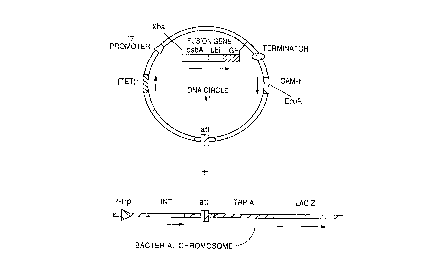
Figure 1 shows steps in the in vitro formation of a chromosomal transfer
DNA, a DNA circle which lacks an origin of replication (and thus is incapable ofself-replication) and is suitable for integration of a foreign gene into the b~ teri~l
chromosome. Until the chromosomal transfer DNA is formed, the foreign gene to be30 t;~ s~ed (here an IGF-I fusion gene) is separated from a functional bacterial promoter
(here the T7 promoter).
CA 02223494 1997-12-04
W O ~ 722 . PCT~US96/~9006
Figure 2 shows a chromosomal transfer DNA ~ormed from the ligation of
two DNA fragments. One of the fragments contains a fusion gene comprising sequences
encoding ~. coli DsbA, yeast ubiquitin (beginning with a Met), and human insulin-like
growth factor I ("dsbA-ubi-IGF") (not beginning with a Met), as discussed in co-owned,
co-pending U.S. patent application no. 08/100,744, filed August 2, 1993. The other
DNA fragment contains a T7 promoter. Both the chromosomal transfer DNA and the
b~ot~ri~l chromosome contain a recombination site from phage lambda, ~ P. The
chromosomal transfer DNA is transformed into E. coli strain B1384, which makes
integrase (INT) under the control of the trp promoter (P-trp). Integrase catalyzes
10 site-specific integration of the chromosomal transfer DNA into the bacterial chromosome
at the ~L site. The trp promoter can be in(~ ed during transformation by adding 1 mM
indole acrylic acid (IAA) to the medium. Cells with integrated chromosomal transfer
DNA sequences are resistant to chloramphenicol (CAM-r, 10 Tglml).
Figure 3 shows a B1384 chromosomal integrant rçsulting from the
15 process described in Figure 2. The integration can be confirmed by amplifying host
chromosomal DNA by PCR with various primer sets (e.g., UBUF x IGFR, 1243 x
T7REV, or TRPPF x 1239), digesting the amplified fragments with the appropriate
restriction enzyme (SacII, HinCII, or BamHI, respectively), and sizing the products by
gel electrophoresis).
Figure 4 shows a Western blot of whole cell lysates of chloramphenicol
resistant W311 ODE3 transductants. Also included are protein size markers (far left lane)
and IGF fusion protein (control).
Figure 5 shows a Western blot of whole cell lysates of kanamycin
resistant trans(l~ t~nt~
Figures 6-9 show diagrarnmatically the general strategy for construction
of chromosomal transfer DNA's. Figure 6 shows a chromosomal transfer DNA
compri~ç~ a single copy of the gene encoding the heterologous protein of interest and
two copies of a second gene which flank the selectable markers, facilit~tinp
chromosomal amplification after integration of the chromosomal transfer DNA. Figure 7
30 shows the "double cassette" system utilized for ~ es~ion of heterologous proteins in
Examples 2 through 6 and Example 8. This embodiment of the chromosomal transfer
CA 02223494 1997-12-04
W 096/40722 . PC.TrUS~6/0~006
DNA comprises two copies of the gene encoding the heterologous protein of interest
fl~nking the selectable markers, facilit~in~ chromosomal amplification of the integrated
DNA. Figures 8 and 9 show ~It~ t.o embo-liment~ of "promoter-less" chromosomal
transfer DNAs. These embotliment~ utilize a DNA sequence homologous to a segment5 of the host cell chromosome. Integration of promoter-less chromosomal transfer DNAs
results in formation of an operably linkage between a host cell promoter and the gene
encoding the heterologous protein of interest and the creation of duplicate DNA
sequences ~nkin~ the selectable markers.
Figures 10- 13 show the plasmid genealogy of chromosomal transfer
10 DNAs.
Figure 14 shows the strategy for construction of the two DNA sources
used in the double c~sette system.
Figures 15 and 16 show the strategy used to construct chromosomal
L,cu.~r~, DNAs for integration and expression of the yeast ubiquitin hydrolase gene.
Figure 17 shows the strategy for constructing the chromosomal transfer
DNA used to integrate and express a gene encoding a DsbA::ubiquitin::IGF-I fusion
protein.
Figure 18 shows the strategy for constructing the chromosomal transfer
DNA used to integrate and express a gene encoding a DsbA::2A::IGFBP-3 fusion
protein.
Figures 19 and 20 show the strategies used to construct chromosomal
transfer DNAs used to integrate and express genes coding for DsbA::2A::IGF-I (Figure
19) and DsbA::3C::IGF-I (Figure 20) fusion proteins.
Figure 21 shows the strategy used to construct the chromosomal L~ r~l
DNA used to integrate and express the gene encoding a DsbA::ubiquitin::TGF-,B2 fusion
protein.
Figure 22 shows the strategy used to construct the chromosomal transfer
DNA used to integrate and express a gene encoding a DsbA::3C::IGFBP-3 fusion
protein.
Figure 23 shows an coomassie blue-stained SDS-PAGE gel of whole cell
lysates of isolates expressing IGF-I fusion proteins. c49222, c49258#46, and c53063
CA 02223494 1997-12-04
W O 96/407Z2 . PCTAUS961'~C~G
express a DsbA::ubiquitin::IGF-I fusion protein (left arrow), which is easily visible.
Surprising, this high level of expression is seen in c49222 and c49258#46, which were
not amplified (i.e. there was no selection for chromosomal amplification of the integrated
DNA). c57264#5 and c57264#28 express a DsbA::3C::IGF-I fusion protein while
S c57265#44 and cS7265i~54 express a DsbA::2A::IGF-I fusion protein. Again, theexpressed fusion protein is easily visible. Densitometric analysis of this gel indicates
that all of the isolates accllmnl~te protein in excess 19% of total cell protein (average
protein accumulation is 25.7% of total cell protein).
Figure 24 shows a Southern blot of chromosomal DNA isolated from
10 c49222, c49258#46, c53063, c57264#5, c57264#28, c57265#44, and c57265#54. Theblot was probed with a DNA fragment encoding ubiquitin fused to IGF-I. The higher
molecular weight band in each lane represents a single copy of the integrated IGF-I
fusion protein gene in each isolate. The lower molecular weight band also .~.ese~ the
integrated IGF-I fusion protein gene, but this fragment can be amplified by chromosomal
15 amplification. Isolates c53063, c57264#5, c57264#28, c57Z65#44, and c57265#54 have
clearly been amplified, showing about 3 to 5 fold amplification.
Figure 25 shows coomassie blue-stained SDS-PAGE gels showing protein
accumulation in isolates carrying integrated genes encoding IGFBP-3 fusion proteins.
A) shows protein accumulation in an isolate ~ es~illg a DsbA::2A::IGFBP-3 fusion20 protein. The right lane shows protein ~x~-c;ssion after induction of T7 RNA polymerase
by addition of IPTG to the culture medium. B) shows protein accumulation in an isolate
~X~l~;S~illg a DsbA::3C::IGFBP-3 fusion protein. As in Figure 23, the bands repr~s~ting
the fusion protein are easily visible. Densitometric sç~nnin~ of these gels found that the
~çcnm~ te-l protein represented 22.6% in Panel A, and the two isolates in Panel B
25 acc--m--l~ted 33.% and 28.2% of total cell protein (left to right, respectively).
Figure 26 shows a coomassie blue-stained SDS-PAGE gel showing
protein accumulation from host cells ~x~essi-lg a gene encoding
DsbA::ubiquitin::TGF-~2. M in(1ic~tt?s molecular weight markers and C indicates a
positive control. The two Plasmid lanes (Lanes 1 and 2) are used as a standard to
30 compare protein accumulation from multicopy number plasmid vectors to protein~ccnnn-ll~tion from genes integrated into the chromosome. Lanes 3 and 4 are whole cell
CA 02223494 1997-12-04
W O 96/~7~2 . PCTAUS96i~~6
lysates of isolates which were negative for T7 RNA polymerase activity when streaked
against phage 4107. Densitometric analysis of this gel showed that the plasmid strain
accllm~ ted protein to 26.4% of total cell protein. Protein accumulation was measured
for isolates 48, 56, 59, 65 and 66, and showed protein accumulation to 36.7%, 33.3%,
32.1%, 29.5%, and 26.7%, respectively.
MOnFS FOR CAR~YrNG OUT THF rNVF~TION
The present invention resides in (a) the creation of an operable linkage
between a promoter and a gene encoding a heterologous protein of interest with the
10 linkage being formed either during the construction of a chromosomal tr~nsfer DNA or
as a result of its integration into the host cell chromosome and (b) the ~imlllt~neous
creation of a means for the ~ iate chromosomal amplification of the integrated gene
of interest.
In the plc~-lcd embodiments, the creation of the chromosomal transfer
15 DNA ~imlllt~neously achieves two goals; (1) the operable linkage ofthe promoter and
the gene of interest and (2) the positioning of duplicate DNA sequences fl~nking a
selectable marker (which can function as a means to f~ilit~te the amplification of the
chromosomal transfer DNA). Another embodiment creates the operable linkage between
the gene and the promoter during creation of the chromosomal transfer DNA, while the
20 means for chromosomal amplification (duplicate DNA sequences fl~nking the
chromosomal transfer DNA) is created as a result of the integration.
Other methods can achieve either or both of these results by integration of
a chromosomal transfer DNA into a suitable site on the chromosome. For example,
integration of a gene of interest near a promoter on the bacterial chromosome can be
25 designed to result in an operable linkage (for example, by integrating a chromosomal
transfer DNA into an operon on the host cell chromosome). The site of integration or
sequences adjacent to the site of integration may facilitate amplification (e.g. where the
site is located in a transposable element, by providing duplicate DNA sequences, or even
by providing a region of DNA sequence homologous to a portion of the chromosomal30 transfer DNA, thus providing duplicate DNA sequences).
CA 02223494 1997-12-04
W O 96/40722 . PCTAJS96/09006
11
The present invention employs "chromosomal transfer DNA" which may
be used to simply, efficiently, and reliably insert a copy of a heterologous gene into the
chromosome of a host cell, e.g., E. coli. A chromosomal transfer DNA is a circular DNA
comprising one or more copies of a gene encoding a heterologous protein of interest, a
5 selectable marker (e.g., an antibiotic resistance gene), a recombination site (e.g., a
site-specific recombination site such as lambda attP or ~B or a DNA sequence
homologous to a segment on the host cell chromosome), and means for facilit~tin~ the
amplification of the chromosomal transfer DNA following recombination into the host
cell chromosome, and lacking an origin of replication or autonomously replicating
10 sequence (ARS). The chromosomal transfer DNA is therefore incapable of replicating
independently when introduced in to the host cell. The chromosomal transfer DNA may
optionally carry a promoter operably linked to the gene of interest.
When a chromosomal transfer DNA carrying a site-specific recombination
site is introduced into a host cell having a chromosome which contains a second, similar
15 recombination site (e.g., another attP or ~B site), expression in the host cell of an
enzyme which is capable of catalyzing the site-specific recombination of the
recombination sites (e.g., integrase) results in the integration of the vector into the host
cell chromosome at the recombination site. This site-specific recombination process is
much more efficient than general recombination systems acting on homologous vector
20 and host chromosomal sequences and results in integrated sequences having greater
stability, particularly when integrase synthesis can be controlled. Integrase may also be
provided by a plasmid or other DNA molecule transiently or stably present in the host
cell at the time when the chromosomal transfer DNA is introduced.
It will be a~a~ ll to one skilled in the art that there are a variety of
25 methods other than the plcrellcd method ll~ili7in~ attP, attB, and INT which may be used
to integrate a chromosomal transfer DNA into the chromosome of a host cell. For
example, non-replicating colE1 replicons, transposable elements, or even naked DNA
carrying sequences homologous to sequences found on the host chromosome may be
used to insert the chromosomal transfer DNA into the host chromosome. The multicopy
30 colicin plasmids ColE1, CloDF13, ColK, and ColA all comprise site-specific
recombination systems including a cis- and trans-acting element. For use in the present
-
CA 02223494 1997-12-04
W O 96/40722 . PCT~US96/09006
12
invention, the cis-acting element from one of these plasmids may be included on the
chromosomal transfer DNA and the kans-acting element may be on the chromosomal
transfer DNA or provided by the host cell. Transposons, such as the insertion sequence
(IS) and Tn3 families of transposons may be used to hlle~,ldL~ DNA into the chromosome
of a host cell. As with the colicin plasmids described above, the ~i~-acting transposon
~lement~ are included on the chromosomal transfer DNA, while the trans-acting factor
may be included on the chromosomal kansfer DNA or provided by the host cell. Thechromosomal transfer DNA may also carry a DNA sequence homologous to a sequence
found on the host cell chromosome, facilit~tinp~ integration of the chromosomal transfer
DNA by homologous recombination. All of these methods fall within the scope of the
invention.
An important feature of this approach is that the gene encoding the
heterologous protein of interest is at no time operably linked to a functional promoter on
a multicopy vector during construction of the ll~lsr~ . DNA. By keeping a functional
l S promoter separated from the gene of interest until immediately before the foreign gene is
introduced into the cell at low copy number, the potential toxic or lethal effects of the
gene product can be minimi7~-1 A toxic foreign gene will not be expressed from amulticopy number plasmid if the gene is not operably linked to a promoter. Othermethods for integrating a gene of interest into the host cell chromosome utilizemulticopy number plasmids carrying a gene of interest operably linked to a promoter
(e.g., Diederich et al. and Weinberg et al.); these genes will be ~ ,sed during the
propagation of the plasmid, making it extremely difficult, if not impossible, to produce
sufficient quantities of the plasmid if the gene of interest is toxic to the host cells in
which the plasmid is propagated.
The operable linkage between the gene encoding the heterologous protein
of interest and the promoter may be created as a result of the formation of the
chromosomal transfer DNA or as a result of integration into the host cell chromosome.
In the case where the operable linkage is formed as a result of the for~nation of the
chromosomal transfer DNA, the linkage is created by circ~ ri7~tion of the chromosomal
transfer DNA. Circ~ ri7~tion may be accomplished by7 for example, ligation of one or
more DNA fr~gment~ to form a circular DNA or by homologous recombination into a
,
CA 02223494 l997-l2-04
W O 96/40722 . PC~J~/0
13
circular DNA, which would result in circularization of the insert. Preferably,
circ~ ri7~tion is accomplished by ligation of one or more DNA fr~gments.
Alternatively, high level expression of less toxic gene products can be
accomplished by multiple integrations or by selection for amplification of integrated
5 genes.
Recomhin~nt ~nNA Methods ~nd Rea~ent~
General techniques for nucleic acid manipulation useful for the practice of
the claimed invention are described generally, for example, in Sambrook et al.,
10 MOLECULAR CLONING: A LABORATORY MANUAL, Vols. 1-3 (Cold Spring Harbor
Laboratory Press, 2 ed., (1989); or F. Ausubel et al., CURRENT PROTOCOLS IN MOLECULAR
BlOLOGY (Green Publishing and Wiley-Interscience: New York, 1987) and periodic
updates. Reagents useful in nucleic acid manipulation, such as restriction enzymes, T7
RNA polymerase, DNA ligases and so on are commercially available from such vendors
15 as New Fngl~n~l BioLabs, Boerhinger Mannheim, Amersham, Promega Biotec, U.S.
Biochemicals, and New Fn~l~n~1 Nuclear.
nefinition.~
"Fore~n" or "heterolo~oll~" or "norl-bacteri~ tive" or
20 "homolo~ous" A "foreign or "heterologous" polypeptide is a polypeptide which is not
normally found in a host cell of a particular species. The nucleic acid encoding such a
polypeptide is also referred to as "foreign" or "heterologous." For example, insulin-like
growth factor (IGF), insulin-like growth factor binding protein (IGFBP), and
transforming growth factor-beta (TGF-,~) are native to m~mm~ n cells and human
25 rhinovirus 3C protease is native to viruses and virally-infected m~mm~ n cells, but
these proteins are foreign or heterologous to E. coli. A "non-bacterial protein" is a
protein or polypeptide which is not naturally found in a bacterial cell. Non-bacterial
proteins include viral and eukaryotic proteins. Non-bacterial, foreign, or heterologous
proteins may also be fusions between non-bacterial, foreign, or heterologous proteins and
30 other proteins or polypeptides. For the embo-1iment~ encomp~c~ed by this invention,
both "heterologous protein" and "non-bacterial protein" may be expressed. As disclosed
CA 02223494 l997-l2-04
W O 96/40722 . PC~nJS~C/O~C~C
14
herein, genes encoding heterologous or non-bacterial proteins of interest do not contain
promoters functional in the host cell. The genes must be linked to a separate promoter
that is functional in the host cell in order to be expressed. A "native" or "homologous"
polypeptide or DNA sequence, by contrast, is commonly found in the host cell. A
5 promoter or other sequence effecting, for example, the transcription or translation of a
gene is also considered "homologous" if it is functional in the host cell. For example, a
T7 promoter is considered "homologous" to an E. coli host cell, since, if T7 RNApolymerase is present in the cell, the T7 promoter is capable of driving the transcription
of a polypeptide-encoding sequence to which it is operably linked.
"Gene~ encodin~ heterolo~ous. forei~n or non-bacterial protein~" "Genes
encoding heterologous, foreign or non-bacterial proteins" contain all of the genetic
elements necessary for the expression of the heterologous, foreign or non-bacterial
protein with the exception of a promoter functional in the host cell. These genes
encompass recombinant genes which may include genetic elements native to the host
cell. Further, the coding regions of these genes may optionally be ~ hlliG~d for the
codon usage of the host cell.
"Fncode" A nucleic acid is said to "encode" a polypeptide if, in its native
state or when manipulated by recombinant DNA methods, it can be transcribed and/or
trAn~l~te~l to produce the polypeptide.
"Op~rably linked" A nucleic acid sequence is operably linked when it is
placed into a functional relationship with another nucleic acid sequence. For example, a
promoter is operably linked to a coding sequence if the promoter affects its transcription
or expression. Generally, DNA sequences which are operably linked are contiguous and,
where necessary, in reading frame.
"RecornbinAnt" A "recombinant" nucleic acid is one which is made by
the joining of two otherwise separated segments of nucleic acid sequence in y~ or by
chemical synthesis.
"Chromosonn~l An~lification" "Chromosomal amplification" refers to the
increase in copy number of a DNA sequence on the host chromosome. Chromosomal
amplification does not refer to extrachromosomal amplification such as replication of
CA 02223494 1997-12-04
W O 96/40722 . PCTAUS96/09006
multicopy number plasmids or in vitro amplification such as the polymerase chainreaction (PCR).
Probes ~n(l primers
S Nucleic acid probes and primers are isolated nucleic acids, generally
single stranded, and, especially in the case of probes, are typically attached to a label or
reporter molecule. Probes are used, for example, to identify the presence of a
hybridizing nucleic acid sequence in a tissue or other sample or a cDNA or genomic
clone in a library. Primers are used, for example, for amplification of nucleic acid
10 sequences, e.g., by the polymerase chain reaction (PCR). The ~ lion and use of
probes and primers is described, e.g., in Sambrook et al., ;~ a or Ausubel et al. ~.
Chemi~l sy~thecic of nucleic acids
Nucleic acids, especially short nucleic acids such as amplification
15 primers, may be produced by chemical synthesis, e.g., by the phosphoramidite method
described by Beaucage and Carruthers (1981) Tetra. T ~ttc 22:1859-1862 or the triester
method according to M~tteucci et al. (1981) J. ~mer. Chem Soc. 103:3185, and may be
p~lrollllcd on automated oligonucleotide syntheci7tors A double-stranded fragment may
be obtained from the single-stranded product of chemical synthesis either by synthesi7ing
20 the complement~ry strand and ~nn~ling the strands together under a~ .iate
conditions or by adding the complement~ry strand using DNA polymerase with an
~plo~liate primer sequence.
Features of chromosom~l tr~ncfer DNA ~n(l of pl~cmi~lc used in their conctruction
Chromosomal transfer DNA comprises a DNA fragment encoding a
selectable marker and a sequence encoding a desired heterologous polypeptide.
Optionally, a chromosomal transfer DNA may also comprise, in an operable linkage to
the sequence encoding the desired heterologous polypeptide, transcription and translation
initiation regulatory sequences and expression control sequences, which may include a
promoter, an enhancer and nf~cPcc~ry procçccing information sites, such as
ribosome-binding sites, and mRNA stabilizing sequences, as well as any necessary
,
CA 02223494 1997-12-04
W O 96/40722 . PCTAUS96/09006
16
secretion signals, where ap~lol,liate, which allow the protein to cross and/or lodge in cell
membranes, and thus attain its functional topology, or be secreted from the cell.
Plasmids used in construction of a chromosomal transfer DNA will also
typically comprise a replication system recognized by the host, including an origin of
5 replication or autonomously replicating sequence (ARS). In the case where a plasmid
used in the construction of a chromosomal transfer DNA carries duplicate DNA
sequences, the plasmid may be propagated in a ~~ host cell. Preferably, rec~ host cells
are used for propagation of plasmids used to create chromosomal transfer DNAs and
plasmids carrying components of chromosomal transfer DNAs when these plasmids
10 carry duplicate DNA sequences, and are not generally utilized as host cells for
integration of chromosomal transfer DNAs.
Chromosomal kansfer DNA may be prepared from such vectors by means
of standard recombinant techniques well known in the art and discussed, for example, in
Sambrook et al., ~a or Ausubel et al. ~upra.
An a~ o~liate promoter and other sequences necessary for efficient
transcription and/or translation are selected so as to be functional in the host cell.
Examples of workable combinations of cell lines and ~ ,ssion vectors are described in
Sambrook et al., ~_ or Ausubel et al., ~; see also, e.g., Metzger et al. (1988)
Nature 334:31-36. Promoters such as the trp, lac and phage promoters (e.g., T7, T3,
20 SP6), tRNA promoters and glycolytic enzyme promoters are useful in prokaryotic hosts.
Useful yeast promoters include the promoter regions for metallothionein,
3-phosphoglycerate kinase or other glycolytic enzymes such as enolase or
glyceraldehyde-3-phosphate dehydrogenase, ~ ylllcs responsible for maltose and
galactose utilization, and other. See, e.g., Hitzeman et al. EP 73,657A. Appropriate
25 m~mm~ n promoters include the early and late promoters from SV40 (Fiers et al.
(1978) ~haL ~:113) or promoters derived from murine Moloney leukemia virus,
mouse m~mm.qry tumor virus, avian sarcoma viruses, adenovirus II, bovine papilloma
virus or polyoma virus. In addition, the construct may be joined to an amplifiable gene
(e.g., DHFR) so that multiple copies of the gene may be made, where desired. For30 a~lo~llate eukaryotic enhancer and other ~ ion control sequences see, e.g.,
CA 02223494 1997-12-04
W O 96/40722 . PCTAJS96~006
ENHANCERS AND EUKARYOTIC GENE EXPRESSION (Cold Spring Harbor Press, New York,
1983).
It is preferable that the promoter driving ~ c;s~ion of the heterologous
gene when integrated in the chromosome of the host is controllable.
Chromosomal transfer DNAs and plasmids employed in their construction
generally comprise a selectable marker, a gene encoding a protein necessary for the
survival or growth of a host cell transformed with the chromosomal transfer DNA or
plasmid. Typical selectable markers (a) confer rç~i~t~nre to antibiotics or other toxic
substances, e.g. ampicillin, neomycin, methotrexate, etc.; (b) complement auxotrophic
10 def1ciencies; or (c) supply critical nutrients not available from complex media, e.g. the
gene encoding D-alanine racemase for BaciJli. The choice of the proper selectable
marker will depend on the host cell.
The chromosome transfer DNAs of the present invention may contain a
site-specific recombination site, such as the phage lambda ~P site. When transformed
15 into a bacterial host strain (such as E. coli B 1384) which makes the enzyme integrase,
integrase recognizes the attP site on the chromosomal transfer DNA and catalyses its
recombination with an ~ site (integrase can catalyze a recombination between two ~P
and ~B or two ~P sites). Bacterial host cells bearing the inL~ dled DNA are selected
for on the basis of a selectable marker carried on the integrated DNA.
Thus, integration lltili7ing site-specific recombination generally involves
expression of an enzyme such as integrase which can catalyze site-specific recombination
and the presence of a site recognized by the enzyme on both the chromosomal transfer
DNA and the b~ctPri~l chromosome. Other site-specific recombination systems
characterized by an "integrase" or similar enzyme and sites specifically recognized by
25 the "integrase" could be used as well.
High level e~.es~ion of a foreign gene integrated into the chromosome of
a host cell in multiple copies is also possible, e.g., by incorporating multiple ~ sites in
the host cell chromosome and introducing multiple chromosomal transfer DNAs into the
host cell. Additionally or alternatively, host cells cont~inin~ multiple copies of the
integrated DNA may be obtained by selecting for chromosomal amplification.
Chromosomal amplification is facilitated when the selectable marker is flanked by
CA 02223494 1997-12-04
W O 96/40722 . 18 PCTAiS96/U9006
duplicate DNA sequences. Preferably, the duplicate DNA sequences flank a first and a
second selectable marker. The first selectable marker is effective at low copy nurnber
and can be used to select for integration of the chromosomal transfer DNA. The second
selectable marker is preferably effective only at high copy number. Following selection
for integration using the first selectable marker, the second selectable marker is then used
to select for host cells which contain multiple copies ofthe i~leo~ DNA.
An important feature of the chromosomal transfer system of the present
invention is that the gene encoding the heterologous protein is not expressed before
integration; it is not operably linked to a promoter until either (a) the transfer DNA is
constructed in vitro or (b) the chromosomal transfer DNA is integr~te~l into the host cell
chromosome. This approach allows one to employ high copy number plasmids as DNA
sources in co~structing the chromosomal transfer DNA. High copy number plasmids
carrying a toxic heterologous gene are often difficult to propagate when the toxic gene is
operably linked to a promoter. Low copy number plasmids are more difficult to work
with in the laboratory. For example, DNA llliniL)r~s may produce inadequate DNA for
in vitro manipulations. The chromosomal ~ r~l DNA is constructed from one or more
DNA sources by circularization of selected DNA fr~gment~
When a single DNA is used to construct the chromosomal transfer DNA,
both the gene encoding the heterologous protein of interest and the promoter are located
on the same DNA, however the gene and promoter are not operably linked. This may be
accomplished by, for example, placing the promoter and gene of interest on either side of
a spacer DNA sequence which blocks any operable linkage (for example, by including a
t~rmin~tc-r sequence). Preferably, this intervening DNA sequence also includes any other
portions of the source DNA which must be removed for creation of the chromosomaltransfer DNA, such as an origin of replication or ARS. The chromosomal transfer DNA
is constructed by deleting the DNA sequence which blocks the operable linkage between
the gene and the promoter, then circul~ri7in~ the rem~ining DNA.
As shown in Figures 7, 8, and 9, chromosomal transfer DNAs may
optionally include a DNA sequence for the ~x~les~ion of E. coli cyclophilin, as described
in U.S. Patent No. 5,459,051.
CA 02223494 1997-12-04
W O 96/40722 . PCT~US96/0~006 19
There are several methods by which one may construct a chromosomal
transfer DNA using two or more DNA sources. In one plerelled embodiment, shown in
Figure 7, also uses two DNA sources. In this embodiment, each of the two DNA sources
carries a copy of the gene encoding the heterologous protein of interest and the promoter,
S but the gene encoding the heterologous protein of interest and promoter are not operably
linked on either DNA source. As with the previously described embodiment, other
necessary sequences may be carried by either DNA source (alternatively the othernecessary sequences may be provided by one or more accessory DNA sources). The two
DNA sources are cleaved, then joined to each other, forming a circular chromosomal
10 transfer DNA which has two copies of the foreign gene, each operably linked to a copy
of a promoter. The promoter from the first DNA source is operably linked to the gene
encoding the heterologous protein of interest from the second DNA source, and the
promoter from the second DNA source is operably linked to the gene encoding the
heterologous protein of interest from the first DNA source.
Chromosomal transfer DNAs may also be designed without promoters
(Figures 8 and 9). These promoter-less chromosomal transfer DNAs are integrated into
target sites on the bacterial chromosome which place the gene encoding the heterologous
protein of interest into an operable linkage with a promoter on the host cell chromosome.
The chromosomal transfer DNA of this embodiment includes a copy of a gene encoding
20 a heterologous protein of interest linked in-frame to a segment of target-site DNA
segment homologous to DNA on the host cell chromosome and a selectable marker.
This target site DNA sequence will typically be the 5' end of a gene located on the
bacterial chromosome downstream from a promoter. Integration of the chromosomal
transfer DNA into the host cell chromosome will place the gene encoding the
25 heterologous protein of interest into operable linkage with a bacterial promoter. The
target sequence on the host cell chromosome may be a naturally occurring sequence or
may be a site which is introduced into the chromosome of the host cell. A target may be
introduced into the chromosome of a host cell ~ltili7in~ a DNA sequence homologous to
- a segment of the host cell chromosome, as described above for integration of the
30 chromosomal transfer DNA. A target site may also be introduced using site-specific
recombination, such as the att~/~~/INT system described above. A target site sequence
CA 02223494 1997-12-04
W O 96/40722 . PCTAJS~6/O~_C
is at least about 10 bases long, preferably at least about 30 bases long, and most
preferably at least about 100 bases long. The DNA sequence on the chromosomal
kansfer DNA and the target site are at least about 80% homologous, preferably at least
about 90% homologous and most preferably at least about 95% homologous. A targetsite is preferably rare in the host cell chromosome and, more preferably, is unique in the
host cell chromosome. Tntt~ tion of the chromosomal transfer DNA using a sequence
homologous to a segment on the host cell chromosome facilitates amplification of the
integrated DNA by placing duplicate DNA sequences fl~nkin~ the integrated DNA (see
Figures 8 and 9).
Introducir~ DNA ;nt- host cells
A variety of methods for introducing nucleic acids into host cells are
known in the art, including, but not limited to, electroporation; transfection employing
calcium chloride, rubidium chloride calcium phosphate, DEAE-dextran, or other
substances; microprojectile bombardment; lipofection; and infection (where the vector is
an infectious agent, such as a retroviral genome). See generally, Sambrook et al., ~L
and Ausubel et al., ~a.
Host cell~
The methods of the present invention are preferably used with prokaryotic
host cells, although they would be applicable to eukaryotic host cells as well. Among
prokaryotic hosts, gram negative bacteria are preferred, especially Fcl~herichiz~ coli
Other prokaryotes, such as Bacillus subtili~ or Pseudomon~ may also be used.
~mmz~ n or other eukaryotic host cells, such as yeast, filamentous
fungi, plant, insect, amphibian or avian species may also be used. See, TISSUECULTURE
(Kruse and Patterson, ed., Ac?~ mic Press, 1973). Useful m~mm~ n host cell linesinclude, but are not limited to, VERO and HeLa cells, Chinese h~m~ter ovary (CHO)
cells, and W138, BHK, and COS cell lines.
Aml~lifir~tion of Tntt-~rated DNA
Amplification of integrated genes can be efficiently accomplished by any
of several methods, for example, chromosomal duplication or replicative transposition.
CA 02223494 l997-l2-04
W O 96/4072Z . PCTAJS96/09006
21
Integrated DNA which contains or is flanked by duplicate DNA sequences of 25 or more
base pairs will form chromosomal duplications (Normark et al. (1977) J. Bacteriol.
1;~:912-922, Edland et al. (1979) Mol. Gen Genet. 173:115-125; Tlsty et al. (1984)
~ 37:217-224; Stern et al. (1984) ~11 37:1015-1026). Selection for duplications
5 (amplification) is greatly facilitated if the duplicate DNA contains a selectable marker,
such as an antibiotic resistance gene or a gene which complement~ a host cell deficiency.
Preferably the integrated DNA includes two selectable markers; a first selectable marker
which is operable at low copy number and is used to select for integrants, and an second
selectable marker which requires high copy number and is used to select for host cells
10 which have arnplified the integrated DNA. Amplification may also be accomplished by
replicative transposition, in the case where the chromosomal transfer DNA contains the
~ro~liate transposon sequences or the chromosomal transfer DNA is integrated into a
transposon. Preferably, amplification is accomplished by selection for chromosomal
duplications.
CA 02223494 1997-12-04
W O 96/40722 . PCTnJS~6/O~OOG
22
Productio~ of non-bactelial protein~
Following integration of the chromosomal transfer DNA into the host cell
chromosome, and optionally following amplification of the integrated DNA, the foreign
gene may be expressed, resulting in the production of the non-bacterial protein of
5 interest. It is preferable that the promoter controlling expression of the integrated gene
be controllable (i.e., inducible), so that any toxic effects of the gene product can be
minimi7e-1 Following expression of the foreign gene, the protein product may be
purified. As will be ~pa~ell~ to one skilled in the art, the purification method used will
depend on the identity of the foreign protein.
The invention will be better lm~1~rstood by reference to the following
examples, which are inten~1ecl to merely illustrate the invention. The scope of the
invention is not to be considered limited thereto.
FX~MPr F!~
Example 1
Tnt~ration of a ~hro~nosoln~l tr;~n~fer DNA comllri.~ir~ a fore~n ~ene int-) thechroInosolne of F coli str~in B 1384
The general strategy for integr~ting a chromosomal l~ reL DNA
20 comprising a foreign gene into the chromosome of ~, coli is depicted schemz~tically in
Figure 1. Two plasmids were constructed: pDM25432 contained a foreign gene of
interest (in this example, an IGF-I fusion gene) lacking an operably linked bacterial
promoter; pDM25423 contained a T7 promoter. By ligating restriction fr~gment~
purified from each of these vectors, a DNA circle lacking an origin of replication - -a
25 chromosomal transfer DNA- - was generated. This chromosomal transfer DNA
contained an antibiotic re~i~t~nce gene which affords resi~t~nee to chlor~mphenicol
(CAM-r) and a site-specific recombination site from phage lambda, attP. This
chromosomal transfer DNA is transformed into a bacterial strain such as coli B1384
(Mascarenhas et al. (1983) Virolo~y 124:100-108) (Figure 2), which makes the enz~rme
30 integrase (INT) under the control of the ~ promoter, which can be in~ ce~l during
transformation by adding 1 mM indole acrylic acid (IAA) to the medium. B1384 also
contains an E~ P in its chromosome. Integrase recognizes the ~ P sites on the
CA 02223494 1997-12-04
W O 96t40722 . PCTAUS9G/~00
23
chromosomal transfer DNA and in the chromosome of B 13 84 and catalyses their
recombination, leading to the site~specific integration of the chromosomal transfer DNA
into the bacterial chromosome at the att P site (Weisberg et al. Comvrehensive Virolo~y.
vol. 8, pp. 197-258 (Plenum, Fraenckel-Conrat and Wagner, eds., New York, NY, 1977).
S Bacterial host cells bearing the integrated DNA are selected for on the basis of their
rçci~t~nce to chloramphenicol.
Chlor~mphenicol-resistant chromosomal integrants were tested as
sllmm~ri7ed in Figure 3. The presence of the integrated chromosomal transfer DNA was
confirrned by arnplifying host chromosomal DNA by PCR with the following primer sets
(e.g., UBUF x IGFR, 1243 x T7REV, or TRPPF x 1239)
IGFR: 5' ... CCC ATC GAT GCA TTA AGC GGA TTT AGC
CGG TTT CAG...3'
#1239: 5'.. GCC TGA CTG CGT TAG CAA TTT AAC TGT
GAT.. 3'
#1243: 5;.. CTG GGC TGC TTC CTA ATG CAG GAG TCG
CAT3'
#1227: 5'.. TAA TAC GAC TCA CTA TAG GGA GA.. 3'
TRPPF: 5'.. GAT CTG TTG ACA ATT AAT CAT CGA ACT
AGT TAA CTA GTA CGC AAG TT...3'
T7REV: 5'... TGC TAG TTA TTG CTC AGC GG.. 3'
CYCF1: 5'...CAG GAT CCG ATC GTG GAG GAT GAT TAA
ATG GCG AAA GGG GAC CCG CAC...3'
CYCR1: 5'..... CAG GAA GCT TAC GGC AGG ACT TTA GCG
GAA AG...3'
UBUF: 5'...GGG GCC GCG GTG GCA TGC AGA TTT TCG
TCA AGA CTT TGA...3'
The amplified fr~gment~ were digested with the ~p,opliate restriction
enzyme (SacII, HinCII, or Bam~II, respectively). The products were sized by agarose gel
electrophoresis. Presence of the in~egr~e~l sequences was demonstrated by amplification
of:
CA 02223494 1997-12-04
W O 96/40722 , PCT~US9~'0~C~C
24
~ chromosomal ubiquitin and IGF sequences, demonstrating
the presence of the relevant foreign gene;
~ chromosomal tet and T7 sequences, demonstrating the
juxtaposition of the T7 promoter and the fusion gene; and
~ adjacent chromosomal trp and tet sequences, demo~ g
insertion of the chromosomal transfer DNA at the expected location.
The chromosomal integration of the chromosomal transfer DNA was also confirmed by
the following evidence:
~ resi~t~nçe ofthe bacterial host to chloramphenicol;
~ no plasmid DNA in DNA lllinipl~s;
~ lack of beta-l~rt~m~e enzymatic activity, co..I~ g the
absence of the parental plasmids (beta-lactamase was assayed using a
chromogenic substrate, 7-thienyl-2-~ret~miclo-3-2-4
n,n-dimethyl~minphenylazopyridi~ ethy~3cephem-4 carboxylic acid
(PADAC), as described in ENZYME INHIBITORs pp. 169-177 (Verlage
Chemie, Broderick, V., ed.); and
~ segregation analysis: Isolates were grown in L broth with
or without 1 mM IAA at 37~ C overnight and plated on LB agar plates.
Single colonies from each culture were tested for retention of
chl~ ,lphellicol re.ci~t~nce. 100% retention was observed from cultures
without L~A, 11 % retention was observed in cultures with IAA.
Six of seven isolates tested showed the expected phenotypes.
B1384 does not contain the gene for T7 RNA polymerase. In order to test
the ~res~ion of the chromosomal constructs, P 1 lysates were prepared on each of the
six strains carrying the integrated DNA and used to tr~n~ ce strain W311 ODE3 tochlor~mph~nicol resi.~t~n~e (A SHORT COURSE IN BACTERIAL GENETICS: A LABORATORY
MANUAL AND HANDBOOK FOR ESCHERICHIA COLI AND RELATED BACTERIA (Cold Spring
Harbor Laboratory Press, Miller, J.H., ed., 1992)). Strain W3110DE3 carries the T7
RNA polymerase gene under the control of the lac promoter. It is also Gal+, unlike
B1384. Tr~n~ ct~nt~ were therefore selected on galactose minim~l plates cont~ininp 20
Tg/ml chloramphenicol. Single colonies from each tr~n~ ction ~x~ llent
(independent donors) were purified and tested further.
CA 02223494 1997-12-04
W O 96/4072Z . PCTAUS9~ ~C
~ The results obtained were identical in all six independent
cases: the chromosomal transfer DNA was transferred with high
efficiency to a new location on the bacterial chromosome, the ~, sites
fl~nkin~ the prophage in W3 1 1 ODE3 . This was confirmed by
~ chlorarnphenicol resistance;
~ no plasmid DNA in DNA minipreps;
~ i21 i",lnlllliLy (DE3 Iysogen; phage lysates were plated on
bacterial lawns by standard techniques);
~ gal+ (i.e. growth on galactose minim~l plates),
~ .,ion of IGF protein under lac conkol (expression
and analysis carried out as described in Example 1 or co-owned, co-
pending U.S. patent application Serial No. 08/101,506, filed August 2,
1993).
Chromosomal DNA from the six skains ("integrants") was digested to
15 completion with BglII and NcoI and a Southern blot of the digested DNA was probed
with a labeled 0.6 kb ~ DNA probe which covers the entire gene sequence coding for
mature DsbA (Bardwell et al. (1991) ~ 67:581-589; see also ~mit~ni et al. (1992)FMRO J 11:57-62). Each of the six integrants cont~ined insertions; the blots
demonskated the existence of several double insertions, one single insertion, and one
(isolate WB3-6) ~p~ ly duplicate double (i.e. kiple) insertion.
The six integrants were tested for expression of the IGF fusion protein
after induction with isopropyl-J-thiogalactopyranoside (IPTG). Cells were induced with
IPTG for two hours and whole cell exkacts for the in~ eecl integrants, as well as size
markers and an IGF fusion protein conkol7 were s~dl~d by 12% SDS-PAGE, Western
blotted, and reacted with polyclonal anti-IGF sera (see Fx~nnple 1 of co-owned, co-
pending U.S. patent application Serial No. 08/101,506, filed August 2, 1993) (Figure 4).
Isolate WB3-6 (Figure 4, lane 6) showed the highest levels of expression of the IGF
fusion protein. An induced band of the same size was also seen on Coomassie
blue-stained gels.
A different binary system was used to generate a chromosomal lla,~
DNA carrying a kanamycin resi~t~nce marker. The plasmids used, pDM25424 and
pDM25427, are described in the figures. The configuration and location of the insert
CA 02223494 1997-12-04
W O 96/40722 r PCT~US96/~9006
26
were confirme-l by PCR, giving results which were virtually identical to those described
above. After transduction into the W311 ODE3 background, several individual isolates
were obtained which expressed the IGF fusion protein at level that could be easily
detected by Western blotting (Figure 5). Procedures used were identie~l to the ones
5 described above for the chloramphenicol-resistant isolates, except that the antibiotic and
resi~t~nce gene were kanamycin instead of chloramphenicol. Purified fusion protein was
the control. Lanes 1 and 2 contain whole cell lysates from two transducted isolates.
The construction of the vectors employed in the two binaly systems is
sllmm~ri7etl in Figures 10-13. The sources for the plasmids employed were: pBR322,
pUC18, pUC19, pKK233-2, ptRC99A, pCHl lO, and pNEO (Pharmacia, Piscataway,
NJ); pLG339HLY (Dr. Barry Holland, Institute de Génetiques et Microbiologie,
Université Paris-Sud); pRcCMV (Invitrogen, San Diego, CA); pACYC177 and
pACYC184 (New England BioLabs, Beverly, MA); pET3b (Studier and Moffat (1986)L
Mol. P~iol. 1~:113-130); pYZ22070 (described in Example 1 of co-owned, co-pending
U.S. patent application Serial No. 08/101,744, filed August 2, 1993).
E. coli K-12 strain W3110 was obtained from B. Ra(~llm~nn, ECGSC,
Yale University. It was lysogenized with the DE3 defective phage as described byStudier and Moffat (1986) J. Mol. Riol. 198: 113- 130. W311 ODE3 was one such
lysogen. The cyclophilin gene was amplified by the polymerase chain reaction (PCR)
20 from W3110 using the primers CYCFl and CYC~l (see above).
Example 2
Chrornosom~ ,rei,sion of a DsbA::ubi~itin-:IGF-I fusion ~Fene
A DsbA::ubiquitin::IGF-I fusion gene was assembled and integrated into
25 the chromosome of bacterial host cells with a chromosomal transfer DNA produced
using the double-cassette binary system. The strategy for constructing the double
cassette binary system vectors is shown in Figure 14. The general strategy for
constructing a chromosomal transfer DNA (CTD) with the double cassette system isshown in Figure 7. The strategy used to create the chromosomal transfer DNA carrying
30 the DsbA::ubiquitin::IGF-I fusion gene is shown in Figure 12. Following chromosomal
inte~ration, the fusion gene was expressed, resulting in extremely high levels of protein
accumulation.
CA 02223494 1997-12-04
W O 96/40722 . PCT~US96/09006
27
The double cassette binary system utilizes two plasmids, pDM25470 and
pDM25465, as shown in Figures 7 and 14. pDM25425 is a pUCl9 derivative carrying a
copies of attP, the T7 promoter, and a copy of the rrntlt2 t~rrninz~tor, from which a 1.6 kb
fragment was deleted by BglII/BamHI digestion. A termin~tor and a sequence encoding
S DsbA (a 1.5 kb NcoI(fill)/NsiI fragment from pDM25463) was added ligated to
EcoRI(fill)/NsiI-digested pDM25459 to form pDM25470 (one of the double cassette
binaries). The other double cassette plasmid, pDM25465, carries two copies of a
tennin~tor, a kanamycin resistance gene, and the cyclophilin gene (the use of the
cyclophilin gene to aid in protein production is described in co-owned, co-pending U.S.
10 patent application Serial Number 08/101,506, incorporated herein by reference in its
ir~ly). The cyclophilin gene was cloned from pER15951 (HinDIII(fill)/XbaI, 0.6 kb
fragment) into pDM25424 (BarnHI(fill)/XbaI, 5.2 kb fr~gment; a pUC 19 backbone
carrying two copies of a t~nnin~tor and a kanamycin resistance gene). The kanamycin
resistance gene in pDM25430 (derived from pDM25424) was insufficiently effective, so
it was replaced with a k~l~llycin resi~nce gene from pLG339hly (PvuII/EcoRI digest),
creating plasmid pDM25443. The T7 promoter was cloned into pDM25443 by ~nne~lin
oligos T7F and T7R and ligating them the EcoRI-digested pDM25443, creating
pDM25465.
Two sets of oligonucleotides were syn~h~si7P~l (1, 2, lR, 2R and 3, 4, 3R,
4R), phosphorylated, denatured, and annealed. The ~nne~ling product of 1, 2, lR, and
2R, which encodes ubiquitin, was ligated into pUC 18 (SphI-BamHI digest). The
~nne~ling product of 3, 4, 3R, and 4R, which encodes IGF-I, was ligated into pUC 18
(EcoRI-BamHI digest). The resulting plasmids were transformed into JM109 and thetransformed host cells were selected on ampicillin plates. Transformants were analyzed
for the presence of the ubiquitin and IGF-I sequences, then sequenced to identify
correctly formed constructs. One isolate from each was selected, and desi~n~ted
pPO39354 and pPO39334, respectively.
5'-CAG ATT TTC GTC AAG ACT TTG ACC GGT AAA ACC ATA
ACA TTG GAA GTT GAA CCT TCC GAT ACC ATC GAG AAC GTT
AAG GCG AAA ATT CAA GAC AAG GAA GGT ATC CCT CCA
GAT CA-3 '
CA 02223494 1997-12-04
W O 96/40722 . PCT~US9~1'0~6
28
5'-ACA AAG ATT GAT CTT TGC CGG CAA GCA GCT AGA AGA
CGG TAG AAC GCT GTC TGA TTA CAA CAT TCA GAA GGA GTC
CAC CTT ACA TCT TGT GCT AAG GCT CCG CG-3'
s
lR
5'-ATA CCT TCC TTG TCT TGA ATT TTC GCC TTA ACG TTC TCG
ATG GTA TCG GAA GGT TCA ACT TCC AAT GTT ATG GTT TTA
CCG GTC AAA GTC TTG ACG AAA ATC TGC ATG-3'
2R
5'-GAT CCG CGG AGC CTT AGC ACA AGA TGT AAG GTG GAC
TCC TTC TGA ATG TTG TAA TCA GAC AGC GTT CTA CCG TCT
TCT AGC TGC TTG CCG GCA AAG ATC AAT CTT TGT TGA TCT
GGA GGG-3'
5'-GAT CCC CGC GGT GGT GGT CCG GAA ACC CTG TGC GGT
GCT GAA CTG GTT GAC GCT CTT CAG TTC GTT TGC GGT GAC
CGT GGT TTC TAC TTC AAC AAA CCG ACC GGT TAC GGT TCC
TCC TCC CGT CGT GCT CCG CAG-3'
5'-ACC GGT ATC GTT GAC GAA TGC TGC TTC CGG TCC TGC
GAC CTG CGT CGT CTG GAA ATG TAC TGC GCT CCG CTG AAA
CCG GCT AAA TCC GCT TAA TGC ATC GAT CTC GAG-3'
3R
5'-AGC ACG ACG GGA GGA GGA ACC GTA ACC GGT CGG TTT
GTT GAA GTA GAA ACC ACG GTC ACC GCA AAC GAA CTG
AAG AGC GTC AAC CAG TTC AGC ACC GCA CAG GGT TTC CGG
ACC ACC ACC GCG GG-3'
4R
5'-AAT TCT CGA GAT CGA TGC ATT AAG CGG ATT TAG CCG
GTT TCA GCG GAG CGC AGT ACA TTT CCA GAC GAC GCA GGT
CGC AGG ACC GGA AGC AGC ATT CGT CAA CGA TAC CGG TCT
GCG G-3'
The ubiquitin and IGF-I sequences were isolated from pPO39354 and
pPO39334 (by SphI-SacII and SacII-NsiI digests, respectively), and cloned into
SphI-NsiI digested pDM25454 (a pUC19-based plasmid carrying a sequence coding for
DsbA), to create a plasmid, ~le~i n~t~-~l pPO39358, cont~inin~ a DsbA::ubiquitin::IGF-I
fusion gene.
CA 02223494 1997-12-04
W O 96/40722 . PCT~US96/09006
29
The fusion gene from pP039358 was ligated into the double-cassette
binary parent vectors pDM25470 and pDM25465 to create pP039377 and pP041623,
respectively. EcoRI-XbaI fr~ nt~ of pP039377 and pP041623 were ligated to form
the chromosomal transfer DNA (Figure 17).
S The chromosomal transfer DNA was transformed into 1~. coli strain
B1384, which contains an attP site as well as a sequence, under the control of the ~
promoter, encoding the enzyme integrase (INT). Indole acrylic acid (l mM) was added
to induce the ~ es~ion of INT and resulted in the integration of tr~lncd~ceA
chromosomal transfer DNAs. Cells were tested for chromosomal transfer DNA
integration by:
Rlue/vellow screenir~ Cells were tested for integrated DNA by
blue/yellow screening with AmpScreen (BRL). Colonies with a blue
phenotype were further screened, yellow colonies were discarded.
~ Cells were tested for properly integrated DNA by amplification of
host cell chromosomal DNA using primer pairs:
T7F1 5'-AAT TGT CGA CAT TAA TAC GAC TCA CTA TAG GGA
GAC CAC AAC GGT TTC CCT GAA TTG TCG ACA TTA ATA CGA
CTC ACT ATA GGG AGA CCA CAA CGG TTT CCC TG-3'
IGFREV 5'-CCC ATC GAT GCA TTA AGC GGA TTT AGC CGG TTT
CAG-3 '
which confirm the presence of the complete fusion gene with its promoter and
T7REV 5'-TGC TAG TTA TTG CTC AGC GG-3'
TRPBR2 5'-AAG GGC TTC ATC ATC GGT AAT AGA CA-3'
which CO~ ll the integration of the chromosomal transfer DNA into the att site of
B1384.
Production of protein from integrated genes requires T7 RNA polymerase
activity, which is lacking in B1384. To test protein production from the integrated gene,
P1 lysates were made using a B1384 integrant. The lysates were then tr~n~ e~ into E~
j strain W311 ODE3 (as described in Exarnple 1), which is Gal+ and carries a copy of
the T7 RNA polymerase gene under the control of the lac promoter. Tr~n~cln~ t~nt~ were
CA 02223494 1997-12-04
W O 96/40722 . PCTAJS96/~9006
selected by plating on galactose minim~l medium plates which contained 10 lg/ml
kanamycin. Single kanr/Gal+ colonies were isolated and reselected on galactose minim~l
medium plates with kanamycin. Kanr/Gal+ colonies were further analyzed by PCR using
primer pairs:
ATT3 5'-GAG GTA CCA GCG CGG TTT GAT CAG-3'
T7RNAP1 5'-CAG CGT TAT CCG CAA CCT CAC C-3'
which showed that the upstream att site fl~nkin~ the prophage in W311 ODE3 is
10 unoccupied; and
T7F1 5'-AAT TGT CGA CAT TAA TAC GAC TCA CTA TAG GGA
GAC CAC AAC GGT TTC CCT GAA TTG TCG ACA TTA ATA CGA
CTC ACT ATA GGG AGA CCA CAA CGG TTT CCC TG-3'
IGFREV 5 '-CCC ATC GAT GCA TTA AGC GGA TTT AGC CGG TTT
CAG-3 '
which confirmed that the fusion gene ~ s~ion cassette was transferred intact.
Individual isolates from the W311 ODE3 transduction were tested for T7
RNA polymerase activity by streaking the isolates against phage 4107, which requires T7
RNA polymerase activity to lyse bacteria (Novagen). An isolate which contained an
intact fusion gene expression cassette and which was positive for T7 RNA polymerase
activity, ~l~si~n~te~l c49222, was used to test protein production. Protein expression was
in~ cecl by the addition of IPTG to the a culture of c49222 for two hours. Protein
production was analyzed by SDS-PAGE of a whole cell lysate on a 12.5% acrylamidegel. Densitometric analysis of and SDS-PAGE gel showed that the
DsbA::ubiquitin::IGF-I fusion protein accllmlll~te~l to 22.3% of total cell protein.
Pl lysates were also used to tr~n~tlllce the integrated gene into E. coli
strain cDM46809 (which is camr, malE deleted, and contains an ~ site introduced into
the lac region). Tran~ ct~nt~ were selected by growth on plates co.~ kanamycin
and chloramphenicol. Integration into the 1~ region was confirmed by PCR using
primer pair:
UBI 1 5'-CAG ATT TTC GTC AAG ACT TTG ACC GGT AAA ACC
ATA ACA TTG GAA GTT GAA CCT TCC GAT ACC ATC GAG AAC
CA 02223494 1997-12-04
W O 96/40722 . PCTAUS~GI'0~C~'
31
GTT AAG GCG AAA ATT CAA GAC AAG GAA GGT ATC CCT
CCA GAT CA-3'
1224 5'-CGC CAG GGT TTT CCC AGT CAC GAC-3'
A Pl lysate was then made f~om an isolate which was kanr/camr and
integrated into the 1~ region. This P 1 was used to tr~n~dllre W311 ODE3. Transductants
were selected for kanamycin and chloramphenicol resistance by growth on selective media.
Kanr/carnr isolates were tested for T7 RNA polymerase activity by streaking against phage
4107 as described above. Two isolates positive for T7 RNA polymerase activity,
deci~ns.te~l c49258#46 and c49258#50, were tested for protein accumulation by induction
with IPTG for two hours. Whole cell lysates were analyzed by SDS-PAGE using 12.5%
acrylamide gels. DsbA::ubiquitin::IGF-I fusion protein accllmul~t~?d to 19.6% of total cell
protein in c49258#46, as measured by densitometry of an SDS-PAGE gel.
Southern blot analysis of chromosomal DNA from c49222 and
c49258#46 was perforrned to check the copy number of the integrated DNA.
Chromosomal DNA from c49222 and c49258#46 was isolated, digested with restriction
endonucleases, l~dnsrt;ll~d to Hybond-N (~mer~h~m), and probed with the a DNA
fragment encoding the ubiquitin and IGF-I portions of the fusion protein. Analysis of the
Southern blot showed that there were approximately two copies each of the
DsbA::ubiquitin::IGF-I gene integrated into the chromosomes c49222 and c49258#46(Figure 24), i.e. a single copy of the integrated DNA). This result was surprising and
unexpected in view of the levels of accurnulation of DsbA::ubiquitin::IGF-I protein
shown by SDS-PAGE (22.3% and 19.6% of total cell protein, respectively). Ordinanly,
it is expected that such high levels of protein accumulation can only be accomplished by
expression of heterologous genes carried by high copy nurnber pl~mi~
DsbA::ubiquitin::IGF-I was also produced by integrating a chromosomal
transfer DNA carrying a gene for tetracycline resistance in addition to the gene for
kanamycin resistance. Pl lysates prepared from a B1384 integrant were used to
tri n~-lrlce W3110DE3 to kanarnycin resistance (see Exarnple 1). Kanr isolates were
checked for properly integrated DNA using primer pairs T7Fl x IGFREV and ATT3 x
T7RNAPl as described above. Isolates were also tested for T7 RNA polymerase activity
CA 02223494 1997-12-04
W O 96/40722 . PCTAUS96/09006
32
by streaking against phage 4107 as described above. Isolates positive for T7 RNApolymerase activity were then selected for amplification of the integrated DNA by
growth on medium co.,~ kanamycin (10 ~lg/ml) and tetracycline (30~1g/ml). Thetetracycline allele incorporated into this construct is effective at high copy number,
5 therefore colonies which are tetracycline resistant may have amplified the integrated
DNA. Kanr/tetr colonies were tested for protein accumulation by induction with IPTG,
as described above. All kanr/tetr colonies produced the fusion protein upon induction.
Example 3
10 ~hromosorn~l e~ression of ubiquitin hydrol~ (UBP- 1)
The construction of plasmids used in this example is described in Figures
10-16. pJT70 was the source ofthe ubiquitin hydrolase. pDM25493 was the source of
the ~ promoter used for this construct. chromosomal tr~n.sfer DNA's for the yeast
UBP-1 gene under the control of the ~ promoter were prepared from pDM46813 and
either pDM25472 or pDM25448. In this example, pDM25472 was used (i.e.
chromosomal transfer DNA# 1 of Figure 16). The fusion gene formed by this
chromosomal transfer DNA encodes an in-frame fusion between a truncated DsbA gene
and a UBP-l cDNA mi~ing the amino-terrninz~l 92 codons.
The chromosomal transfer DNA was introduced into 13l384 as in
20 Example 2. Integrants were selected for with kanamycin (10 ~Lg/ml). Isolated colonies
were tested in a diagnostic PCR reaction using primers TRPPF and 1239 (as described in
Example 1). All isolates were positive by this test. All isolates were also ampicillin
sensitive.
One colony was selected for further characterization. Pl lysates were
25 prepared of this isolate and used to tr~n~ ce W311 ODE3 to kanamycin resistance as
described in Example 1. Kanamycin resistant colonies were further tested by PCR using
primers ATT3 and T7RNAP1, as described in Example 2. All isolates showed the
expected location at the ~ or ~ sites fl~nking the DE3 lysogen.
The isolates were tested for protein expression by testing for ubiquitin
30 hydrolase activity. Isolates were grown in c~mino acid minim~l mediurn, harvested
and lysed by sonication. The soluble fraction was assayed for activity by incubation with
CA 02223494 1997-12-04
W O 96/40722 . PCTAJS96/09006
33
DsbA::ubiquitin::IGF-I fusion protein substrate at 37~ C for one hour. Cleavage was
monitored by SDS-PAGE. All isolates (WBD311, 312, 313, 314, 331, and 332) showedgood levels of enzyme activity (i.e. complete cleavage of the substrate under assay
conditions).
Example 4
F~2ression of an insulin-like ~rowth factor b;n~1ir~ protein-3 (IGFBP-3) fusio~ protein
A chromosomal transfer DNA carrying a fusion protein comprising DsbA,
a linker including a human rhinovirus 2A protease site, and IGFBP-3
(DsbA::2A::IGFBP-3) was created using the double cassette method. Construction of
the fusion gene and chromosomal transfer DNA are shown in Figure 18. DsbA was from
pDM46905, the 2A protease site was created by ~nn~ling primers V2ATA and V2ATB,
and IGFBP-3 was PCR amplified from pYZ42580 using primers BP3RZ and NBP3F.
The IGFBP-3 gene used to create the DsbA::2A::IGFBP-3 fusion was
created by ~nn~?~ling and ligating a number of synthetic oligonucleotides, which, when
fully assembled, code for IGFBP-3 protein. The oligonucleotides were assembled in
three segment~; 5', 3', and middle. Oligonucleotides
F1-1 5'-AGC TTG GTG CTT CTT CTG CTG GTC TTG GAC CAG
TTG TTC GTT GTG AAC CAT GTG ATG CAC GAG CTT TAG CTC
AAT GTG CTC CAC CAC CAG CTG TT-3',
F1-2 5'-TGT GCT GAA TTA GTT CGA GAA CCA GGT TGT GGT
TGT TGT TTA ACT TGT GCT TTA TCT GAA GGT CAA CCA TGT
GGT ATT TAT ACT GAA CGT TGC GG-3',
F1-3 5'-TAG TGG TTT GCG TTG TCA ACC AAG CCC AGA TGA
AGC TAG GCC TTT ACA AGC ATT ATT AGA TGG TCG AGG TCT
GTG TGT TAA TGC GTC CGC TGT TTC TCG ATT GCG CGC G-3',
Cl-1 5'-TCG ACG CGC GCA ATC GAG AAA CAG CGG ACG CAT
TAA CAC ACA GAC CTC GAC CAT CTA ATA ATG CTT GTA AAG
~ GCC TAG CTT CAT CTG GGC TTG GTT G-3',
C1-2 5'-ACA ACG CAA ACC ACT ACC GCA ACG TTC AGT ATA
AAT ACC ACA TGG TTG ACC TTC AGA TAA AGC ACA AGT TAA
ACA ACA ACC ACA ACC TGG TTC TC-3',
CA 02223494 1997-12-04
W O 96/40722 . PCTAUS~6/0~006
34
and
C 1 -3 5'-GAA CTA ATT CAG CAC AAA CAG CTG GTG GTG GAG
CAC ATT GAG CTA AAG CTC GTG CAT CAC ATG GTT CAC AAC
GAA CAA CTG GTC CAA GAC CAG CAG AAG AAG CAC C-3'
were ~nne~led and ligated to form the 5' segment of the IGFBP-3, then cloned into
pUC18 (HinDIII-SalI digest); this construct was ~lecign~t~-l pYZ37437. The 3' section
of the gene was created by annealing and ligating oligonucleotides
F-1 5'-TCG ACG TGA GAT GGA GGA TAC CTT AAA CCA TTT
AAA ATT TTT GAA CGT TTT ATC CCC GCG TGG CGT TCA TAT
CCC GAA TTG CGA T-3',
F-2 5'AAA AAA GGC TTC TAC AAA AAG AAA CAA TGC CGT
CCG AGT AAG GGT CGT AAA CGA GGT TTT TGT TGG TGC GTT
GAC AAA TAC GGT-3',
F-3 5'-CAA CCG TTG CCG GGT TAT ACT ACT AAA GGC AAA
GAA GAT GTT CAT TGT TAT TCT ATG CAA TCT AAA TAA TGC
ATC TCG AG-3 ',
C-1 5'-AAT TCT CGA GAT GCA TTA TTT AGA TTG CAT AGA
ATA ACA ATG AAC ATC TTC TTT GCC TTT AGT AGT ATA ACC
CGG C-3',
C-2 5'-AAC GGT TGA CCG TAT TTG TCA ACG CAC CAA CAA
AAA CCT CGT TTA CGA CCC TTA CTC GGA CGG CAT TGT TTC
TTT TTG TAG AAG-3',
and
C-3 5'-CCT TTT TTA TCG CAA TTC GGG ATA TGA ACG CCA
CGC GGG GAT AAA ACG TTC AAA AAT TTT AAA TGG TTT AAG
GTA TCC TCC ATC TCA CG-3',
followed by cloning into SalI-EcoRI digested pUC18 (fle~i~n~tecl pYZ37405).
pYZ374100 contained the middle segment of the IGFBP-3 gene and was created by
~nn~ ing and li~ting oligonucleotides
MFl 5'-CGC GCT TAT TTA TTA CCT GCC CCA CCG GCA CCG
GGT AAC GCC TCC GAA A-3',
CA 02223494 1997-12-04
WO 96/40722 . PCT/US96/09006
MF2 5'-GCG AAG AGG ATC GTT CTG CGG GTT CCG TTG AAT
CTC CAA GTG TGA GTT CTA CCC ATC GAG TTA GCG ACC CGA
AA-3~,
MF3 S'-TTT CAT CCG TTG CAC TCT AAA ATC ATT ATT ATT
AAA AAG GGT CAC GCA AAG GAT TCT CAA CGT TAT AAG
GT-3 ',
MF4 5'-GGA TTA TGA AAG CCA ATC TAC CGA CAC TCA AAA
TTT TAG TAG TGA AAG TAA ACG TGA AAC CGA GTA CGG CCC
GTG-3 ',
MB 1 5'-TCG ACA CGG GCC GTA CTC GGT TTC ACG TTT ACT
TTC ACT ACT AA-3',
MB2 5'-AAT TTT GAG TGT CGG TAG ATT GGC TTT CAT AAT
CCA CCT TAT AAC GTT GAG AAT CCT TTG CGT GAC CCT TTT
T-3',
MB3 5'-AAT AAT AAT GAT TTT AGA GTG CAA CGG ATG AAA
TTT CGG GTC GCT AAC TCG ATG GGT AGA ACT CAC ACT TGG
AGA TT-3',
and
MB4 5'-CAA CGG AAC CCG CAG AAC GAT CCT CTT CGC TTT
CGG AGG CGT TAC CCG GTG CCG GTG GGG CAG GTA ATA
AAT AAG-3 ',
digesting the ligated DNA with BssHII and SalI, end filling with Klenow then cloning
into Klenow-filled, XbaI-digested pUC 18.
PCR ~mplifir~tion of a segment of pYZ37490 was used to add a SacII site
and repair a cloning artifact. Primer pairs
pFl 5'-GGT TGT TGT TTA ACT TGT GCT TTA TCT GAA GGT
CAA CCA TGT GGT ATT TAT ACT GAA CGT TGC GGT AGT GGT
TTG CGT TGT CAA CCA AGC CCA GAT GAA GCT AGG-3'
1233 5'-AGC GGA TAA CAA TTT CAC ACA GGA-3'
and
pR1 5'-TAA AGC ACA AGT TAA ACA ACA ACC ACA ACC TGG
TTC TCG AAC TAA TTC AGC ACA AAC AGC TGG TGG TGG AGC
~ 40 ACA TTG AGC TAA AGC TCG TGC ATC ACA TGG T-3'
1224 5'-CGC CAG GGT TTT CCC AGT CAC GAC-3'
,
CA 02223494 l997-l2-04
W 096/40722 . PCTAJS96/09006
36
were used to introduce the restriction site and repair the defect. The two PCR amplified
fragments were then mixed and amplified to form a single DNA using primer pair 1233 x
1224. The resulting DNA segment was cloned into HinDIII-SalI digested pYZ37437,
creating pYZ37490.
S PCR amplification was also used to introduce an additional SacII site, to
facilitate later cloning steps. Primers
SpMP 5'-GAC TGC AAG CTT CCG CGG TGG TGG TGC TTC TTC
TGC TGG TCT TGG A-3'
and
1233 5'-AGC GGA TAA CAA TTT CAC ACA GGA-3'
were used to amplify a Segm~nt of pYZ37490, which was then ligated into HinDIII-SalI
digested pYZ~37490, forming pYZ42519.
The IGFBP-3 gene was assembled from the three segment.~ in a three-way
15 ligation reaction. pYZ42519 (HinDIII-BssHII digest), pYZ374100 (l~ssHII-SalI digest)
and pYZ37405 (SalI-EcoRI digest) were ligated into HinDIII-EcoRI digested pUC18. A
CL1Y assembled clone was identified by restriction mapping and sequencing.
Cloning artifacts were repaired using PCR. BPFIXl was created by
amplifying pYZ42509 with primers
YZM1 5'-CTC GAT TGC GCG CTT ATT TAT TAC C-3'
and
YZM2 S'-TCT CAC GTC GAC ACG GGC CGT ACT CGG TTT CAC
GTT TAC TCA GTA CTA AAA T-3',
and cloning the resulting fr~gment (BssHII-SalI digested) into a BssHII-SalI digest of
pYZ42509. A HinDIII-BssHII digest of BPFIXl was ligated with a HinDIII-BssHII
digest of pYZ42519 to create pYZ42529. A second repair was made using primer pairs
71 SFl ' 5'-TGT TGG TGC GTC GAC AAA TAC GGT C-3'
1233 S'-AGC GGA TAA CAA TTT CAC ACA GGA-3'
and
715R' S'-GAC CGT ATT TGT CGA CGC ACC AAC A-3'
1224 5'-CGC CAG GGT TTT CCC AGT CAC GAC-3'.
CA 02223494 1997-12-04
W O 96/40722 . PC~AJS96/09006
37
This repaired a cloning defect and added a SalI site. Two DNA fr~m~ntc were
amplified from pYZ42529 using primer pairs 715Fl' x 1233 and 715R' x 1224. Thesetwo fr~gment~ were mixed and PCR amplified into a single DNA fragment using 1233 x
1224. This single fragment was digested with BssHI and SalI, then ligated into aBssHI-SalI digest of pYZ42529, creating pYZ50559.
pYZ42580, the donor construct for the IGFBP-3 gene, was created by
ligation of EcoRI-SacII fr~gment~ from pYZ50559 and pDM25497.
The chromosomal transfer DNA carrying the DsbA::2A::IGFBP-3 fusion
gene were transfected into E. coli strain B1384, which was grown in the presence of 100
lM IAA to induce the expression of INT and the integration of the chromosomal transfer
DNA. Integrants were selected with kanamycin. All isolates were also ampicillin
sensitive.
Isolates were further characterized by diagnostic PCR amplification of the
host cell chromosome. PCR amplification with primer pairs
1227 5'-TAA TAC GAC TCA CTA TAG GGA GA-3'
BP3-607 5'-GGG ATA TGA ACG CCA CGC GGG GAT AA-3',
INT107 5'-GCG GAG AAA CCA TAA TTG CAT CTA CTC-3'
BP3-559 5'-CGT GAA ACC GAG TAC GGC CCG TGT C-3,'
20 and
T7REV 5'-TGC TAG TTA TTG CTC AGC GG-3'
TRPBR2 5'-AAG GGC TTC ATC ATC GGT AAT AGA CA-3'
confirmed the proper hlle~ lion of the intact chromosomal transfer DNA into the
25 chromosome at the att site.
Pl lysates were prepared from a single isolate and used to tr~n~ çe
W3110DE3 to kanamycin resi~t~n~e (as described in Example 1). K~ul~lly~;hl resistant
isolates were assayed for T7 RNA polymerase activity by streaking against phage 4107,
as described in Example 2. Isolates with T7 RNA polymerase activity were then tested
30 for expression of the fusion gene by induction with IPTG, followed by analysis of protein
expression by SDS-PAGE of whole cell Iysates on 12.5% polyacrylarnide gels.
Densilollletric analysis of whole cell lysates indicated that the DsbA::2A::IGFBP-3
fusion protein acclln~ te~l to a level of 22.6% (Figure 25 A).
CA 02223494 1997-12-04
W O 96/40722 . PCTAUS9G/O~vOG
38
Example 5
Production IGF-I fusion proteins
Fusion proteins including DsbA and IGF-I linked by a sequence including
a site for either human rhinovirus 2A or 3C protease were produced using the double
S c~sselte binary system. Construction of binary plasmids and chromosomal transfer
DNAs is diagramed in Figures 19 and 20.
For expression of DsbA::2A::IGF-I, EcoRI/XbaI digest fr~nent~ of
pP053096 and pP057211 were ligated to form the chromosomal transfer DNA
(CTD-DsbA::2A::IGF-I). EcoRI/XbaI fr~gmentc of pP053097 and pP057210 were
ligated to form a chromosomal transfer DNA carrying a gene encoding DsbA::3C::IGF-I
(CTD-DsbA::3C::IGF-I). CTD-DsbA::3C::IGF-I and CTD-DsbA::2A::IGF-I were each
transfor~ned into B 1384 cells in the presence of indole acrylic acid (to induce INT
expression). Transformants were grown on media co~ lg kanamycin to select for
integrants. Nine individual kanr colonies from each ll~l~rollllation were tested for
ampicillin sensitivity. All tested colonies were ampicillin sensitive.
Isolates were tested for correctly integrated DNA by PCR amplification
with primer pairs T7Fl x IGFREV and T7REV x TRPBR2 to confirm the presence of the
intact fusion gene and integration into the ~1, site of B 1384, as described in Exarnple 2.
P 1 Iysates were ~,c~ d from one of the B 1384 integrants from each
transfonnation and used to tr~n~ ce W311 ODE3 to kanamycin r~ci~t~n-~e Kanr/gal+isolates were tested for the presence of T7 RNA polymerase activity as described in
Example 2. Isolates positive for T7 RNA polymerase activity were further tested by
PCR using primer pairs T7F l x IGFREV and ATT3 x T7RNAP l to confirm ~ iate
integration of the intact fusion gene, as described in Example 2.
Two isolates from each tr~n~ ction (c57265#44 and c57265#54 for
DsbA::2A::IGF-I; c57264#5 and c57264#28 for DsbA::3C::IGF-I) were then grown on
medium co~ i--p both kanamycin and tetracycline. Both DsbA::3C::IGF-I and
CTD-DsbA::2A::IGF-I carry a tetracycline resistance allele which confers resistance
when the gene is in high copy number. Growth in the presence of tetracycline selects for
amplification of the int~gr~t~-l DNA. Both isolates from each transduction were kanr/tetr.
The isolates were then-tested for ~l,lc;s~ion of the fusion proteins by induction with
-
CA 02223494 1997-12-04
W O 96/40722 . PCTAJS96,'09006
39
IPTG. Protein expression was assayed by SDS-PAGE of whole cell Iysates.
Densitometric sc;~nning of a SDS-PAGE gel showed that the two isolates expressing
DsbA::3C::IGF-I fusion protein accumulated the fusion protein to 20% and 20.1% of
total cell protein and the two isolates ~x~ s~ing DsbA::2A::IGF-I accllm~ t~l the
S fusion protein to 25.7% and 38% of total cell protein.
Example 6
Chrolnnsotns~l ~xl~ession of TGF-~2 USiT~ the double ~ tt~ bin~-y systern
A chromosomal transfer DNA encoding a fusion protein comprising
DsbA, ubiquitin, and human TGF-J2 (DsbA::ubiquitin::TGF-,B2) was created using the
double cassette method. Construction of the fusion gene and chromosomal transfer DNA
are shown in Figure 21. DsbA::ubiquitin was from pDM25497, and TGF-J2 was PCR
amplified from pPC-21 (Madisen et. al. (1988) DNA 7:1-8) using primers
UBTGFJ2F 5'-GGG GCC GCG GTG GTG CTT TGG ATG CGG CCT
ATT GCT TTA GA-3'
and
TGFJ2R 5'-GGG GAA TTC TTA GCT GCA TTT GCA AGA CTT TAC
A-3'.
pDM25497 was digested with SacII-EcoRI and the 4.3 kb fragment
cont~ining pUC18 and DsbA::ubiquitin sequences was isolated. The 0.35 kb PCR
product rçelllting from the amplification of pPC-21 encoding the last 112 amino acids of
human TGF-J2 was purified and digested with SacII-EcoRI. These two fr~gment~ were
ligated to create pDP26, a pUC 18 derivative co~ g a DsbA: :ubiquitin: :TGF-J2
fusion gene. pDP26 was the donor construct for assembly of the binarv plasmids used to
make the chromosomal transfer DNA.
The fusion gene from pDP26 was ligated into the double-czl~et~e binary
vectors pDM25470 and pDM25465 to create pC9DP and pA6DP, respectively. Briefly,
- pDM 25470 was digested with BamHI-SmaI and the 4.2 kb fragment was isolated.
pDP26 was digested with EcoRI, blunt ended with the Klenow fragment of DNA
polymerase, and then digested with BamHI. The 1.1 kb fragment from this digest was
isolated. The two fr~gment~ described above were ligated to create pC9DP.
CA 02223494 1997-12-04
W O 96/40722 . PCTAUS9G~O~G
pDM25465 was digested with BamHI, blunt ended with Klenow, digested
with XbaI and the 7.1 kb fragment was isolated. pDP26 was digested with EcoRI, blunt
ended with Klenow, digested with BamHI, and the 1.2 kb fragment was isolated. This 1.2
kb fragment was ligated to the 7.1 kb fragment from pDM25465 to create pA6DP.
An additional binary plasmid co~ ;.lg a tetracycline resistance
selectable marker was created using the 7.2 kb fragment isolated from pDM46932
following XbaI-XhoI digestion, and the fusion gene from pA6DP (2.7 kb XbaI-XhoI
fragment). These two fr~gment~ were ligated to create pA6DPIIT. EcoRI-XbaI fragments
of pC9DP (2.2 kb) and pA6DPIIT (6.4 kb) were ligated to form the chromosomal
10 transfer DNA (Figure 21).
The chromosomal transfer DNA was transformed into ;E~ çoli strain
B1384, which was grown in the presence of 500 ,um IAA to induce the e~ ssion of INT
and the integration of the chromosomal transfer DNA. Integrants were selected with 10
,ug/ml kanamycin. All isolates were found to be ampicillin sensitive.
Isolates were further characterizéd by diagnostic PCR amplification of
host cell chromosomal DNA. PCR amplifications with primer pairs
1227 5'-TAA TAC GAC TCA CTA TAG GGA GA-3'
,1321079 5'-GGA AAT GGA TAC ACG AAC CC-3',
and
INT107 5'-GCG GAG AAA CCA TAA TTG CAT CTA CTC-3'
6HE,B2 5' GGG GGA TCC GAT CGT GGA GGA TGA TTA AAT GCA
- CCA CCA CCA CCA CCA CGA CGA CGA CAA AGC TTT GGA
TGC GGC CTA T-3'
and primers T7REV and TRPBR2, described previously (see Example 2), conrllllled the
proper integration of the intact chromosomal trarlsfer DNA into the chromosome at the
site.
Pl lysates were prepared from a single isolate and used to tr~n~ ce
W3110DE3 to kan~llycill resistance, as described previously. Amplification of the
integrated fusion gene is accomplished by growth of kanamycin resistant isolates on
medium co~ kanamycin and tetracycline (3011g/ml). Kanamycin/tetracycline
resistant isolates were assayed for T7 RNA polymerase activity by streaking against
phage 4107, as described in Example 2. Isolates with T7 RNA polymerase activity were
then tested for ~x~lt;s~ion of the fusion gene by in~luction with IPTG, followed by
CA 02223494 1997-12-04
W O 96/40722 . PCT~US96/09006
41
analysis of protein expression by SDS-PAGE of whole cell lysates on 10%
polyacry-lamide gels (Figure 26). Protein accumulation in chromosomal integrants was
comparable to the levels seen in host cells co..~ p a multicopy number plasmid
l~tili7ing the same T7 promoter linked to the a copy of the gene encoding the
S DsbA::ubiquitin::TGF-,~2 fusion protein. Densitometric analysis showed that protein
accllm~ tinn in chromosomal integrants was as high as 36.7% of total cell protein.
F~mrle 7
F~ression of a heteroloL~ous prote;n ~ a promoter-less CTD
This example shows the use of a chromosomal transfer DNA which does
not carry a promoter. The chromosomal transfer DNA carries a segment of DNA
homologous to a bacterial gene (in this exarnple, 1~ or DsbA) linked in-frame to a
DNA sequence encoding a heterologous protein of interest (in this case the
DsbA::3C::IGF-I fusion protein of Example 5), as well as selectable marker genes. The
15 homologous DNA encodes the ~' region of the bacterial gene. The chromosomal transfer
DNA is introduced into the host cell, where it integrates into the homologous gene on the
chromosome of the host cell, forming an operable linkage between the homologous
gene's promoter and the DNA sequence encoding the heterologous protein of interest.
Integrants are selected for using the selectable markers carried on the chromosomal
20 transfer DNA. The heterologous protein of interest is expressed through the homologous
gene's promoter (Figure 8).
The DNA encoding the DsbA::3C::IGF-I fusion protein is constructed as
described in Example 5. This fusion gene is then placed in frame to a DNA segment
encoding the first 100 amino-terrninz~l amino acids of the l~Ç~ gene, forming a
25 ~ç~DsbA::3C::IGF-I gene. The cyclophilin, kanalllycin resistance, and tetracycline
resistance genes utilized in Example 5 are also cloned into the plasmid carrying the
l~Z/DsbA::3C::IGF-I gene. This plasmid is then cleaved with restriction endonucleases
to remove the plasmid origin of replication, the ampicillin resistance gene and other
non-essential sequences, then re-ligated to form a circular chromosomal transfer DNA.
30 The chromosomal transfer DNA is transformed into E. coli host cells and the
transforrned host cells are grown on media cont~ininp kanamycin (10 ,ug/ml).
CA 02223494 1997-12-04
W O 96t40722 . PCTAUS96/~9006
42
Kanamycin resistant isolates are tested for ampicillin sensitivity, to show that the host
cells carry integrated DNA, not plasmid DNA. Kanr isolates are also tested using PCR.
PCR primers from the lacZ promoter and the DNA sequence encoding DsbA are used to
confirm integration of the intact chromosomal transfer DNA. Amplification of the5 integrated fusion gene is selected for by growth of kanr isolates on medium cont~ining
kanamycin and tetracycline (30 ~Lg/ml). Expression of the integrated fusion gene is
inrlllce~l by growth of kanr/tetr isolates in the presence of IPTG. Protein expression is
assayed by SDS-PAGE.
Promoter-less chromosomal transfer DNAs may also be integrated into
10 other sites on the host cell chromosome (Figure 9). Specialized host cells may be
constructed which carry a chromosomal copy of an inducible promoter (in this case the
T7 promoter~ linked to a particular gene (in this case DsbA). This host cell is made by
tran~r ~ lhlg a variant chromosomal transfer DNA (carrying a copy of the 1~ gene, the
T7 promoter operably linked to the 5' end of the DsbA gene and the chlor~mphenicol
15 rçei~t~n~e gene) into the host cell(in this case W31 lODE3, which also carries a copy of
the gene encoding T7 RNA polymerase). Tntegr~tion of the chromosomal transfer DNA
is selected for by growth of transformed W3 1 lODE3 cells on m~ lm co- 1~;1il .; --
~chloramphenicol. The integration of the chromosomal transfer DNA produces a
W3 1 l ODE3 host cell co. .~ g the T7 promoter linked to the 5' portion of the DsbA
20 gene. This integrated DNA then becomes the target for integration of a chromosomal
transfer DNA carrying a DNA sequence encoding the heterologous protein of interest.
A chromosomal transfer DNA carrying the DNA sequence encoding the
heterologous protein of interest is constructed (in this case the DsbA::3C::IGF-I fusion
gene described above and in Example 5). The cyclophilin, kanamycin resistance and
25 tetracycline rçsi~t~nce genes are also cloned onto the plasmid. This plasmid is then
cleaved with the a~plo~liate restriction enzymes to remove the plasmid origin ofreplication, ampicillin resistance gene, and other non-essen~i~l sequences, and re-ligated
to form a circular chromosomal transfer DNA. The chromosomal l~ r~l DNA is
transformed into the T7-DsbA W3 1 lODE3 host cells described above. Integrants are
30 selected by growth on medium col~ g chlolc~llpllellicOl and kanamycin. Kanr/camr
isolates are checked for integration of the intact chromosomal transfer DNA by PCR.
-
CA 02223494 1997-12-04
W O 96/40722 . PCTAUS9G/O~C~
43
PCR arnplification of host cell chromosomal DNA using primer pairs T7F 1 x IGFREV
confirms the integration of the intact chromosomal transfer DNA. Integrants are checked
for T7 RNA polymerase activity by streaking against phage 4107, as described in
Example 2. Amplification of the integrated DNA is selected for by growth of T7 RNA
polymerase-positive isolates on kanarnycin, chloramphenicol, and tetracycline. Resistant
isolates are assayed for protein ~yles~ion by induction with IPTG. Protein ~lession is
assayed by SDS-PAGE.
Example 8
10 F~ression of a DsbA::3C::IGFBP-3 fusio~ protein u~ the double (~ ttt? system
A gene encoding DsbA::3C::IGFBP-3 fusion protein was expressed using
the double cassette binary system shown in Figure 7. The DsbA sequence was originally
isolated by PCR amplification of the DsbA gene from the E. cnli chromosome, plasmid
pDM25454 was used as the source of the DsbA sequence for this fusion gene. The site
15 for 3C protease was created by synth~ci7ing two oligonucleotides,
RV3CTA 5'-CCCGATTCTCTGGAAGTTCTGTTCCAA-3'
and
RV3CTB 5'-TTGGAACAGAACTTCCAGAGAATCGGGCATG-3',
which were annealed to form a double stranded DNA fragment encoding a 3C protease
20 cleavage site. The IGFBP-3 gene was constructed by ~nn~ling and ligating synthetic
oligonucleotides, as described in Example 4. The IGFBP-3 sequence used for
construction of the gene encoding the DsbA::3C::IGFBP-3 fusion protein was a PCRamplified DNA fragment made using primers BP3RZ and NBP3F and template
pYZ42580. Cloning of the two DNA sources used to make chromosomal transfer DNA
25 carrying the gene encoding the DsbA::3C::IGFBP-3 fusion protein, pDM46947 and pDM46948, is shown in Figure 22.
The chromosomal transfer DNA was constructed using EcoRI/XbaI
fragments from pDM46947 and pDM46948. The chromosomal transfer DNA was
transformed into B 1384 cells grown in the presence of indole acrylic acid (to induce the
~res~ion of INT). Integrants were selected for by growth of tr~n~folmall~ on media
c~ g kanamycin. All kanamycin resistant isolates were also ampicillin sensitive.
CA 02223494 1997-12-04
W O 96/40722 . PCTAJS96/09006
44
Kanamycin resistant isolates were checked by PCR using primer pairs 1227 x BP3-607,
INT107 x BP3-559, and T7REV x TRPBR2, as described in Example 4.
P1 lysates were prepared from one of the kanamycin resistant isolates and
used to tr~n~ ce W311 ODE3 to kanamycin resistance. Kanamycin transductants wereS tested for the presence of T7 RNA polymerase activity by streaking against phage 4107,
as described in Exarnple 2. Kanamycin resistant/T7 RNA polymerase positive isolates
were selected for chromosomal amplification by growth on media co~ ;--;--g kanamycin
and tetracycline. One kanr/tetr isolate was selected and checked for protein expression by
induction with IPTG. Protein accumulation was assayed by SDS-PAGE (Figure 25 B).10 Densitometric analysis of an SDS-PAGE gel showed that the DsbA::3C::IGFBP-3 fusion
protein accum~ te~l to an average of 27.4% of total cell protein.
All publications, patents and patent applications cited in this specification
are incorporated herein by reference to the same extent as if each individual publication,
patent, or patent application was specifically and individually indicated to be
15 incorporated by reference.
It should be a~alel,l that one having ordinary skill in the art would be
able to surmise equivalents to the claimed invention which would be within the spirit of
the description above. Those equivalents are to be included within the scope of the
present invention.