Note: Descriptions are shown in the official language in which they were submitted.
CA 02224169 2004-07-27
WO 96/41481 PCT/U596J10496
I
IMAGING SYSTEM WITH INDEPENDENT PROCESSING
OF VISIBLE AND INFRARED I~GHT ENERGY
FIELD OF TFiE INVENTION
A
The present invention relates to methods and
apparatus for imaging the site of an operation as well as
various body parts in the region of an operation and more
particularly to simultaneous or alternate display of the
site of the operation as well as organs, passages, etc.,
in the region of the operation to avoid inadvertent
damage to such organs, passages, vessels and the like.
CA 02224169 2005-02-25
WO 9b/41481 PCTNS96/10496
2
~CKGROUND OF T~E~1I VENTION
In prior applications of one of the present
inventors, there are disclosed various methods and
apparatus for illuminating, primarily, though not
necessarily, with infrared, various body parts in the
region of a body invasive procedure which body parts are
to be protected against inadvertent cutting or other
damage or trauma. Infrared light energy is preferred
since such energy penetrates surrounding tissue to a
significantly greater extent than visible light.
In one such exemplary method of the use of the
infrared light energy in surgery, a catheter is inserted
into the ureter of a patient and a light guide is
inserted into the catheter. The light guide is modified
such that a predetermined length of the distal end of the
guide will, when the proximate end is connected to an
infrared light source, emit infrared light energy
generally transverse to the length of the guide. Various
means may be used to detect the infrared light energy and
thus locate the body member to be protected.
The various means for detecting the infrared
light energy may include a video camera sensitive to such
energy, means for display of an image thus produced on a
monitor along with images of the site of the operation, a
detector that provides an audible or visual indication of
CA 02224169 2004-07-27
WO 96/41481 PGT/US96/10496
3
the location of the body member to be protected or a
combination of both approaches.
In the systems as presented in the prior
applications the visual and infrared images are processed
through the same signal channels, it was not possible
with the equipment disclosed therein to independently
manipulate the signals to selectively enhance one set of
signals relative to the other or to apply various digital
techniques to both signals to enhance viewing of the site
of the procedure. Further since infrared and visual
light do not normally focus at the same distance from an
imaging lens one of the images may be slightly blurred
relative the other.
An additional problem that has developed is in
the use of an endoscopic light source. The source
introduces infrared light into the region of the surgery
or of investigation. Such additional infrared light
reduces the gain of the system to infrared light.
Further the removal of the IR filter from the
laparoscopic camera reduces certain color compensation
provided by such filter and, for instance, causes dried
blood to look almost black instead of dark red.
SUMMARY OF THE INVENTION
In one aspect, the present invention provides a
method of prctecting body members from damage during
surgery or other invasive body procedures from
CA 02224169 2004-07-27
WO 96/41481 PCTNS96/10496
9
accidental trauma by producing images of the body
members in the surgical site in a first spectrum and
producing images in the surgical field of the body
members to be protected that are not physically in the
surgical field and are hidden therefrom in a second
spectrum and processing the images of one of said
spectra differently frown the images of the other of
said spectra comprising the steps of:
illuminating the surgical site to produce visible
images in a first spectrum,
causing images in a spectrum not visible to the
human eye to be emitted by the body member to be
protected and to appear in the surgical site,
producing images in both spectra along a common
optical path,
separating the images of the two spectra,
producing signals each developed from the images
of a different spectrum,
processing the signals produced by the signals of
at least one of the spectra to enhance its image, and
selectively displaying the images representative
of the two spectra.
Another aspect of the invention provides a system
for preventing damage to body members adjacent to but
not visible in or located at a site of a body invasive
procedure due to intervening tissue. The system
includes an imaging system with independent visual and
infrared display, means for transmitting an image of
said body members into the site of the procedure by
transmitting infrarEd light energy through the
intervening tissue and a prism having s filter lying at
CA 02224169 2004-07-27
PCTNS96/10496
WO 96141481
an angle to a light path containing visible and
infrared light energy. The filter transmits visible
light energy and reflects infrared light energy. The
system also includes a first video camera sensitive to
and positioned to receive a visible light energy and
rendered insensitive to infrared light energy. A
second video camera is also provided and is sensitive
to and positioned to receive infrared light energy.
Each video camera produces signals indicative of the
light energy directed thereto. The system further
includes different means for processing each of the
signals and means capable of visually displaying the
signals together after processing.
In another aspect, the invention provides a system
for protecting body members from damage during surgery
or other invasive procedures from accidental trauma by
producing images of the body members in the surgical
site in a first spectrum and producing images in the
surgical field of the body members to be protected that
are not physically in the surgical field and are hidden
therefraan in a second spectrum and processing the
images of one of the spectra differently from the
images of the other said spectra. The system includes
means for illuminating a surgical site, including a
source of broad spectrum light energy, means for
introducing the light into the surgical site, a filter
located between said source and said means for
introducing, and means for viewing the site. The
filter removes infrared light energy from the light
introduced into the surgical site and provids color
compensation to provide color corrected light to the
means for viewing.
CA 02224169 2004-07-27
WO 96/41481 PCTNS96/I0496
5a
In accordance with one embodiment of the
present invention, independent visual light and infrared
light paths are provided whereby processing of the
w
signals from an imaging lens may be accomplished
independently of one another. Specifically, light from
an imaging lens or endoscopic coupling lens is directed
to a beam splitter prism having a dichroic filter
oriented at 95° to the direction of the propagation axis
of the light (optical axis). The visible light proceeds
directly through the prism to a standard color CCD camera
chip mounted at the exit region of light from the prism
,.
l
CA 02224169 1997-12-08
WO 96!41481 PCT/US96/10496
6
along the optical axis. An infrared blocking filter is
normally placed in front of the CCD of the standard video
color camera and in this situation it is removed from the
camera and placed in the visual light path to eliminate
any infrared light that may have passed through the
dichroic filter. The signal from the visual light CCD
may be processed conventionally or various enhancement
techniques such as edge enhancement may be employed.
The infrared light energy is reflected from the
dichroic filter at right angles to the optical path and
directly to an infrared sensitive monochrome CCD camera
chip. This chip is also mounted on an edge of the prism
without or with a visible light blocking filter so as to
eliminate any visual light that may have been reflected
by the dichroic filter. Appropriate adjustment may be
independently made in the length of the paths of the two
light spectra through the prism to correct for the
different focal lengths of the two light spectra.
The signal produced by the now infrared light
sensitive CCD may be processed in a number of ways: gain
enhancement, digital edge detection, addition of pseudo-
color, etc. Further by adjusting the controls manually
or electronically it is possible to display one or the
other light image, alternate the displays or display both
images at once. The ability to independently control
gain of the images permits enhancement of one relative to
CA 02224169 1997-12-08
WO 96/41481 PCT/US96/10496
7
the other when displayed concurrently or to provide equal
intensity of display.
In an alternative embodiment of the present
0
invention there is provided a method and system that does
permit in a single channel independent displays and
processing of visual and infrared light energy signals.
In this latter embodiment an infrared blocking filter and
a visual light blocking filter are arranged on a slide,
rotatable disk or the like (hereinafter "slide") that by
moving the slide inserts one or the other of the filters
in the light path to an infrared sensitive color video
camera. The original processing of the individual
signals may be as in the preferred embodiment by
switching various processing circuits in and out
depending upon the position of the slide. The slide may
also compensate for path length and the camera must be
able to sense infrared light energy as well as visible
light energy. Simultaneous display of light and infrared
images is not directly achievable without storage in a
system employing such a system but by employing for
instance a rotating disk synchronized with the
electronics of the system a display of great clarity of
both images is possible. If storage of signals is
employed, the signals of both images may be displayed at
the same time, combined and displayed as a single set of
signals or displayed separately.
CA 02224169 1997-12-08
WO 96/41481 PCT/US96/10496
8
In a still further system, a rotating disk has
red, green and blue transmitting filters as well as an
infrared transmitting filter all arranged in a circular
path along the disk. The camera is a monochrome video
camera and signal processing circuits synchronized with
the electronics of the system produce the required color
mix to reproduce the colors in the field of view. When
the IR filter is in front of the camera, any desired
visible color, such as purple or a very bright green, may
be electronically substituted so that the body member to
be protected shows up differently from the other areas of
the surgical site and body members in the area. The
infrared filter has compensating optics to correct for
the different IR focal length of the common imaging
optics.
It should be noted that the rotating wheel
embodiment has advantages over the split prism approach
in that there is no image inversion, it provides full
motion video, has no registration errors and has a cost
advantage as a result of the availability of off-the-
shelf hardware.
Instead of the use of a rotating disk a liquid
crystal shutter may be employed such as a Varispec RGB
filter. The advantages of such are obvious because
length of time of display of a single color is readily
controlled. For instance, in a given situation the
surgeon may find that a green only and IR display with
CA 02224169 1997-12-08
WO 96/41481 PCT/US96/10496
9
false color provides him with the detail he desires. In
this latter system (and in the rotating disk system if,
for instance, a servomotor is employed) the surgeon has
complete (and uncomplicated) control over the display.
He can readily have a red false color display of the IR
signal and thus have a red-green display of the different
elements in the view. As indicated immediately. above,
the same effect is achievable with a rotating disk by
moving only between a fixed color and IR segments using
servo control. A bi-directional stepper motor may also
be employed but does not provide quite the same
flexibility as a servo control. It is also of interest
that the liquid crystal filter can be used with the slide
discussed above and with control of the crystal, a very
simple but highly flexible system can-be provided. In
such a structure red, green and blue liquid crystal
filters may be aligned in series in the optical path with
each filter selectively energized by applying a voltage
thereacross. Such a filter is available from Cambridge
Research and Instrumentation of Cambridge, Massachusetts
under the name "Varispec".
As indicated above the standard endoscopic
camera has an IR filter over the silicon CCD; this filter
also supplying color compensation to the light received
from the site of the procedure. According to the present
invention this filter is removed from the camera and
placed in the path of the light from the endoscopic light
CA 02224169 1997-12-08
WO 96/41481 PCT/US96/10496
source. This procedure produces several results in
numerous benefits. It results in rendering the camera
sensitive to infrared light while preventing the
endoscope from introducing infrared light energy into the
5 site of the procedure which would reduce the response of
the camera to the infrared light from the IR source.
Further the filter removed from the camera has color
compensation included in it so that the color display on
the monitor is more realistic and approximates the color
10 rendition previously produced by the filter when located
in front of the CCD of the camera.
In accordance with the invention, the light
cable from an endoscopic light source to an endoscope
houses a filter that blocks infrared from the light
source and adds a cyan color to the light. The Hoya
CM500 light filter is cyan in color, blocks near infrared
light and adds color to the light illuminating the
surgical field. To the naked and unaided eye, the light
exiting the light cable appears cyan in color. However,
this cyan filtered light that illuminates the surgical
field corrects or compensates for reflected light from
organs and instruments during an endoscopic procedure
that is captured by the laparoscopic camera. The net
effect is an improvement in the color fidelity of the
imaged field using the aforesaid camera.
The following must be accomplished in order for
a camera to render an image of true color fidelity.
CA 02224169 1997-12-08
WO 96/41481 PCT/US96/10496
11
1. The CM500 infrared and color compensating
- filter must be removed from the camera and replaced with
a filter that is transparent to visible and infrared
light.
2. The CM500 compensating filter or other
appropriate filter is placed between the endoscopic light
source and the surgical field. Note, in the typical
endoscopic camera, the CM500 filter is located between
the surgical field and the CCD.
3. The light incident in the body cavity
during endoscopic procedures using an endoscopic cable
with the CM500 color compensating filter is free of
infrared and is cyan colored.
4. Other color compensating filters can be
used on other than xenon and metal halide light sources
to correct for cameras that are set up for other light
sources.
The above and other features, objects and
advantages of the present invention, together with the
best means contemplated by the inventor thereof for
carrying out the invention will become more apparent from
reading the following description of various embodiments
of the invention and perusing the associated drawings in
' which:
CA 02224169 1997-12-08
WO 96/41481 PCT/US96/10496
12
F3RIEF DESCRIPTION OF THE DRAWINGS
Figure 1 of the accompanying drawings .
illustrates a beam splitter and following circuitry
employed in practice of the present invention;
Figure 2 is a block diagram of the signal
processing circuits;
Figure 3 illustrates a slide containing an
infrared and a color filter to permit such signals to be
processed in a single channel;
Figure 4 illustrates a viewing system employing
a single channel for independently processing color and
infrared light energy signals;
Figure 5 illustrates a rotatable disk for use
in the system of Figure 3;
Figure 6 illustrates a color separation system
employing LCD filters;
Figure 7 illustrates a prism system for
separating infrared light and the red, green and blue
light signals of a visible light spectrum;
Figure 8 is a graph of the sensitivity of the
laparoscopic cameras) to visible and infrared light
energy;
Figure 9 is a view of the endoscope with a
color correcting and infrared blocking filter attached '
thereto; and
Figure 10 illustrates a system employing the
endoscope of Figure 9.
CA 02224169 1997-12-08
WO 96/41481 PCT/US96/10496
13
DETAILED DESCRIPTION OF THE PRESENT INVENTION
Referring specifically to Figure 1 of the
accompanying drawings there is illustrated an imaging
y
system according to a first embodiment of the present
invention. A beam splitter prism 2 has a.dichroic filter
4 extending at approximately 45° from the upper left hand
corner of the prism to the lower right hand corner.
Light from an imaging lens enters the prism from the left
as viewed in Figure 1 and visual light proceeds directly
through the filter along the optical axis of the light to
the right edge of the prism. A charge coupled device
(CCD) color camera chip 6 is secured to the right
vertical surface (as viewed in Figure 1) of the prism 2.
The chip 6 is equipped with the standard infrared
blocking filter (6a) so that any infrared light energy
that does penetrate the dichroic filter is blocked at the
CCD. The output signal from the chip is applied via
signal processing electronics 8 and display electronics
10 to a color TV monitor 12 where the color images may be
displayed.
Infrared light energy entering the prism 2
along the optical path is deflected, by the dichroic
filter, in this instance 90°, so as to proceed at right
angles to the optical path and impinge upon a second CCD
14 of a camera. The CCD 14 has had the conventional
infrared light energy blocking filter omitted so that
this camera is sensitive to such light energy. If
CA 02224169 1997-12-08
WO 96/41481 PCT/US96/10496
14
convenient a visible light blocking filter 14a to
eliminate visible light that may have been deflected by
the filter 4 may be employed.
The infrared image is reversed relative to the
visible light image. This problem can be corrected by
the use of corrective lenses or by use of a prism
employing an even number of reflections or by digitizing
all signals and employing conventional digital techniques
to reverse the infrared image. Such an approach requires
an ~/D converter and a store that can reverse the digits
on interrogation such as disclosed in U.S. Patent No.
3,756,231 to Faustini.
The CCD 14 is a monochrome sensitive chip with
high IR sensitivity. The output signal from the chip 14
proceeds via signal processing electronics 16, and the
display electronics 10 to the monitor 12.
The signals from the signal processing
electronics 8 and 16 are combined in the display
electronics 10 so that the display on the monitor 12 is a
composite of the two signals. Normally as a result of
chromatic aberration visible light and infrared light do
not focus at the same distance from an imaging lens
resulting in a partially blurred image of either the
visible light or infrared light image. This problem is
readily corrected in accordance with the present
a
invention by making the prism rectangular so that one
path is longer than the other to the extent necessary to
CA 02224169 2004-07-27
WO 96141481 PCT/US96/10496
correct focal length or by inserting a filter of the
~ proper depth. Specifically, the path of the infrared
light is made longer than that of the visible light.
The imaging Lens may be an endoscopic imaging
5 lens. Such a lens is also used in U.S. Patent No.
5,517,997. Alternatively the lens may be that of the
optical instrument illustrated in Fig. 4 of U.S. Patent
No. 5,423,321. Such lenses are available from Universe
Kogaku or F Prime Optics and others. The designations
of right, left, up and down refer to the objects
10 illustrated in Figure 1 and are not limiting since the
location of the lens, prism, CCDs, etc. may readily be
changed as long as the relative location of the
elements to the optical axis resoain the same.
The circuitry of signal processing electronics
are essentially standard signal processing circuits and
simplified system is illustrated in block diagram form in
Figure 2.
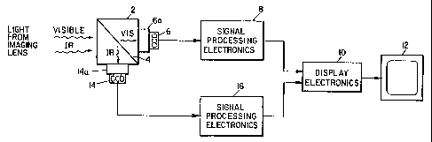
Referring to Figure 2 of the accompanying
drawings, the signal processing electronics includes and
reference is made only to electronics 8 since the
electronics of channels 8 and 16 may be identical, a
CA 02224169 1997-12-08
WO 96/41481 PCT/US96/10496
16
preamp 18, correlated double sampler 20, and an analog-
to-digital converter 22 for developing signals for .
processing by digital signal processor 24. The
processing is controlled by user selected processing
programs stored in memory 26. The program may include
facility for edge enhancement, gain control, image
coring, gamma control and the like. In the case of the
element in processor 16 corresponding to element 26,
color may be added to the infrared derived signal. It
should be noted that the preamp 18 and other elements are
employed in the other two embodiments of the invention.
The display electronics 10 includes all
standard elements including, for instance, a frame buffer
memory in which the signals of the two channels are
stored frame by frame for synchronized transmission to a
digital-to-analog converter where the signals are
combined and fed to a video amplifier, sync generator and
deflection control circuits and thence to a color
monitor.
The elements employed are all standard items
and the programs are relatively simple by today's
standards.
Referring now specifically to Figure 3 of the
accompanying drawings, there is illustrated a slide for
use in a single channel system. An image carrying light
guide 28 introduces light to a lens 30 that focuses light
on a color video camera CCD 32 through a slide 34. The
CA 02224169 1997-12-08
WO 96/41481 PC.'T/US96/10496
t
17
slide includes a color pass filter 36 and an IR pass
. filter 38 and is biased to an upward position as
illustrated in Figure 3 by a compression spring 40. The
slide is configured to be operated by a surgeon or
his/her assistant; the view can be changed by merely
depressing the slide.
The CCD 32 has the IR blocking filter omitted
so that it is sensitive to infrared light energy which
when the filter 38 is depressed is passed to the CCD 32.
The CCD 32 feeds its signals to a preamp, such as preamp
18 of Figure 2, and thence through the circuits 8 or 16
of Figure 2.
The slide 34 has a notch 42 or other detectable
physical characteristic (magnet, mirror, etc.) that is
detectable by a sensor 44. The sensor sends a signal to
circuitry in communication with User Selected Processing
Programs, such as stored in element 26 of Figure 2 to
select which program is to be in use, one for color - one
for infrared. The two sets of signals may be displayed
individually or stored and combined for concurrent
display.
Another single channel system is illustrated in
Figures 4 and 5 of the accompanying drawings. This
system employs only a monochrome CCD video camera with
the IR blocking filter omitted and all color is provided
by processing circuits.
CA 02224169 1997-12-08
WO 96/41481 PCT/US96/10496
18
Specifically, a lens 50 that receives light
from a source via, for instance, an image carrying light -
guide, focuses light on a monochrome video CCD camera 52
through a circular filter wheel 54. The filter wheel 54,
see Figure 5, has red, green, blue and infrared pass
filters disposed in a circular array about the filter
wheel; the red, green and blue colors constituting the
additive color primaries employed in video to process the
complete visual spectrum. The filter wheel has an index
notch 56 in its periphery for purposes described
subsequently.
Returning to Figure 4, the filter wheel 54 is
rotated by a motor 58 under control of a motor controller
60. The periphery of the wheel 54 is rotated through a
slot 62 in an index sensor 64 that produces a
synchronizing signal for a specific position of the
wheel. The signal from the index sensor is processed
through the motor controller, where the angular position
of the motor is controlled, and thence to a write
controller 66.
The video camera 52 also supplies its output
signals to the write controller which distributes signals
to dual port frame memory circuits 68, 70 and 72 as
determined by the position of the filter wheel. Thus,
when a red filter is disposed between the lens 50 and the
camera 52 the signal produced by camera 52 is gated to
the circuit 68. Likewise green and blue signals are
CA 02224169 1997-12-08
WO 96/41481 PCT/US96/10496
19
gated sequentially to circuits 70 and 72. In customary
fashion these signals are converted to digital signals,
applied to a lookup table and a signal of an intensity
determined by the amplitude of, for instance, the
incoming red signal, is made available to the "read" or
output circuit of the write-read circuit 68. Similarly
the signal produced when the IR filter disposed between
the lens and camera is applied to IR write-read circuit
74 having its own lookup table.
The write controller 66 supplies indexed output
control signals to system controller 76. The controller
76 outputs signals to a read controller 78. This element
appropriately times the output of the system and also
permits selection of which signals are to be displayed:
color, infrared or both. Thus when a read circuit of say
the red circuit is gated to the monitor, the read
controller synchronizes this with impingement of the
electron beam of monitor 80 on the red CRT phosphor.
As in Figure 3, processing of the individual
signals may take place as desired and may be accomplished
in the read controller 78, the write-read circuits or
both but most appropriately in the system controller 76.
This controller may have input from a keyboard 82, RS232
input or rotary controls on a front panel. Control may
be over color mix to highlight a particular element of
the view, adding color particularly to the IR signal, or
produce true color or an increase in color intensity and
CA 02224169 1997-12-08
WO 96/41481 PCT/US96/10496
shading or providing "false" colors. Also the wheel 54
may be stopped so that a particular color element may be
viewed for an extended time. There are no constraints on
flexibility.
5 The same flexibility is available from the
system of other designs, particularly the system of
Figure 1, the same degree of control being available from
standard circuits employed in Figure 3. In any event the
system of Figure 4 provides a single channel system using
10 a monochrome camera with extreme flexibility and
reasonable cost. The use of a single camera reduces cost
and avoids the image inversion and registration problems
of a prism based system. The physical components can be
quite small particularly if they are to be used in an
15 operating room or the like. The motor-disk structure may
readily be smaller than illustrated in Figure 4 so that
the entire physical system produces no problems in an
operating room.
The monochrome camera is available from ELMO
20 TSE-270, the dual port frame memory may be a Fidelity 100
or Vision-EZ from Data Translation and others, the image
software stored in the system controller 76 is available
from NOESIS as Visilog or Image-Pro from Media
Cybernetics and others. A circuit for processing the
monochrome images to produce color is available from
Cambridge Research & Instrumentation, Inc. under the name
Varispec. The precision motor is available from Globe or
CA 02224169 1997-12-08
WO 96/41481 PCT/US96/10496
21
Micro-Mo. The write controller via keyboard 82 or other
input controls, if desired, may control all of the
display functions; color, other processing such as edge
enhancement, etc. as set forth above, all in conventional
manner using conventional programs.
As indicated previously the color wheel may be
replaced by a series of LCD color filters (red, green and
blue) aligned in series and energized sequentially by
well known techniques such as a rotary switch. The
switch may be an electronic switch for rapid processing
of signals and/or manually operated or keyboard
controlled to permit the surgeon or an attendant to
select a single color or even two of the three colors.
The advantage of such a system is size and no mechanical
inertia.
The system is illustrated in Figure 6 and is
quite simple. It employs four LCD filters 81, 83, 85 and
87, filter 81 for IR and each of the others for a
different color. A voltage control switch 89 illustrated
as a mechanical switch for simplicity controls the
ability of a filter to pass light of its color. A color
or monochrome CCD 93 is also employed.
Each filter passes all light from IR through
the visible spectrum except when energized. When
energized it passes only the color for which it is
designed. Thus when it.is desired to pass IR only the
filter 81 is energized and only infrared is passed
CA 02224169 1997-12-08
WO 96/41481 PCT/US96/10496
22
through the system. Each of the other filters 83, 85 and
87 are energized in sequence so the red, green and blue
are passed in sequence: the IR filters being in the
sequence also. Thus a stationary color sequential system
is provided with no moving parts.
The advantage of the apparatus of Figure 6 is
the elimination of the IR separation prism and the image
reversal, path length and mechanical problems with some
of the other embodiments.
Referring now specifically to Figure 7 of the
accompanying drawings, there is illustrated another
method of producing separate red, green and blue signals
for subsequent processing.
A beam splitter prism 82 employs a dichroic
filter 84 to separate visible light energy from infrared
light energy. As in the embodiment of Figure 1 the
infrared light energy is reflected from the filter 84
through a visible light blocking filter 86 to a CCD 88
associated with a monochrome camera sensitive to infrared
light energy and thence to processing circuits.
The visible light proceeds along the optical
path through an IR blocking filter 90 to a prism set 92,
94, 97 that splits the visible light into red, green and
blue light energies. The green light energy proceeds
directly along the optical axis and through a trim filter
96 to a CCD 98. Blue light energy is deflected from
prism 94, back through prism 92, thru through a trim
CA 02224169 1997-12-08
WO 96/41481 PCT/US96/10496
23
filter 102 to a CCD 104. Red light energy is deflected
from the rear and then front surface of prism 94, through
a trim filter 108 to a CCD 110.
The CCDs 88, 98, 104 and 110 are monochromatic
and may be processed as discussed relative to the
embodiment of Figure 4.
Reference is now made to the feature of the
invention that provides color correction and increases
the gain of the apparatus to infrared light energy
emitted from the ureter.
From the standpoint of spectral sensitivity,
all commercially available endoscopic cameras use either
single or three chip silicon photodiode CCDs. The
typical current responsivity of silicon CCDs ranges from
300 nm to 1,150, peaking at approximately 900 nm. The
endoscopic camera uses a single chip silicon CCD, and
therefore is confined to the limitations of the silicon
CCDs, i.e., in the present system wavelengths from 300 nm
to 1,150 nm.
The imaging system of the present invention
employs a different light filtering scheme. This
significant modification is important when attempting to
identify infrared transilluminated structures and allow
true fidelity color imaging of the surgical field. The
camera detects visible light in the same range as other
commercially available single chip CCD endoscopic
cameras. As a result of removal of the IR filter from
CA 02224169 2004-07-27
WO 9W41481 PCTiU596110496
24
the camera however; the camera detects infrared (see
Figure 8) as well as visible light. The IR filter is
replaced with a sapphire window that readily passes IR
light energy as well~as visible light. Thus, the camera
can efficiently detect the infrared transilluminated
ureters when used with the endoscope light sensor whereas
typical endoscopic cameras cannot (Figure 8). The only
difference between the camera of the present invention
and the commercially available camera employed herein is
replacement of the IR blocking and color compensating
filter with a sapphire filter that passes light in the
range of 300 - 2700 nm.
Referring to Figure 9 a light cable houses a
filter 119 that blocks infrared light from an endoscopic
light source and adds a cyan color to the light
illuminating the surgical field. To the naked and
unaided eye, the light exiting the light cable appears
cyan in color. However, this cyan filtered light that
illuminates the surgical field corrects or compensates
for reflected light from organs and instruments during an
endoscopic procedure that is captured by the camera. As
previously indicated, the net effect is an improvement in
the color fidelity of the imaged field, produced by the
camera in accordance with the invention.
Referring specifically to FIG. 10 of the
G~cvmgar:~~_r~c crav:i~:~~, a i~gt~t source i16 supplies light
energy via a light :~abie 11& tc ar. endoscope IeO. The
CA 02224169 2005-02-25
WO 96J414$1 25 PCTNS96J10496
' w cable includes, a filter as illustrated in FIG. 9 and
thus infrared light energy does not enter the endoscope.
The endoscope 120 enters the body on which a procedure is
being performed via a trocar 122 and illuminates the
region of the procedure. Light from this region proceeds
back through the endoscope, an optical coupler 124 to a.
laparoscopic camera 126 sensitive to both visible and
infrared light energy in the range of light energy as
depicted in FIG. 8. Signals produced by the camera 126
axe supplied via a camera control unit 127 to a monitor
128 for viewing.
Infrared light energy is supplied by an infrared
source and detector 129 to a light guide 130 that is
located in a catheter 132 inserted into the ureter 134.
The region of the light guide located in the ureter is
conditioned to emit infrared light energy into the body
cavity subject to the procedure. This light is detected
by both the laparoscopic camera 126 and a probe 136
coupled to the source and detector 129.
Once given the above disclosure, many other
features, modifications and improvements will become
apparent to the skilled artisan. Such.features,
modifications and improvements are, therefore, considered
to be a part of this invention, the scope of which is to
be determined by the following claims.