Note: Descriptions are shown in the official language in which they were submitted.
CA 02225626 1997-12-23
WO 97/01339 - ~ - PCT/US96/1041i8
METHOD OF USING (2-IMIDAZOLIN-2-YLAMINO)
QUINOXALINES IN TREATING OCULAR NEURAL INJURY
Background of the Invention
The present invention relates to methods for the protection of the
optic nerve and the retina of mammalian eyes from noxious
provocations including damage by compressive (mechanical) effect,>
of elevated intraocular pressure caused by glaucoma or other
etiologic factors and impaired blood flow to these nerves.
Glaucoma is a disease of the eye characterized by increased
intraocular pressure. On the basis of its etiology, glaucoma has been
classified as primary or secondary. Further, primary glaucoma in
adults may be either chronic open-angle or chronic angle-closure.
Secondary glaucoma results from pre-existing ocular diseases such as
uveitis, intraocular tumor or enlarged cataract.
The underlying causes of primary glaucoma are not yet well known.
Increase intraocular pressure is due to obstruction or aqueous humor
outflow. In chronic open-angle glaucoma, the anterior chamber and.
its anatomic structures appear normal, but drainage of the aqueous
humor is impeded. In acute and chronic angle-closure glaucoma, the
anterior chamber is shallow, the filtration angle is narrowed and the
iris may obstruct the trabecular meshwork at the entrance to the
canal of Schlemm. Dilation of the pupil may push the root of the iris
forward against the angle or may produce pupillary block and thus
precipitate an acute attack of elevated intraocular pressure. Eyes with
narrow anterior chamber angles are predisposed to acute angle-
closure glaucoma attacks of varying degrees of severity.
Secondary glaucoma is caused by any interference with the flow of
aqueous humor from the posterior chamber into the anterior
chamber and, subsequently, into the canal of Schlemm.
Inflammatory disease of the anterior segment may prevent aqueous
escape by causing complete posterior synechia in iris bombe, and may
plug the drainage channel with exudates. Other common causes are
CA 02225626 2002-03-05
!.
WO 97/01339 FCT/US96110468
-2-
intraocular tumors, enlarged cataracts, vE~ntral retinal vein occlusion,
trauma to the eye, operative procedures and iTitraocular hemorrhage.
Considering all types together, glaucoma occurs in about 2% of all
persons over the age of 40 and may be asymptomatic,for years before
progressing to ral ~id loss of vision. In cases where surgery is not
indicated, topical beta-ac:lrenoceptor antagonists have been the drugs
of choice for treating glaucoma. However; alpha adrenergic agonists
are awaiting approval for use in the treatment of elevated intraocular
pressure and will probably become mainstays in the treatment of this
disease once they become available.
Various quinoxaline derivatives having alpha2 agonist activity have
been suggested as therapeutic agents by, fc~r example, Danielewicz, et
al. in U.S. Patent No.s 3,890,319 and 4,029,792. They disclose
compounds as regulators of the cardiovascular system which have
the following formula:
H x
,,N \
~N , . I R
C._
y z
where the 2-imidazolin-2-ylamino group may be in any of the 5-, 6-,
7- or 8-position of the quinoxaline nucleus; x, y and z may be in any
of the remaining 5-, 6-, 7- or 8-positions and may be selected from
hydrogen, halogen, lower alkyl, lower alkoxy or trifluoromethyl; and
R is an optional substituc3nt in either the 2- or 3-position of the
quinoxaline nucleus and may be hydrogen, lower alkyl or lower
alkoxy. The presently useful compounds may be prepared in
accordance with the procedures outlined by Danielewicz, et al,
In "Ocular Effects of a Relatively Selective Alpha-2 Agonist (UK-
14,304-18) in Cats, Rabbit, and Monkeys" jJ.A. Burke, et al., Current
Eye Rsrch., 5, (9), pp. 665-676 (1986)] the quinoxaline derivative was
CA 02225626 1997-12-23
WO 97/01339 PCT/US96/10468
-3-
shown to be effective iri reducing intraocular pressure in rabbits, cat:>
and monkeys. Compounds in this study were administered topically
to the eye of the study animals.
N' ' 'NH
Br
N
H / N\
It has long been know that one of the sequelae of glaucoma is damage
to the optic nerve head. This damage, referred to as "cupping",
results in depressions in areas of the nerve fiber of the optic disk.
Loss of sight from this cupping is progressive and can lead to
blindness if the condition is not treated effectively.
Unfortunately lowering intraocular pressure by administration of
drugs or by surgery to facilitate outflow of the aqueous humor is not
always effective in obviating damage to the nerves in glaucomatous
conditions. This apparent contradiction is addressed by Cioffi and
Van Buskirk [Sure. of Ophthalmol., 38, Suppl. p. S107-16, discussion
S116-17, May 1994) in the article, "Microvasculature of the Anterior
Optic Nerve". The abstract states:
The traditional definition of glaucoma as a disorder of
increased intraocular pressure (IOP) oversimplifies the
clinical situation. Some glaucoma patients never have
higher than normal IOP and others continue to develop
optic nerve damage despite maximal lowering of IOP.
Another possible factor in the etiology of glaucoma may be
regulation of the regional microvasculature of the anterior
optic nerve. One reason to believe that microvascular factors
are important is that many microvascular diseases are
associated with glaucomatous optic neuropathy.
Subsequent to Cioffi, et al., Matusi published a paper on the
"Ophthalmologic aspects of Systemic Vasculitis" [Nippon Rinsho. 52
(8), p. 2158-63, August 1994) and added further support to the
CA 02225626 1997-12-23
WO 97/01339 PCT/US96/10468
-4-
assertion that many microvascular diseases are associated with
glaucomatous optic neuropathy. The summary states:
Ocular findings of systemic vasculitis, such as polyarteritis
nodosa, giant cell angitis and aortitis syndrome were
reviewed. Systemic lupus erythematosus is not categorized
as systemic vasculitis, however its ocular findings are
microangiopathic. Therefore, review of its ocular findings
was included in this paper. The most common fundus
finding in these diseases is ischemic optic neuropathy or
retinal vascular occlusions. Therefore several points in
diagnosis or pathogenesis of optic neuropathy and retinal
and choroidal vaso-occlusion were discussed. Choroidal
ischemia has come to be able to be diagnosed clinically, since
fluorescein angiography was applied in these lesions. When
choroidal arteries are occluded, overlying retinal pigment
epithelium is damaged. This causes disruption of barrier
function of the epithelium and allows fluid from choroidal
vasculatures to pass into subsensory retinal spaces. This is a
pathogenesis of serous detachment of the retina. The retinal
arterial occlusion formed non-perfused retina. Such hypoxic
retina released angiogenesis factors which stimulate retinal
and iris neovascularizations and iris neovascularizations
may cause neovascular glaucoma.
B. Schwartz, in "Circulatory Defects of the Optic Disk and Retina in
Ocular Hypertension and High Pressure Open-Angle Glaucoma"
[Sure. Ophthalmol., 38, Suppl. pp. S23-24, May 1994]. discusses the
measurement of progressive defects in the optic nerve and retina
associated with the progression of glaucoma. He states:
Fluorescein defects are significantly correlated with visual
field loss and retinal nerve fiber layer loss. The second
circulatory defect is a decrease of flow of fluorescein in the
retinal vessels, especially the retinal veins, so that the
greater the age, diastolic blood pressure, ocular pressure and
visual field loss , the less the flow. Both the optic disk and
CA 02225626 1997-12-23
WO 97/01339 PCT/US96/10468:
-5-
retinal circulation defects occur in untreated ocular
hypertensive eyes. These observations indicate that
circulatory defects in the optic disk and retina occur in
ocular hypertension and open-angle glaucoma and increase
with the progression of the disease.
Thus it is evident that there is an unmet need for agents that have
neuroprotective effects in the eye that can stop or retard the
progressive damage that occurs to the nerves as a result of glaucoma
or other ocular afflictions.
5ummarv of the Invention
A new method of protecting the optic nerve and retina of the
mammalian eye from damage by glaucoma and other noxious
provocations has been discovered. This method comprises
administering to the mammal either systemically or by intrabulbar
injection an effective amount of one or more of certain aryl-imino-2--
imidazolidines (as defined herein), salts thereof and mixtures
thereof. This new method is particularly effective when
administered as a prophylactic treatment, i.e. before damage to the
nerve takes place, or before long-term progression of the disease,
such as glaucoma, has taken place.
Detailed Description of the Invention
The drawings will first be briefly described.
Drawings
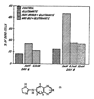
Figure 1 is a bar graph showing the percentage of cells killed by
treatment with glutamate plotted by number of days since glutamate
treatment. A control which was not treated with glutamate has been
included to determine cell death which occurred without any such
treatment. Also shown are measurements taken after treatment with.
both AGN191103 and glutamate, and treatment with MK-801 and
glutamate. MK-801 is a well known neuroprotective agent in the art.
The numbers beneath the bars for glutamate; AGN191103 +
glutamate; and MK-801 + glutamate show the concentrations of
CA 02225626 1997-12-23
WO 97/01339 ' PCT/US96/10468
-6-
glutamate and drug used in each case. At day 8, AGN 191103 and MK-
801 show comparable effects in protecting cells from glutamate
induced neurotoxicity. Experimental procedures followed in
generating the data for this figure are detailed in Example 1.
Figure 2 shows plots of compound action potentials (CAP) measured
for optic nerve fibers: in the left-hand frame, measured at 2 weeks
post injury (i.e. after nerve crush) for optic nerve treated with AGN
191103 (the upper line) and for an untreated nerve used as a control
(lower line); and in the right-hand frame a comparison CAP of non-
injured optic nerve. The scales of the plots are given for each of the
frames. The post-injury abscissa scale is 25 X the scale of the non-
injured plot. (Units: millivolts and milliseconds). The value of the
compound action potential is calculated as the integral of the area
under each curve. The irregularity of the curve is a feature of the
dispersion of the compound response; some nerve cells conduct
more rapidly than others and so amplitude of the measured voltage
varies with time.
Figure 3 is a bar graph showing the maximal CAP amplitude in
microvolts (~.V) for cells injured by a optic nerve crush in rats and
treated with: 1) vehicle alone; 2) clonidine and 3) AGN191103. Each of
the drugs was tested at four different concentrations (administered as
a multiple of body weight for the test subject) and is represented by a
bar on the chart. Clonidine was chosen as a benchmark a2 agonist
compound with very well defined pharmacology to compare against
the test compound AGN 191103. While clonidine did show some
neuroprotective activity over vehicle alone, it showed about half the
maximal CAP response of AGN191013.
Figure 4 is a graphic plot of the Visual Evoked Potential Response
and shows the electrical potential activity evoked at the surface of the
visual cortex (comparable to an electroencephalogram) as a result of
visual (light) stimulus. The test is performed in live rats and is a
measure of the integrity of the whole visual system from the retina
through the optic nerve into the lateral geniculate nucleus and
ultimately to the visual cortex located in the back of the brain. The
CA 02225626 1997-12-23
WO 97/01339 PCT/US96/104f~8
-7-
left-hand frame shows the response without nerve crush injury and
the right-hand frame shows the responses measured at 2 weeks post-
injury for rats treated with AGN191103 above (labeled positive) and
control rats below (labeled negative) prior to nerve crush. The scale
in ~.V vs. milliseconds for both plots is shown below the ordinate
axis.
For a discussion and bibliography regarding the nerve crush model
and its significance in evaluating nerve damage and recovery see:
"Functional Recovery and Morphological Changes after Injury to the
Optic Nerve", Sabel, B.A. and Aschoff, A., Neuropsvchobiolog~, 28,
pp. 62-65 (1993).
Injury to the mammalian optic nerve, as in any other parts of the
mammalian central nervous system (CNS), leads to axonal
degeneration followed by a loss of cell bodies, with failure of axonal
regrowth from the surviving neurons. Initially, degeneration of the
injured nerve is probably attributable to direct neuronal damage.
However, the associated physiological and biochemical events
occurring in the nerve immediately after injury are probably
responsible for the subsequent progressive degeneration, not only of
the directly injured axons, but also of those that escaped the primary
damage and largely determine the long-term functional outcome.
The immediate injury-induced response strongly influences the
subsequent degenerative response. Treatment that reduces or
attenuates the immediate response is therefore likely to achieve
optimal prevention or delay of the secondary degenerative processes.
For monitoring of the immediate response, it is obviously preferable
to employ a noninvasive technique. An adaptation of the
nicotinamide adenine dinucleotide (NADH) monitoring technique
to enable measurement of the earliest post-traumatic events has
proved to be a valuable non-invasive approach. Use of the technique
allows the immediate effect of the injury to be evaluated in real time
and on-line before and after a well-controlled crush injury in
inflicted on the adult rat optic nerve in vivo. In this experimental
paradigm, measurement of the metabolic activity of the injured optic
CA 02225626 1997-12-23
WO 97/01339 PCT/LTS96/10468
-g_
nerves represent the activity of both injured axons and their
associated non neuronal cells, and thus evaluate the potential ability
to cope with injurious stresses. The model is also useful for
monitoring the activity of various agents that may overcome or
mitigate nerve cell damage or death from such stresses.
The earliest injury-induced response is a decrease in the energy state
of the nerve, under conditions where ischemic events can be
completely ruled out. The reduction in the energy state may be
related to: 1) postinjury elevation in free fatty acid levels, which may
interfere with mitochondria) function and result in uncoupling of
electron transport; and 2) a marked rise in intracellular free Ca2+. It is
known that axonal injury is generally followed by an increase in
extracellular potassium ions, which stimulate the uptake of Ca2+ via
either voltage sensitive channels (L, T or N type) or receptor-operated
Ca2+ channels. A marked rise in intracellular free Ca2+ can accelerate
processes that are inimical to cell survival, including those
involving Ca2 -dependent enzymes, mainly lipases, proteases and
endonucleases, that may cause mitochondria) damage and lead
eventually to cell death. The cell, in order to overcome these events,
needs more energy to actively restore ionic homeostasis. The
combination of increased energy demands and decreased energy
conservation resulting from mitochondria) dysfunction at the site of
injury may be the major reason for the subsequent irreversible nerve
damage and nerve degeneration following injury. Early
measurement of metabolic activity could therefore indicate the fate
of the axon, its associated glial cells and its non-neuronal cell bodies.
It follows from the above that restoration of the mitochondria)
activity may be critical in preventing the degenerative process
occurring in the nerve after injury.
Since the injury inflicted on the nerve in the nerve crush model is a
well-controlled, calibrated and reproducible lesion, it is possible to
correlate early post-traumatic metabolic deficits and possible
mitigation of these by drug or other treatments with long-term
morphological and physiological effects.
CA 02225626 1997-12-23
WO 97/01339 PCT/US96/1046~8
-9-
From the foregoing figures and discussion it is apparent that
neuroprotection is conferred on nerve cells to both glutamate-
induced toxicity and physical insult in the nerve crush model.
It has now been discovered that neuroprotection is conferred upon
ocular nerve cells by administration of a drug of formula I to the
optic nerve and/or retina of a mammal within a period prior to or
following an insult to ocular nerve cells but prior to cell death
H x
N
,.
~ R
y Z N
formula I
wherein the 2-imidazolin-2-ylamino group may be in either the 5- or
6-position of the quinoxaline nucleus; x, y and z may be in any of the
remaining 5-, 6-, 7- or 8-positions and are selected from hydrogen,
halogen, lower alkyl, lower alkoxy or trifluoromethyl; and R is an
optional substituent in either the 2- or 3-position of the quinoxaline
nucleus and may be hydrogen, lower alkyl or lower alkoxy.
Definitions
The compound identified as AGN 191103 has the chemical structure
as
HNn shown. It is also known by the chemical
~N nomenclature 6-methyl-(2-imidazolin-2
HN y3 ylamino) quinoxaline.
N\
25~
-N
The neuroprotective agent identified as MK-801 is also known by the
name dizocilpine and has the following chemical structure:
It is additionally identified and described in the
1 ~ HN p / 11th edition of the Merck Index at monograph
number 3392.
' ~3
Human dosag_e_ and administration
CA 02225626 1997-12-23
WO 97/01339 PCT/LJS96/10468
-10-
The methods of this invention are useful in treating any mammal,
including humans.
According to this invention, mammals are treated with
pharmaceutically effective amount of a neuroprotective agent for a
period of time and at a time such that noxious provocations to the '
optic nerve and retina do not kill or permanently damage the nerve
cells. Protective agents may be administered orally or by any other
appropriate means of delivery described below or known in the art.
In accordance with this invention, pharmaceutically effective
amounts of a protective agent can be administered alone to treat
nerve injury or to prevent nerve cell death. Alternatively a
protective agent may be administered sequentially or concurrently
with an antiglaucoma drug, e.g. a beta-blocker, an alpha2 agonist , a
muscarinic agent such as pilocarpine, a carbonic anhydrase inhibitor
(CAI), or another drug useful in maintaining intraocular pressure
(IOP) at normal levels or in lowering elevated IOP. The most
effective mode of administration and dosage regimen of protective
agent will depend on the type of disease to be treated, the severity and
course of that disease, previous therapy, the patient's health status,
and response to the drug and the judgment of the treating physician.
Generally, the neuroprotective agent should be administered in a
dose to achieve a serum or intravitreal concentration of 0.01 nM to 50
nM. Preferably the neuroprotective agent is administered prior to
injury to the nerve, but can be administered injury has occurred with
lessened effect.
Conventional modes of administration and standard dosage
regimens of protective agents, e.g. MK-801, can be used. Optimal
dosages for co-administration of a drug, e.g. an IOP-lowering drug,
with a neuroprotective agent can be determined using methods
known in the art. Dosages of neuroprotective agents may be adjusted
to the individual patient based on the dosage of the drug with which
the agent is co-administered and the response of the patient to the
CA 02225626 1997-12-23
WO 97/01339 PCT/US96/1046~8
-11-
treatment regimen. The protective agent may be administered to thc~
patient at one time or over a series of treatments.
An agent that cannot pass the blood/brain barrier, e.g. MK-801, may
be administered locally, e.g. intravitreally by intrabulbar injection, or
intrathecally. Agents which are capable of crossing the blood/brain
" barrier, e.g. AGN191103 can be administered systemically, e.g., orally,
or intravenously, or by injection.
The composition used in these therapies may also be in a variety of
forms. These include, for example, solid, semi-solid, and liquid
dosage forms, such as tablets, pills, powders, liquid solution or
suspension, liposomes, suppositories, injectable and infusible
solutions. The compositions also preferably include conventional
pharmaceutically acceptable carriers which are known those of skill
in the art.
The following non-limiting examples describe assays and
measurements used in 1) determining protection of nerve cells from
glutamate induced toxicity and 2) methods of determining neural
protection conferred by neuroprotective agents in a nerve crush
model of mechanical injury.
Example 1:
Experimental procedure for measuring neural vrotection in a model
Qf glutamate induced excitotoxic effects on nerve cells:
Low-density rat hippocampal neuronal cultures were prepared by the
procedure of Goslin and Banker. Coverslips were cleaned and
sterilized in porcelain racks in such a way that they did not stick to
one another (Cohen cover glass staining racks, Thomas Scientific).
Coverslips (13 mm) were placed in staining racks, rinsed in distilled
water (four rinses, 1 min. each) to remove dust and transferred to
concentrated HN03 for 36 hours. Coverslips were rinsed in distilled
water (four changes over 3 hours) and sterilized with dry heat
(overnight at 225° C). The coverslips were transferred to 24-well
dishes, one coverslip per well. To support the coverslips above the
CA 02225626 1997-12-23
WO 97/01339 PCT/US96/10468
-12-
glia during coculturing, paraffin dots were placed on~ dishes, and UV
irradiation (30 min.) was applied before the coverslips were
introduced. One mg/mL of poly-L-lysine hydrobromide (PLL)
(Sigma) (MW 30,000-70,000) was dissolved in borate buffer (0.1 M, pH
8.5), filtered, sterilized and used to cover each coverslip overnight.
The PLL was removed, coverslips were rinsed in distilled water (two
washes, 2 hrs. each), plating medium [Eagle's MEM with Earle's salts '
containing extra glucose (600 mg/L) and 10% horse serum] was added
and the dishes were stored in an incubator. Astroglial cultures were
prepared from the brains of neonatal rats by a method similar to that
described by Levinson and Mc Carthy, except that they were plated at
a lower density so that they contained predominantly type 1 astroglia.
105 cells were plated in each well. Glial cultures were fed with plating
medium twice a week and were used after reaching confluence, about
2 weeks after plating . One day before use, the plating medium was
removed, neuronal maintenance medium (MEM containing N2
supplements) was added, and incubation continued. 3 X 10⁴ of viable
rat hippocampal nerves (E18 embryos) were plated on the PLL-treated
coverslips kept in plating medium. After 3-4 hrs, when most of the
neurons were attached, the coverslips were transferred to the dishes
containing the glial cell in maintenance medium in such a way that
the neuronal side was facing the glia, which support neuronal
survival and development. To reduce glial proliferation, cytosine
arabinoside (1-b-D-arabinofuranosylcytosine)(Calbiochem)(5 X 10 M
final concentration) was added to the cultures 2 days after plating. At
day 6 in culture, cells were treated with 1mM glutamate or with
glutamate together with either AGN-191103 - 0.1 nM (MW = 200) or
MK-801 - 10 nM (2-3 coverslips were used to each treatment).
After 24 hrs. of incubation, cells were stained with trypan blue. Live
and dead neurons were counted from randomly selected culture
fields (5 fields from each coverslip) . Percentage of dead cells was
calculated.
CA 02225626 1997-12-23
WO 97/01339 ~ ~ PCT/US96/1046li
-13-
Example 2:
Procedure for nerve crush i~ njury and measurements of compound
action potentials CAP subseduent to injury.
Part A.
Metabolic Measurements
Animal utilization was according to the ARVO Resolution on the
use of animals in research. Male Sprague-Dawley (SPD) rats weighing
300-400 g were anesthetized with sodium pentobarbitone
(intraperitoneally, 35 mg/kg). A cannula was introduced into the
trachea for artificial ventilation when required. With the animal's
head held in place by a head holder, a lateral canthotomy was
performed under a binocular operating microscope and the
conjunctiva was incised lateral to the cornea. After separation of the
retractor bulbi muscles, the optic nerve was identified and a length o~f
3 0 3.5 mm was exposed near the eyeball by blunt dissection. The dura
was left intact and care was taken not to injure the nerve. The first
part of a light guide holder (see below) was inserted under the optic
nerve and the nerve was gently eased into the light guide canal. The
second part was then fixed in place in such a way that the light guide
was located on the surface of the optic nerve 1 mm from the site at
which the injury was to be administered.
Surface fluorometry-reflectometry
Monitoring of the intramitonchodrial NADH redox state was based
on fluorescence of NADH at 366 nm, resulting in the emission of
blue light with a peak intensity at 450 nm, which is unlike its
oxidized form, NAD+, which lacks this fluorescence. The source of
the 366 nm excitation is a 100-W air-cooled mercury lamp equipped
with a strong 366-nm filter (Corning 5860 (7-37) plus 9782 (4-96)). A
flexible Y-shaped bundle of optic fibers (light guide) is used to
transmit the light to and from the optic nerve, thus making in vivo
measurements technically feasible. Excitation light is transmitted
through the bundle of excitation fibers to the nerve. The light
emitted from the nerve, after being transmitted through a second
CA 02225626 1997-12-23
WO 97/01339 PCT/US96/10468
-14-
bundle of fibers, is split in a ration of 90:10 for measurement of the
fluorescent light (90%) at 450 nm and the reflected light (10%) at 366
nm by two photomultipliers connected to a one-channel direct
current fluorometer-reflectometer. In order to minimize variations
among animals, standard signal calibration procedures were applied
at the start of the recordings. Changes in the fluorescence and
reflectance signals during the experiment are calculated relative to
the calibrated signals. This type of calibration, although not absolute,
has nevertheless been found to yield reliable and reproducible results
from various animals and among different laboratories.
Changes in reflected light were correlated with changes in tissue
absorption caused by hemodynamic effects and movements of the
optic nerve secondary to alteration in arterial blood pressure and
nerve volume. The fluorescence measurements are found to be
adequately corrected for NADH redox state measurements by
subtraction of the reflected light (366 nm) from the fluorescent light
(1:1 ratio) to obtain the corrected fluorescence signal.
Metabolic Measurements
Animals which were still anesthetized were allowed to recover for 30
min. from the surgical procedures described above and were then
exposed to anoxic and hyperoxic conditions. An anoxic state was
achieved by having the rat breathe in an atmosphere of 100%
nitrogen for 2 min., after which it was returned to air. Whenever
animals did not return spontaneously to normal breathing, they
were ventilated by blowing twice into the trachea. A hyperoxic state
was induced by having the animal breathe 100% oxygen for 6-10 min.
In order to evaluate the metabolic activity of the optic nerve, the
relative changes in reflected and fluorescent light intensities in
response to anoxia and to hyperoxia were measured before and after
crush injury.
CA 02225626 1997-12-23
WO 97/01339 PCT/US96/104fi8
-15-
Experimental Protocol For Metabolic Measurements
Using calibrated cross-action forceps, a well-calibrated moderate crush
injury was inflicted to the nerve between the eye and the light guide
holder at a pressure corresponding to 120 g for 30 sec.
Part B.
Physiological Measurements
Experimental setup for recording compound action potential (CAP):
Prior to removal of optic nerves for electrophysiological
measurement, the rats were deeply anesthetized with 70 mg/kg
pentobarbitone. The skin was removed from the skull and the optic
nerves were detached from the eyeballs. Subtotal decapitation was
performed and the skull was opened with a rongeur. The cerebrum
was displaced laterally, exposing the intracranial portion of the optic'
nerve. Dissection at the level of the chiasm enabled removal of the
whole length of the nerve, which was transferred to vials containing
fresh, cold Krebs solution, consisting of: NaCI (125 mM), KCl (5 mM),
KH2P04 (l.2mM), NaHC03 (26 mM), MgS04 (0.6 mM), CaCl2 (24
mM), D-glucose (11 mM), aerated with 95% 02 and 5% C02. The
nerves were kept in this solution, in which electrical activity
remained stable for at least 3-4 h. After 1 h of recovery, nerves were
immersed in Krebs solution at 37° C. Electrophysiological recording
were obtained from the nerve distal to the crush lesion, since the
nerves were too small to allow measurement on both sides of the
crush. The nerve ends were then connected to two suction Ag-AgCl
electrodes immersed in the bathing solution. The stimulating pulse
was applied through the electrode at the proximal end and the action
potential was recorded by the distal electrode. A Grass SD9 stimulator
was used for electrical stimulation (2 V, 50 ~,s). The signal was
transmitted to a Medelec PA63 preamplifier and thence to a Medelec
MS7 electromyograph and AATT amplifier. The solution, stimulator
and amplifier had a common ground. The maximum amplitude of
eight averaged CAPs was recorded and photographed with a Polaroid
camera. The left nerves (uninjured) were used to measure the
reference values of normal nerves and to calibrate the crush forceps.
CA 02225626 1997-12-23
WO 97/01339 ~~ PCT/LTS96/10468
-16-
Recording, of Visual Evoked Potential VEP Response
Injured drug-treated rats were examined in 2 weeks after the injury
for assessment of their functional recovery. In this set of
experiments, the pattern of filed potentials in response to light
stimulation was recorded from the primary visual cortex. The
potential evoked by the light originates in the retina and is
propagated along the surviving axons to reach their final target, the
visual cortex. Only those axons that survived the primary and
secondary degenerative processes are capable of conducting an action
potential. A comparative analysis of the pattern of field potentials in
treated and untreated animals will reveal the effect of the treatment
on axonal survival.
Anesthetized rats (Rumpon, Ketalar) were placed in a small animal
sterotaxic instrument. After exposure of the skull, two holes were
drilled with a cylindrical drill bit, with the dura kept intact to
minimize cortical damage. One hole, drilled above the nasal bone,
was used as a reference point. The second hole was in the area OC1
with the coordinates Bregma # 8 mm, lateral # 3 mm. A gold contact
pin connected to a screw was used as the electrode, which was
screwed into the holes and glued by acrylic cement to the skull. The
field potential was evoked by stroboscopic stimulation, with an
average of 90 sweeps per minute. The flash-evoked potential was
analyzed by the use of the Lab View data acquisition and
management system. The field potentials were digitized and stored
for off line analysis.
Part C.
Measurement of effects of drug tests for neural protective properties
The first set of experiments involved metabolic measurements. Each
drug was injected intraperitoneally at several different
concentrations. Each drug was tested in a group of 8 animals, together
with 8 controls (injured animals treated with the buffer vehicle). In
each case, metabolic measurements were obtained on-line before
CA 02225626 1997-12-23
WO 97/01339 PCT/US96/10468
-17-
injury, 0.5 h after injury and every hour for 4-6 h thereafter. The data
obtained were analyzed by ANOVA.
Measurement of long term effects. Ph~ siological Activities.
CAPS
Immediately after injury, the drug to be tested was injected into 10
animals, and 10 control animals were injected with vehicle. Two
weeks later the CAPs of each nerve were recorded in vitro, using
suction electrodes. The contralateral side was used as an internal
control. The results indicated whether the examined drug had any
potential effects on the rescue of spared axons and / or slowing of
degeneration. Positive results led to efforts at determining the
optimal dosage for each promising drug.
VEP response
Electrodes were implanted in the cortex of naive SPD rats in two age--
and sex-matched groups. Immediately after implantation, the VEP
response was recorded from the left side while a light was flashed
into the right eye, with the left eye covered. A well-controlled crush
injury was then inflicted on the optic nerve and the drug was
immediately administered at the previously determined optimal
dosage. Control animals were handled in the same way except
vehicle was administered rather than drug. The VEP response for
each animal was recorded 1 day, 1 week, 2 weeks and 4 weeks after
operation.
While this invention has been described with respect to various
specific examples and embodiments, it is to be understood that the
invention is not limited thereby and should only be construed by
interpretation of the scope of the appended claims.