Note: Descriptions are shown in the official language in which they were submitted.
,r-~ CA 02230527 2006-09-26 ~~
DOCKET: 1849 Canada
CATHETER HAVING VALVE MECHANISM
BACKGROUND
1. Technical Field
This application relates in general to catheters having slit valves to permit
ingress and egress of fluids through the catheter into and out of the body of
a patient.
1o 2. Discussion of the Prior Art
The use of catheters in intravenous procedures and for intravenous
therapies is well known in the medical community. Catheters typically are
implanted
into various vessels in the patient's body to provide for the ingress and/or
egress of
fluids, such as blood and other bodily fluids, and as well for the infusion of
medication
15 or other medical solutions for both specific treatment of the patient and
to facilitate other
treatments and diagnoses. The use of catheters may be for short term
procedures, and
are also commonly used in long term nrocedutes wherein the catheter is
imrlantPrl in the
body and left in place for an extended Period of time tn facilitate long term
trP.,~tment of
the patient.
z o Catheters typically take the forth of an elongated tube constructed of a
biocompatible surgical grade material which is flexible to permit guiding or
steering of
the catheter through blood vessels or anatomical passages. Initially,
catheters generally
included an open ended tube which was positioned during the surgical
procedure, and
was capped at its proximal end (i.e., the end positioned outside the body) to
provide a
2 s port for the infusion or withdrawal of fluids. The distal end of the
catheter remained
open inside the vessel within the patient's body, and allowed for ready
withdrawal or
infusion of fluids through the catheter. These catheters were typically used
in short
term procedures, such as surgical procedures in which the catheter would be
removed
after completion of the surgical procedure. Leaving a catheter of the open-
ended type in
3 o the vessel of the patient subjected the catheter to a number of potential
problems,
including the formation of blood clots which would obstruct the end of the
catheter.
Open-ended catheters are thus flushed regularly, typically with a saline
and/or
anticoagulant solution, to keep the distal end of the catheter open.
~'- CA 02230527 1998-02-25 ' '
-2-
Catheters intended to remain in the body for a longer term have been
developed and generally include a closed distal end and a valve adjacent the
distal end to
perrrut the infusion or withdrawal of fluids. Typically, these valves operate
by reacting
to the pressure differential within the tube as compared to the vessel (or
other anatomical
location) in which the catheter is placed. Generally, increasing the pressure
within the
cathE;ter provides fot infusion of fluids through the valve and into the
vessel, while a
pressure decrease in the catheter provides for withdrawal of the fluids from
the site in
which the catheter is placed.
A challenge associated with closed end catheters having valves adjacent
their distal end is the performance of the valve based on a pressure
differential.
Although efforts have been made to optimize the performance of such valued
catheters,
e.g., by chemical weakening the are of the catheter tube adjacent to the valve
or other
localized treatment (see e.g., US. Patent Nos. 4,549,879 and 4,701,166 to
Groshong
et al.; 4,995,863 to Nicholas et al.; and 5,147,332 to Moorehead), a need
remains to
further optimize the fabrication andlor performance of existing valued
catheters.
S~ _._UM_MARY
The present catheter device includes an elongated flexible tube which has
an open end, a closed end and which is fabricated from a surgical grade
material. The
2 o cathcaer tube has a wall which is defined by an inner and outer surface of
the tube,
where the inner surface of the tube is defined by a lumen which extends the
length of
the tube. In one preferred embodiment, when viewed in cross-section at two
different
longitudinal points, at least a portion of the tube at the more distal point
has a reduced
thickness with respect to the tube when viewed at a more proximal point, and
at least
25 one valve is positioned solely in the portion of reduced thickness so as to
communicate
the lumen with the exterior of the tube. The valve is oriented at an angle to
the
longitudinal axis of the tube.
The reduced thickness portion of the catheter tube, in a further
embodiment, is the result of the lumen of the catheter tube being offset and
parallel to
the longitudinal axis of the tube, and in another embodiment is the,result of
the lumen .
having an oval cross-section such that the major axis of the oval defines the
portions of
reduced thickness in the wall of the tube. In each of these cases, the valve
is provided
d'
.-~ CA 02230527 1998-02-25
-- 3 -
in the portion or portions of reduced thickness, and does not extend into the
areas of
incret~sed thickness so that the operation of the valve is consistent along
its length.
Xn an alternate embodiment of the present catheter, the slit valves
comprise at least one pair of slits which are parallel to each other but are
positioned at an
s angle to the longitudinal axis of the catheter tube. Preferably, the slits,
when formed
through the tube, are cut at different angles relative to the catheter tube
wall surface to
facilitate the infusion or withdrawal of fluids.
In each of the embodiments, it is preferred that the valves are positioned
at an angle to the longitudinal axis of the catheter in an area of reduced
thickness to
to increase the size of the opening for the ingress and egress of fluids.
~F,F DESCRIPTION OF THE DRAWINGS
The features of the present catheter will become apparent from the
detained description set forth below, taken with reference to the accompanying
~s drawings, in which:
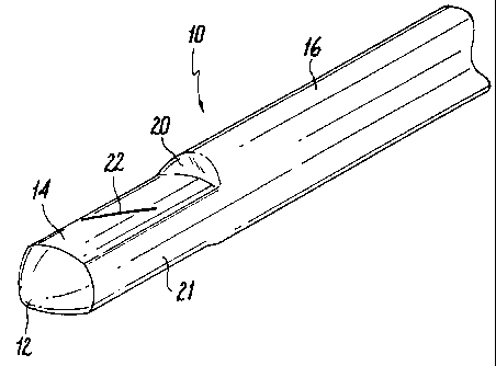
Fig. 1 is a perspective view of the present catheter according to a first
embodiment;
Fig. 2 is a top plan view of the present catheter of Fig. 1;
Fig. 3 is a side elevation view of the present catheter of Fig. 1;
2 o Fig. 4 is a side cross-sectional view of the catheter of Fig. 1 taken
along
lines 4-4 of Fig. 2;
Fig. 5 is a perspective view of the present catheter according to a second
embodiment;
Fig. 6 is a side elevation of the present catheter of Fig. 5;
25 Fig. 7 is a side cross-sectional view of the catheter of Fig. 5 taken along
lines 7-7 of Fig. 6;
Pig. 8 is a front elevation view of the catheter of Fig. 5;
Fig. 9 is a perspective view of the present catheter according to a third
embcxiiment;
3 o Fig. 10 is a top plan view of the present catheter of Fig. 9;
Fig. 11 is a cross-sectional view of the catheter of Fig. 9 taken along
lines 11-11 of Fig. 10 showing a circular lumen;
CA 02230527 1998-02-25
-4-
lumen;
embodiment;
Fig: 12 is a cross-sectional view similar to Fig. 11 showing an oval
Fig. 13 is a perspective view of the present catheter according to a fourth
s Fig. 14 is a side elevation view of the catheter of Fig. 13; and
Fig. 15 is a cross-sectional view of a catheter similar to Fig. 13, except
that the two slits of each valve lie in planes which intersect;
Fig. 16 is a perspective view of the present catheter according to a fifth
embodiment;
1 o Fig. 17 is a side cross-sectional view of the catheter of Fig. 16 taken
along lines 17-17 of Fig. 16;
Fig. 18 is a cross-sectional view of the catheter of Fig. 16 taken along
liuas lE-18 of Pig. 1G;
Fig. 18A illustrates a top view of the slit when opened during
15 applications of suction;
Fig. 19 is a perspective view of the present catheter according to a sixth
embodiment;
Fig. 20 is a side cross-sectional view of the catheter of Fig. 19 taken
along; lines 20-20 of fig. I9; and
2 o Fig. 21 is a cross-sectional view of the catheter of Fig. 19 taken along
lines 21-21 of Fig. 19.
DETAILED DESCRIPTION OF THE ILLUSTRATED EMBODIMENTS
Referring now to the drawings, in which like reference numerals
25 represent similar or identical elements throughout the several views, there
is illustrated
in Fig. 1 the present catheter 10 having a valve 22 positioned in an area of
reduced
thickness relative to proximal portions of catheter 22 which in combination
with its
orientation discussed below, facilitates the operation of the valve to open
and close for
infusing or withdrawing fluids. Catheter 10 preferably is constructed of a
flexible,
3 o biocompatible surgical grade material and terminates in closed distal end
12, which may
take the form of an end cap 13, as seen in Figs. 2-4, or may be molded as part
of the
catheter body 16.
CA 02230527 1998-02-25
-5-
Body 16 has a first diameter which corresponds to a first thickness 28,
as seen in Fig. 4, of the wall of the catheter 10. A transition region 20 is
provided
which Leads to a region 14, which is preferably substantially planar and which
has a
second reduced thickness 26 that is reduced relative to first thickness 28, as
best seen in
Fig. 4. The reduced thickness adds flexibility to the slit valves 22, 23
thereby
facilitating opening and closing of the valves.
Slit valves 22, 23 open in response to increased or decreased pressure
within the lumen 24 to permit infusion and egress of fluids into or from the
catheter into
the vessel in which the catheter is positioned. In the embodiment shown in
Figs. 3 and
l0 4, a pair of slit valves 22, 23 are cut or otherwise configured in such a
manner so as to
provide for infusion through one valve, i.e. valve 22, and egress through a
second
valve, i.e. valve 23. That is, in this embodiment, valve 22 opens in response
to
increased pressure in lumen 24 and valve 23 opens in response to decreased
pressure in
lumen 24. Planar region 14 facilitates opening and closing of the valves
through the
reduced thickness 26 of the wall of the catheter, and it can be seen that
valves are
positioned exclusively within the area of reduced thickness 26. In an
alternate
embodiment, the slit valves 22, 23 are identical and the ingress and egress of
fluids is
through both valves.
Preferably, planar region 14 is formed in wall 28 on diametrically
2 0 opposite sides of catheter 10. As seen in Fig. 4, however, the reduction
in wall
thickness does not impact on the diameter of lumen 24, which is maintained
substantially constant throughout the length of catheter 10. As seen in Fig.
2, the outer
diameter of the catheter 10 remains constant along sides 21. Alternately, the
thickness
of wall 28 can be reduced circumferentially about the end of catheter 10
distal of
z s transition region 20, with the wall thickness being constant at this
distal end of catheter
10 and the diameter of the lumen remaining constant throughout the catheter
length.
Figs. 1 and 2 show the valve 22 oriented at an angle to the longitudinal
axis of catheter 10. Thus, valve 22 lies in a plane oriented at an angle to
the longitudinal
axis. , Positioning the valve 22 at an angle within the reduced wall thickness
results in a
30 larger opening for the ingress and egress of fluids. When suction is
applied, the
reduced thickness wall will want to collapse so it will twist. Thus the slit
opens into an
eye-shaped opening as shown for example in Fig. 18A. A preferred angular
orientation
CA 02230527 1998-02-25
-6-
of valve 22 relative to the longitudinal axis is 30 degrees, although
differing angles, and
particularly greater angles, will provide the desired advantage.
Figs. 5-8 illustrate a second embodiment of the present catheter 30, in
which the reduced wall thickness 34 is located at the distalmost end of the
catheter 30.
Valve 36 is provided in the tapered closed distal end 34 and permits the
infusion or
egress of fluids in response to increased or decreased pressure, respectively,
in the
lumen of the catheter. Opening 38 permits the ingress or egress of fluids
through the
distal end 34.
In order to facilitate manufacturing of the catheter 30, the valve 36 may
1 o be provided on a tip 30a of the catheter as shown in Figs. 6-8. Tip 30a
includes a
catheter entrance 40 which accommodates the distal end of an open ended
catheter
which slips into tip 30a at entrance 40 and abuts against catheter abutment
42. Lumen
52 of tip 30a communicates with the lumen of the catheter as seen in Fig. 7.
Catheter
tip 30a includes a wall 44 having a first thickness and a reduced wall
thickness 46 at
valve 36, so that valve 36 is positioned exclusively within the area o.f
reduced thickness
46 and in a plane which is at an angle to the longitudinal axis of the
catheter, in this case
perpendicular. In this embodiment of Fig. 7, valve 36 further includes a hinge
portion
48 which facilitates opening and closing of the valve 36, and a seal SO which
seals the
opening 38 at the distal end of the catheter tip. Valve 36 will flex outwardly
to permit
2 o the infusion of fluids from the catheter into the vessel in which the
catheter is positioned
in response to increased pressure within the lumen 52, and valve 36 will flex
inwardly
to permit the withdrawal of fluids from the vessel and into the lumen 52.
Turning now to Fig. 9, there is illustrated an additional embodiment of
the catheter 60 in which a pair of valves 64 are provided in the body 62 of
the catheter
2s 60, adjacent the closed distal end 66. Valves 64 are each positioned at an
angle to the
longitudinal axis of the catheter 60, and prefexably at a 30° angle.
Optionally, the valves
64 may be provided at angles which are opposite to each other. Preferably each
such
valve is positioned at an angle of approximately 30° to the
longitudinal axis. Thus, in
an embodiment wherein the two valves are oriented opposite to each other, the
angles
3 o would be plus and minus 30 degrees relative to the longitudinal axis,
respectively.
As seen in Figs. 10-12, valve 64 is positioned exclusively within the
reduced thickness portion 76 of the catheter wall 74, and is positioned at an
angle to the
_ ..-.--~--r'
CA 02230527 1998-02-25 '
longitudinal axis 70. The reduced wail thickness 76 is a result, as seen in
Fig. 11, of
extrudsng the catheter tubing so as to have a lumen 68 which is offset from
the
longitudinal axis 70 of the catheter 60. In the embodiment shown in Fig. 11,
lumen 68
has a longitudinal axis 72 which is offset from the longitudinal axis 70 of
the catheter
60. Wall 74 has a greater thickness than reduced thickness portion 76, and
valve 64 is
positioned exclusively within the reduced thickness portion 76.
Fig. 12 illustrates a further manner of extruding the catheter 60 in order
to provide for the positioning of valves 64 in the reduced thickness portion
76. In this
embodiment, the lumen 68 has an oval cross-section, such that its longitudinal
axis is
to aligned with longitudinal axis 70 of the catheter 60. The reduced thickness
portions 76
are located at the ends of the major axis 78 of the oval shaped lumen 68, and
valves 64
are provided at the end of the major axis 78.
Figs. 13-15 illustrate further embodiments of the catheter 80, in which
the valves 82, 83 comprise a pair of slits 84, 84 and 86, 86. In the
embodiment of
Figs. 13 and 14, the slits of each pair are placed side by side and the planes
of the slits
of each pair are substantially parallel. Ingress and egress of fluids occur
through both
valves 82, 83.
The embodiment of Fig. 15 is similar to Figs. 13 and 14 in that each
valve 82' 83' has a pair of slits 84', 84', 86', 86', however, the planes of
the slits of
2o each pair intersect. In this embodiment, as best seen in Fig. 15, the slits
84' are
positioned side by side, spaced equidistantly along their lengths, and are cut
at an angle
from the outer surface 88' through wall 90' to inner surface 92' such that one
of the
slits 84' is cut in the direction towards the other slit 84'. Slits 84'
intersect interiorly
within the catheter 80 within lumen 94. When cut in this manner, valve 82'
opens
outwardly in response to increased pressure in the lumen 94 to permit the
infusion of
fluids from the lumen 94 of the catheter into the vessel in which the catheter
is
positioned.
As further seen in Fig. 15, slits 86' of valve 83' are cut at an angle from
the outer surface 88' to the inner surface 92' through wall 90' away from each
other,
3 o are positioned side by side, and spaced equidistantly along their lengths.
As can be
seen from Fig. 15, slits 86 will intersect exteriorly to the catheter 80. This
valve opens
-., CA 02230527 1998-02-25
_g_
inwardly in response to decreased pressure in the lumen 94 of the catheter 80
to permit
the withdrawal or aspiration of fluids from the vessel into the catheter.
In addition, it can be seen in Fig. IS that increased pressure in lumen 94
will force valve 83' outwardly against wall 90, further sealing valve 83' to
facilitate
infusion through valve 82'. Likewise, decreased pressure in lumen 94 forces
valve 82'
inwardly against wall 90, further sealing valve 82' to facilitate aspiration
through valve
83.
Figs. 16-18 illustrate another alternate embodiment in which a separate
valve assembly 100 is mounted e.g., by insert molding, on the tip of catheter
l0I to
to form the catheter for insertion into the body. Valve assembly 100 includes
a reduced
thickness area 102 around the entire circumference. Nose 104 is configured for
easier
penetration, is glued to the valve assembly, and seals the distal end of the
catheter and
assembly 100. As shown, the reduced thickness area 102 is formed by reducing
the
thickness of wall 105, thereby maintaining the diameter of lumen 106 constant
so as not
i5 to effect flow. Note that walls 120a-120d are slightly radiused with
pot~tions 107a-d of
increased wall thickness to increase stability. The transition areas 108, 109
preferably
slope at an angle of about 8 to about 12 degrees to maintain stability of the
catheter. A
pair of diametrically opposed slits 110, 112 are angled with respect to the
longitudinal
axis (illustratively at an angle of about 24 degrees) and function as
described above with
2 o respect to the embodiment of Fig. 1. Thus, slit valves 110, 112 open into
eye-shaped
openings as shown in Fig. 18A.
Length L between nose 104 and transition 108 is selecaed to optimize
valve performance and preferably in a 9 French catheter ranges from about .1
to about
.2 inches and more preferably about .144 inches.
25 Valve assembly 240 illustrated in Figs. 19-21 is identical to valve
assembly 100 of Figs. 16-18 except that the reduced thickness area 202 is
circular in
cross section. As shown, area 202 is formed by reducing the thickness of wall
205
without effecting the internal diameter of lumen 206. Nose 204 is affixed in
the same
manner as nose 104. Slits 210, 212 are illustratively angled at about 24
degrees. As
3 o with the aforementioned embodiments, other angles are contemplated.
.~ CA 02230527 1998-02-25
_g_
As noted above, the combination of an angled slit disposed on a region
of reduced thickness results in a larger opening. Fig. 18A illustrates by way
of example
the resulting eye shaped opening O which can be achieved.
While the catheter has been particularly shown and described with
reference to the illustrated embodiments, it will be understood by those
skilled in the art
that various modifications and changes in form and detail may be made therein
without
departing from the scope and spirit of the novel aspects of the above-
described catheter.
Accordingly, modifications such as those suggested above, but not limited
thereto, are
to be considered within the scope of the appended claims.
c
t.
),