Note: Descriptions are shown in the official language in which they were submitted.
- CA 02231305 1998-03-OS
-1-
IMPROVED ANALYZER THROUGHPUT FEATURING
THROUGH-THE-TIP ANALYSIS
Field of the Invention
This invention relates to a new use of old
apparatus, and a new dispensing station, that allow
spectrophotometric analysis to be done on blood samples
before they are conventionally tested in a dry or wet
assay.
Background of the Invention
Spectrophotometric analysis is commonly applied
to many liquids to determine the contents. Such analysis
is particularly useful if done with near infrared
radiation, due to the latter's ability to discriminate
between a target analyte and other substances.
That such analysis is possible to ascertain
hemoglobin, glucose, albumin, lipids, and many other sera
components is evident from, e.g., Clin. Chem., Volume 38,
Pages 1623-1631 (1992).
Problems have existed, however, in applying
such analysis to blood samples to determine the contents
or quality of such samples. It has been difficult, for
example, to apply it to samples as they are obtained
initially, namely in primary patient collection
containers. These are usually tubes of varying size that
have been centrifuged to separate the liquid serum or
plasma from the cellular phases. Such tubes therefore
have a) a patient-identification label, b) varying and
unpredictable locations of the sera to be analyzed, and
c) a large amount (milliliters) of sample required. As
to the varying locations, the gel barrier used to
separate the liquid phase from the cellular phase, if
scanned instead of the liquid phase, no doubt will
produce an incorrect evaluation.
Thus, it has been the practice, when dealing
with tubes of liquid of unpredictable height, to aliquot
into a secondary tube, with added exposure and time, or
ascertain where the liquid phase is, such as by LED
,- CA 02231305 1998-03-OS
."".
-2-
scanning of the tube contents, as shown, for example, in
Fig. 3 of EPA 185,330. Such requirements introduce
additional equipment expenses and process delays. This,
coupled with the difficulties of spectrophotometrically
scanning through the patient label, has rendered such
scanning of primary collection containers problematic and
expensive.
On the other hand, conventional clinical
analyzers using dried slide test elements to test for
target substances, require usually at least five minutes
to conduct an assay of the target substance, given the
need for incubation. With these incubator times, it
becomes difficult to obtain throughputs much greater than
about 1000 tests per hour. A technique that would allow
for much higher throughput in such analyzers is sorely
needed.
Thus, there has existed prior to this
invention, the need to provide an inexpensive and simple
method of spectrophotometric scanning of biological
liquids such as blood sera or plasma separated from whole
blood, that is, one which eliminates the need to locate
the liquid's position in whatever container is used, and
the need to scan through an identification label. There
is further a need to enhance the throughput of tests in
an analyzer that assay target substances.
Summarx of the Invention
In accordance with one aspect of the invention,
there is provided a method for improving throughput in a
clinical analyzer, the analyzer comprising a dispensing
station and at least one test station for detecting a
target substance in a patient sample, the dispensing
station comprising: an aspirator probe; a tip mounted on
the probe, for collecting a biological liquid from a
primary collection container and for dispensing at least
a portion of the collected liquid onto or into a test
element; and means for creating a partial pressure or
partial vacuum within the probe and the tip. This method
comprises the steps of:
,- CA 02231305 1998-03-OS
-3-
a) aspirating a biological liquid into one of
the tips mounted on the probe,
b) while the liquid is within the tip,
detecting one or more target substances in the liquid by
transmitting light of near infrared and adjacent visible
radiation wavelengths through the tip and
spectrophotometrically analyzing the portion of the light
transmitted through the tip, by correlating the
transmitted light with the concentration of one or more
target substances in the liquid;
c) dispensing a portion of the liquid from
the tip onto a test element; and
d) testing at the test station, the test
element plus liquid, for target substances other than the
one or more target substances;
so that throughput is increased by the amount
of time not required to test the one or more target
substances at the test station.
In accordance with another aspect of the
invention, there is provided a new use of tips used in
analyzer aspirators to collect a biological liquid for
dispensing into or onto a test element, comprising the
steps of
a) aspirating a known quantity of the liquid
from a supply of the same, into a disposable tip mounted
on an analyzer aspirator;
b) inserting the tip with the aspirated
liquid, while still mounted on the aspirator, into a
light-tight enclosed container;
c) passing through the tip while enclosed, a
beam of light of near infrared and adjacent visible
wavelengths; and
d) spectrophotometrically analyzing the
portion of the light transmitted through the tip, by
correlating the transmitted light with the concentration
of one or more target substances in the liquid.
In accordance with yet another aspect of the
invention, there is provided a dispensing station for use
in a clinical analyzer, comprising:
- CA 02231305 1998-03-OS
-4-
an aspirator probe;
a tip mounted on the probe, for collecting a
biological liquid from a primary collection container and
for dispensing at least a portion of the collected liquid
onto or into a test element;
means for creating a partial pressure or
partial vacuum within the tip;
a spectrophotometer emitting near infrared and
adjacent visible radiation and generating a signal
responsive to portions of the radiation absorbed by any
medium the radiation passes through;
a light-tight enclosure defining a cavity sized
to receive the tip while mounted on the probe; and
passageways defining radiation paths to and
from the enclosure from and to the spectrophotometer, the
passageways being constructed to deliver and receive,
respectively, the radiation for transmission through the
tip when the tip is in place in the cavity, so that
liquid in the tip can be irradiated by the radiation to
determine concentration of target substances therein.
Accordingly, it is an advantageous feature of
the invention that throughput of assays of target
substances in an analyzer is increased.
Yet another advantageous feature is that
results are achieved in less time since no incubation
time is required for the spectrophotometric analysis.
Other advantageous features will become
apparent upon reference to the following description,
when read in light of the attached drawings.
Brief Description of the Drawings
Fig. 1 is a fragmentary isometric view of an
analyzer aspirator probe, illustrating the location of
the scanning block of Figs. 2A and 2B in the rest of a
conventional analyzer;
Figs. 2A and 2B are alternative fragmentary
elevational views in mid-section of the apparatus
providing two alternative embodiments of the invention;
Fig. 3 is an isometric view, partially broken
away at its mid-section, of the station 82 of Fig. 2A,
CA 02231305 1998-03-OS
-5-
showing the air vent that prevents both a pulse of upward
pressure from occurring when the tip is inserted, and
suction on the end of the tip when the tip is removed;
Fig. 4 is a fragmentary isometric view of
another alternative embodiment of the invention;
Fig. 5 is a plan view in section of a portion
of the structure of Fig. 4, illustrating a mechanism for
locating tip 48A;
Figs. 6 through 11 are regression plots of test
samples scanned and analyzed spectrophotometrically in
accordance with the invention, for levels of hemoglobin,
lipemia, or icteric nature of the sample;
Fig. 12 is a plot of spectral transmissions
detected by the invention, showing a sample of each of
icterus, hemoglobin and lipids;
Figs. 13 and 14 are fragmentary elevational
views similar to that of Fig. 2A, but of alternative
light-tight enclosure embodiments; and
Figs. 15 and 16 are regression plots of test
samples scanned and analyzed spectrophotometrically in
accordance with the invention, for levels of bilirubin.
Description of the Preferred Embodiments
The invention is hereinafter described in
connection with preferred embodiments, in which a
preferred (and conventional) translucent disposable tip
is used on a preferred (and conventional) analyzer
aspirator, and a preferred light-tight enclosure
connected to the spectrophotometer by passageways using
fiber optics, to analyze for targets representing patient
sample quality in blood serum or plasma. Additionally,
however, the invention can be used regardless of the type
of translucent or transparent tip, aspirator, liquid, or
light-tight enclosure that is used, regardless of the
optical system providing passageway of the light to and
from the spectrophotometer, and regardless of the target
substance being detected, so long as the target has
sufficient NIR and adjacent visible radiation absorption.
That is, the target substance can be a traditional
substance tested for concentration in an analyzer
CA 02231305 1998-03-OS
-6-
heretofore on a slide test element, for example, albumin
or glucose.. The liquid can be whole blood, urine or
cerebral spinal fluid as well. Also, the tip can be
permanent rather than disposable, and an open lens system
could be used in place of fiber optics, to focus the
light to the light-tight enclosure and then to the
detecting station.
Not shown herein nor described in any detail is
the spectrophotometer used with the invention. The
reason is that any spectrophotometer is useful, provided
it generates and detects via transmission, radiation
emitted in the near infrared and adjacent visible light
regions with sufficient spectra precision. As used
herein, "near infrared and adjacent visible" means,
radiation between about 400 and 2500 nm, and most
preferably, between about 475 and 1075 nm. These
wavelengths are advantageous as they provide sufficient
spectral penetration of the disposable tip as well as
sufficient spectral absorption from target analytes. 475
nm is considered to be particularly useful for bilirubin
detection by this invention. Useful materials for the
tips that allow desired spectral penetration are those
commonly used to manufacture disposable tips
(polypropylene or polyethylene).
Also as used herein, "spectrophotometric" means
a technique that captures the spectral response over a
range of wavelengths and correlates a response for each
wavelength in the range. In contradistinction,
"photometric" means an analysis of light radiation to
correlate a response to only a particular wavelength. A
"spectrophotometer" then is the apparatus that does this
spectrophotometric analysis.
Also, as used herein, "primary patient
collection container" means, a container in which patient
biological liquid, usually blood, is placed initially,
with a label, and processed to prepare the desired sample
liquid for testing. In the case of whole blood, such
processing includes phase separation in which liquid
serum or plasma is separated from the cellular phase
- CA 02231305 1998-03-OS
_7_
comprising, the blood cells, usually with a gel separation
barrier.
Further, as used herein, a "test element" means
any reaction vessel in which at least one reagent has
been pre-supplied, for example so-called dried slide test
elements such as are described in, e.g., U.S. Patent No.
3,992,158; or a cup or well having a cavity pre-coated
with one or more antibodies, such as is described in U.S.
Patent No. 5,441,895, or an uncoated cavity to which
reagent is added.
Further as used herein, "light tight" means,
effective to exclude ambient light by an amount such that
no more than about 10 percent of the detected light is
due to the exterior ambient light.
Still further as used herein, "icteric" means
the condition wherein high levels of bilirubin and/or
biliverdin are present in the sample.
No details are provided as to the mathematical
analysis involved in correlating the amount of
transmission of the near infrared and adjacent visible
radiation through the biological liquid, with the
concentration of the target substance. The reason is
that such is well-known, as is evident from Canadian
Patent No. 2,019,511; the article in Clin. Chem.,
Volume 38, Pages 1623-1631 (1992); and the tutorial
articles in Anal. Chem., Volume 59, Number 17,
Pages 1007A-1017A (9/1987) and Anal. Chem., Volume 66,
Number 15, Pages 795A-804A (8/1994).
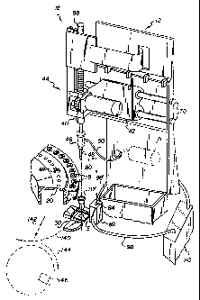
Fig. 1 illustrates a conventional analyzer 12
utilizing the current invention. It is conventional to
utilize a dispensing station 18 to collect by aspiration,
a sample of biological liquid, e.g., serum or plasma,
from a supply comprising primary collection containers 19
in tray 20, into a disposable tip 48 mounted on aspirator
probe 46. The sample liquid is subsequently dispensed
onto a slide test element E held at a slide distributor
30 and obtained from a source of test elements, not
shown. Control of the dispenser 40 providing probe 46 is
via the mechanisms such as vertical drive 44 and carriage
CA 02231305 1998-03-OS
_g_
42 mounted on support rods 70, all as described in, e.g.,
U.S. Patent No. 4,340,390. A conventional pump 71 of any
kind is used as the means for creating a partial vacuum
or partial pressure within tip 48.
In accordance with one aspect of the invention,
a new use is made of tips 48 besides simply, the
collection by aspiration, of liquid from containers 19,
and then subsequent dispensing onto slide test elements
E. Tip 48 carrying the sample liquid aspirated into it,
is moved, arrow 80, Fig. 1, to a test station 82 prior to
placing it in holder 117 for dispensing. Station 82
comprises, as is more clearly shown in Figs. 2A, 2B, and
3, a scanning block that is an effective light-tight
enclosure having a cavity 84 sized to receive a tip 48.
Preferably, cavity 84 comprises an upper portion 86, Fig.
3, a lower portion 88 of smaller inside diameter than
portion 86, a ledge 90 of demarcation between the upper
and lower portions, an air vent 92 in ledge 90, a conical
exit port 94 extending from the lower portion away from
the upper portion, and two passageways 96 adapted to
receive fiber optics 98,98' to and from portions of a
spectrophotometer. Exit port 94 is shaped generally with
the shape of the exit orifice portion of tip 48, hence
its conical shape for this preferred tip 48. An optional
air tube 100 is connected to exit port 94 to reduce the
potential of pumping fluid out of the tip. If the tube
is also opaque, an option, then it also helps to
eliminate light leakage up into the tip.
Fiber optics 98,98' are connected to a
spectrophotometer, Fig. 1, comprising a light source 110
and a detector combined into a single unit 110, which is
conventional.
For maximum efficiency, station 82 is
effectively light-tight as defined herein so that the
light passing to the detector is at least 90% of that
transmitted through tip 48 from fiber optic 98. There
are several ways in which this can be achieved.
First, for a station 82 as shown in Fig. 2A,
comprising block 83 having an upper surface 90 that acts
- CA 02231305 1998-03-OS
_g_
as the support shoulder for the enlarged upper portion
111, and hence. goes no higher than that, and a side
clearance of about 0.5 mm between tip 48 and block 83,
the light leakage that occurs is corrected for by taking
a blank reading (with the fiber 98 delivering no light)
at the same ambient light conditions as is used when NIR
and adjacent visible radiation is delivered by fiber
optic 98. The blank reading is then subtracted from the
sample reading and reference reading.
Alternatively, if a subtracted blank reading is
not to be used, and the side clearance is still the same
as noted above, the same light tightness can be achieved
by extending the height of block 83 up to at least the
height of the top surface 113 of upper portion 111 of tip
48, Fig. 2B.
Because the seating of tip 48 on shoulder
surface 90 is an effective seal, it is preferable that
some air release be provided between upper and lower
portions 86 and 88 as tip 48 is inserted and withdrawn.
That is the function of vent 92, Fig. 3. This vent
allows the release of the increase in pressure created
when a tip is inserted into station 82, so that a bubble
of air is not forced into the liquid of the tip to
possibly interfere with the light-scanning of the liquid.
Likewise, when tip 48 is withdrawn after having been
light-scanned, vent 92 prevents a vacuum being created
such as could draw out of tip 48, a portion of the sample
liquid which then contaminates the station 82 for
subsequent tips and samples.
To further assist in centering tip 48 within
cavity portion 88 between fiber optics 98 and 98',
locator bumps 140 can be disposed, Fig. 3, near the
bottom of portion 88 above passageways 96.
In use, tip 48 is inserted into station 82
before insertion into holder 116. While at station 82, a
beam of NIR and adjacent visible wavelengths as defined
above, is passed through the tip and its liquid so that
transmitted radiation is spectrophotometrically analyzed
at spectrophotometer 110. The signal produced by the
- CA 02231305 1998-03-OS
-10-
detector is then correlated with the concentration of
target substances. A preferred set of target substances
is those that measure sample quality, specifically those
selected from the group consisting of hemoglobin, lipids,
bilirubin (BR), and biliverdin (BV), as shown in the
examples below. However, any target substances capable
of spectrophotometric detection by its absorption
spectra, can be correlated and detected by this
invention. More specifically, certain assays that
heretofore have been conducted in slide test element E,
can be conducted spectrophotometrically through the tip,
as described hereinafter.
Thereafter, the tip is withdrawn and inserted
into holder 116 at which point the sample liquid is
dispensed onto slide test element E conventionally
containing one or more reagents to ascertain the
concentration of an analyte in the sample liquid, as is
well-known.
As will be readily evident, the tips 48 used
herein allow transmission of NIR and adjacent visible
radiation, and most preferably 475 to 1200 nm, and
preferably are free of labels, since any labeling is done
exclusively on primary containers 19. Materials useful
for this purpose include polypropylene and polyethylene.
It is not necessary that test station 82 be
constructed as a solid block with only a cavity for the
disposable tip and apertures for the fiber optics, or
that the tip be lowered into the same. Instead, side
walls of station 82 can be opened and closed, to provide
a slot that allows pass-through of the tip, as shown in
Figs. 4A and 5. Parts similar to those previously
described bear the same reference numeral, to which the
distinguishing suffix "A" has been appended.
Thus, station 82A comprises two fixed, opposed
segments 109,112 spaced a distance apart. Each segment
has an opposing face, 116,116' that defines a slot 115
between them. Top surface 117 of faces 116,116' provide
a guide rail and seat for upper portion 111A of tip 48A.
Segment 109 has a fiber optic 98A penetrating it from a
- CA 02231305 1998-03-OS
-11-
light source, not shown, whereas segment 112 has a sensor
114 in face 116 that is connected to a spectrophotometer
built into or connected with segment 112.
The opposing faces of segments 109 and 112
define slit 115 with a spacing distance that allows a
disposable tip 48A to slide through, arrow 120. Those
opposing faces can be spaced apart a fixed distance for
the sliding of tip 48A.
Because segments 109 and 112 create slot 115
for the through-passage, arrow 120, of tip 48A, with an
aspect ratio much smaller than that described above for
the vertical aperture 84, it is preferred to close slot
115 for the spectrophotometric measurement. To that end,
pivoting doors 130,132 are hingedly attached at 134 to
opposite edges of segment 109, of sufficient width to
close off slot 115 when they are pivoted, arrows 136,138
to their closed positions (not shown). (Door 132 is
shown in phantom for clarity only.) To pivot the doors,
preferably the pintle of hinges 134 is attached or
affixed to a rotating drive shaft (not shown), of
conventional motors 136.
Alternatively, doors 130 and 132 can be omitted
by lengthening slot 115 so that it has an aspect ratio in
the horizontal direction that is comparable to the
vertical aspect ratio stated for cavity 84 above.
To assist in stopping the lateral movement,
arrow 120, of tip 48A just precisely at fiber optic 98A
and detector 114, Fig. 5, a spring-biased detent 210 is
preferably located in face 116, cooperating with a fixed
projection 212 on opposite face 116'. Detent 210 is
pushed by the tip into face 116 when it is time, after
the reading, to move tip 48A out of slot 115 in the
direction of arrow 120. As noted in the previous
embodiment, tip 48A allows transmission of the NIR and
adjacent visible wavelengths used.
Alternatively, Fig. 4B, segment 112B can be
movably mounted on plate 122B to close off light leakage.
Parts similar to those previously described bear the same
reference numeral, to which the distinguishing suffix "B"
- CA 02231305 1998-03-OS
-12-
is appended. Thus, station 82B comprises plate 122B
forming with faces 116B and 116'B a U-shaped slot that
allows a tip 48B to slide through, arrow 120B, while
supported on top surfaces 117B. Fiber optic 98B delivers
light through stationary segment 130B, and sensor 114B in
stationary face 116B delivers light to a
spectrophotometer, not shown.
To close the light leakage that can occur
through the U-shaped slot of plate 122B and faces 116B
and 116'B, segment 112B is mounted to slide on plate 122B
as driven by a rack 162 and a drive pinion 164, arrow
168, thus opening or closing off the slot. When closed,
face 116B and tip 48B occupy the space 172 within segment
112B, and wall portion 169 closes off slot 115B.
In addition to testing for patient sample
quality, any target substance that is analyzable
spectrophotometrically using NIR and adjacent visible
wavelengths, can be analyzed by spectrophotometer 110
while the patient sample is in tip 48A. These include
hemoglobin, albumin, and glucose, among others. By
testing these target substances in the tip, it is not
necessary, and indeed the analyzer preferably skips,
further assays for them when the sample is deposited onto
slide test element E. This enhances greatly the total
throughput of the analyzer, inasmuch as the
spectrophotometric detection through the tip requires
only about 4 seconds for all the target substances so
analyzed, compared to about 4 seconds for each separate
assay done on a slide test element E. "Time to result"
is also drastically improved by the spectrophotometric
analysis through the tip - 4 seconds for through-the-tip,
compared to 5 minutes on a slide test element.
As an example of the enhanced throughput, the
following is a calculation of the advantages that can be
achieved on an analyzer such as is available from Johnson
& Johnson Clinical Diagnostics under the trademark
"VITROS 950" analyzer. This assumes 1) that dispensing
of sample liquid onto a slide test element is the
limiting step in the analysis, and that this involves 8
- CA 02231305 1998-03-OS
-13-
seconds to, aspirate, 4 seconds to dispense onto a test
element and load the element into the distributor of the
VITROS 950 analyzer, and that all, and only, colorimetric
analysis is done in the tip by this invention.
If the mix of chemistries to be run is zero
potentiometric, 7 colorimetric and zero rates, then
without the invention the throughput is 300 test elements
per hour. With the invention, it can be shown to be
about 2100 per hour, which is a 7-fold increase. If on
the other hand there are only 5 colorimetric tests, and
either 2 rate or 2 potentiometric tests to be conducted,
then the throughput without the invention should be about
420 per hour, and about 1050 per hour with the invention,
for a 2.5-fold increase. Still further, if the mix of
seven chemistries is such that there are only 3
colorimetric and 4 potentiometric tests to run, there is
no increase in throughput obtained by doing this
invention (525 tests per hour in both cases.)
Testing of such analytes in this manner while
in the tip is preferably done with some kind of
temperature control of the sample liquid. This need not
be done only by controlling the temperature at test
station 82, but can also be done by heating or cooling
the sample liquid in containers 19, Fig. 1, or while the
liquid is in the tips 48, etc.; but not at station 82.
Nevertheless, there will still be some assays
that require the use of slide test element E. The
process is schematically illustrated in Fig. 1. Tip 48
is inserted into holder 117 and a portion of the patient
sample is dispensed onto slide test element E.
Thereafter, distributor 30 is rotated, arrow 140, to a
position in which test element E is linearly transferred,
arrow 142, to an incubator (not shown) within which it
rotates, arrow 144, until it is read or detected at a
test station 146, all as is well-known and conventional.
Test station 146 conventionally comprises a colorimetric
or potentiometric detector, in contrast to the
spectrophotometer 110 used with tips 48,48A.
- CA 02231305 1998-03-OS
-14-
Although as noted above, tests conducted at
station 146 preferably skip those done through the tip,
it is also possible to repeat at station 146 such
spectrophotometric assays, to obtain a "check" on the
accuracy of the latter.
It is also contemplated that the order of
testing can be reversed - that is, a portion of the
sample liquid can be deposited on a test slide as
described above, before doing the measurements through-
the-tip at the NIR and adjacent visible wavelengths.
Examples
The following non-exhaustive tests were run to
demonstrate the invention:
The apparatus of Fig. 2 was used, in which a
disposable tip available from Johnson & Johnson Clinical
Diagnostics, Inc., under the trademark "Vitros",
heretofore known as the "Ektachem" disposable tip, was
used. The optical fibers were 0.2 mm single fibers,
connecting station 82 via the fibers 98 and 98', to a "TC
2000" dual beam, in-time spectrophotometer that uses a
linear diode array detector, available from CME
Telemetrix, using a tungsten-halogen light bulb light
source 110 as detector 112. Diffraction gratings were
used at detector 112 to allow only radiation of 580 to
1100 nm to be detected. (The reference beam portion of
the spectrophotometer has been omitted for clarity.) The
amount of liquid aspirated into tip 48 was 50 ~L, so that
the liquid level was well above the pass-through arrow
200, Fig. 2. Testing has demonstrated that only 30 ~L is
needed.
The liquids tested were, first as calibrators,
a randomized set of liquids comprising known amounts of
hemoglobin, IntralipidT"" (a fat emulsion which mimics
naturally occurring chylomicrons) available from
Pharmacia, Inc., and biliverdin all spiked onto a human
serum matrix.
- CA 02231305 1998-03-OS
-15-
The following Table 1 sets forth the levels of
Hb, IL, and BV in serum after spiking. "Hb" means
hemoglobin, "IL" means Intralipid, "BV" means biliverdin
dihydrochloride, and "BR" means bilirubin.
TABLE 1
Sample g/L g/L mg/dL
Number Hb IL BV
1 0.56 0.00 0.00
2 0.83 0.00 0.00
3 1.11 0.00 0.00
4 1.38 0.00 0.00
5 1.65 0.00 0.00
6 1.91 0.00 0.00
7 2.17 0.00 0.00
8 2.43 0.00 0.00
9 2.69 0.00 0.00
2.95 0.00 0.00
11 1.19 0.00 0.00
12 1.77 0.00 0.00
13 2.35 0.00 0.00
14 2.93 0.00 0.00
3.50 0.00 0.00
16 4.06 0.00 0.00
17 4.62 0.00 0.00
18 5.17 0.00 0.00
19 5.71 0.00 0.00
6.26 0.00 0.00
21 0.54 1.00 0.83
22 0.79 1.97 0.41
23 1.01 2.83 1.17
24 1.22 1.14 3.77
1.50 1.63 2.32
26 1.73 2.30 1.91
27 2.03 1.42 1.57
28 2.25 0.47 2.70
29 2.46 0.68 3.03
2.54 3.00 3.21
- CA 02231305 1998-03-OS
-16-
'r~RT.F! 1 (C'nnt i ntled _ _ _ )
Sample g/L g/L mg/dL
Number ~ IL BV
31 1.14 2.00 1.66
32 1.69 3.94 0.82
33 2.13 5.61 3.10
34 2.59 2.27 7.55
35 3.19 3.26 4.63
36 3.68 - 4.61 3.82
37 4.36 2.86 2.37
3g 4,78 0.93 5.40
39 5.22 1.37 6.06
40 5.40 6.01 6.41
A second set of 21 liquids similarly prepared,
were prepared to have the components of Table 2, and
treated as unknowns.
TABLE 2
Sample g/L g/L mg/dL
Number Hb IL BV
1 0.34 2.05 3.40
2 0.50 2.44 4.06
3 0.66 2.83 4.69
4 0.80 3.19 5.30
5 3.77 3.27 5.43
6 1.08 3.88 6.44
7 1.35 1.56 3.33
g 1.56 1.15 1.15
9 5.73 3.04 2.21
1.80 3.04 1.80
11 4.75 3.54 1.31
12 2.12 2.60 2.16
13 2.18 4.13 2.74
14 2.58 0.46 0.76
5.26 1.55 4.50
- CA 02231305 1998-03-OS
-17-
TABLE 2 (Continued ...)
Sample g/L g/L mg/dL
Number Hb IL BV
16 2.68 1.26 5.56
17 2.83 0.83 6.23
18 0.00 2.38 0.00
19 1.79 0.00 0.00
20 0.00 0.00 3.95
21 0 0 0
The first set of liquids was irradiated as
described above to create a calibration algorithm using
conventional spectrophotometric practice, and the values
of Hb detected in this measurement were plotted against
the actual values, Fig. 6, to obtain a regression plot.
A variety of calibration algorithms is useful. The
following equations are exemplary only:
1) Hb (g/1) - C1 (dAsoo/d~soo) ' C2 (~663/d~663) ' C3
2) IL(g/1) - C4(dA874/d~874) +
3) BV (mg/dL) - C6 (dA~24/da'724) C7 (~803/d~803) + C8
where A6oo is the absorbence at 600 nm, ~soo is the 600 nm
wavelength, and so forth for the other A + ~, values,
(dAi/d~.i) is the first derivative of absorbence versus
wavelength and C1, ... C9 are constants preferably having
the following values:
C1 15.892 CS = 0.244
=
C2 = 15.882 C6 = 98.068
= 0.21 C~ = 122.732
C4 = 252.155 C8 = 0.0685
The regression correlation coefficient R2 in the
case of Fig. 6 was 0.991.
- CA 02231305 1998-03-OS
-18-
The second set of liquids was then irradiated
as described above and the predicted values plotted
against their known results, Fig. 7, using the
calibration algorithm derived from the first set of
liquids, Fig. 6. The R2 value of 0.982 was excellent.
This accuracy is adequate to allow the results to be
relied upon for clinical assay of Hb in unknown samples,
in place of testing on a slide test element.
In a like manner, the spectra detected as noted
above was evaluated for IL. The calibration results
appear in Fig. 8, and the prediction results in Fig. 9.
R2 in this case was, respectively, 0.9941 and 0.9878.
Again, the spectra noted was evaluated, but
this time the analysis was for BV. Fig. 10 shows the
calibration results, and Fig. 11 the prediction results
with R2 being as indicated.
A new, third set of liquids was prepared to
illustrate the invention in the detection of bilirubin,
and the calibrator version of that set was composed as
1
fol
ows
Sample BR Hb IL BV
Number mg/dL g/L gL mg/dL
1 8.33 0.65 0.00 0.00
2 8.33 0.65 0.00 0.00
3 0.00 1.92 0.00 0.00
4 0.00 1.92 0.00 0.00
5 34.79 0.91 0.00 0.00
6 34.79 0.91 0.00 0.00
7 23.41 1.53 0.00 0.00
g 23.41 1.53 0.00 0.00
9 31.49 0.31 0.00 0.90
10 31.49 0.31 0.00 0.90
11 37.33 1.17 0.00 1.72
12 37.33 1.17 0.00 1.72
13 22.15 0.00 0.93 0.00
14 22.15 0.00 0.93 0.00
15 0.00 0.00 2.15 0.00
- CA 02231305 1998-03-OS
-19-
lrnnrinued _..)
Sample BR Hb IL BV
Number mg/dL g/L gL mg/dL
16 0.00 0.00 2.15 0.00
1g 17.02 0.00 0.00 8.74
19 17.02 0.00 0.00 8.74
20 33.31 0.00 0.00 1.80
21 33.31 0.00 0.00 1.80
22 25.02 0.00 0.00 5.34
23 25.02 0.00 0.00 5.34
24 29.13 0.00 0.00 3.58
25 29.13 0.00 0.00 3.58
26 13.59 0.00 0.00 7.18
The calibration algorithm used for this test
was as follows:
4) BR(mg/dL) - C9 ~dA49s/d~49s~ + C10 ~~s12/da'512~
Cll Ws~e/d~s~s~ - Clz wherein the constants were as follows
C9 = -24.878
Clo = 201. 61
Cll = 44 . 9 8
C12 = 6 . 475
A fourth set of liquids was similarly prepared
to check for prediction of the bilirubin values, and that
set was comprised as follows:
Sample BR BV
Number mg/dL HB IL mg/dL
1 19.86 1.25 0.00 0.00
2 19.86 1.25 0.00 0.00
3 26.59 0.60 0.00 4.38
4 26.59 0.60 0.00 4.38
5 6.10 0.00 2.35 0.00
6 6.10 0.00 2.35 0.06
7 10.31 0.00 1.19 0.00
g 10.31 0.00 1.19 0.00
9 15.53 1.07 0.00 3.58
15.53 1.07 0.60 3.58
CA 02231305 1998-03-OS
.~.
-20-
The spectra was evaluated as in the previous
examples. Fig. 15 shows the calibration results, and
Fig. 16 the prediction results with Rz being as indicated.
For all four experiments (Hb, IL, BV and BR)
the results showed excellent correlation such that the
results are sufficient to use in place of testing on a
slide test element, should any of these be considered a
desired assay. In any event, the results clearly allow
the biological liquid's sample quality to be ascertained
so that the sample can be rejected if determined to be
outside the scope of acceptable quality.
As an example of other calibration algorithms
that can be used, the following is an alternative to
equation #2 above, for IL:
2 ~ ) IL(g/1) - C13 (~999/d~999) + C14 (~1051/d~1051) ClSi
where C13 = 166.068, C14 = 92.352, and Cls = 0.693. When
this is used on the first and second set of liquids noted
above, R2 becomes 0.988 for the calibration and 0.984 for
the prediction. (The actual plots are not shown.)
Fig. 12 is a plot demonstrating that, in fact,
the first derivative of absorbence values in the NIR and
adjacent visible spectra does produce sufficient
separation, at useful wavelengths, of a sample having
either IL, BV, or Hb components present, to allow for
independent detection. That is, curve 200 is a sample
having none of those components, curve 202 is a sample
having only 1.79 g/1 of Hb, curve 204 of a sample having
only 2.38 g/1 of IL, and curve 206 of a sample having
only 3.95 mg/dL of BV. Thus, the Hb contributes
primarily to the 580-605 nm region of the NIR, IL to the
896-1051 nm region and preferably 896-939 nm, and BV to
the 680-750 nm region.
In yet another embodiment, the tip is unchanged
from conventional tips, but more than a single pass of
the NIR and adjacent visible radiation is achieved
through the tip before the absorption spectra is received
by the spectrophotometer, Fig. 13. Parts previously
CA 02231305 1998-03-OS
- ,~"..~ ~....
-21-
described are referred to by the same reference numeral,
to which the distinguishing suffix "D" is appended.
Thus, tip 48D is mounted in cavity 84D as
before, for irradiation by NIR and adjacent visible
radiation emanating from fiber optic 98D, to be received
by fiber optic 98'D for processing. However, unlike
previous embodiments, receiving optic 98'D is not
directly opposite transmitting optic 98D, nor in position
to receive the "first pass" radiation. Instead at least
one, and preferably three pairs) of mirrors (230,232;
240,242; and 250,252) are disposed to re-pass the
radiation back through tip 48D as many times as there are
mirrors. (Six mirrors of the three pairs retransmits the
radiation through the tip six times.)
In yet another embodiment, it is not necessary
that optics 98,98' (or other versions thereof disclosed
above) pass NIR and adjacent visible light through only
the thickest part of the tip. Instead, the light can be
transmitted through the narrower neck portion. (Parts
similar to those previously described bear the same
reference numeral, to which the distinguishing suffix "E"
is appended.)
Thus, Fig. 14, illuminating fiber optic 98E is
positioned in the block of station 82E so as to
illuminate conical neck portion 300 of tip 48E, that has
a decreasing diameter compared to diameter "d" of main
body portion 224E. The light then transmitted through
the tip to receiving fiber optic 98'E passes through much
less of the sample. This is desirable if the analyte to
be detected is one of high density or has a higher
extinguishing coefficient for the NIR and adjacent
visible wavelengths in question. In the most extreme
cases, fiber optics 98E and 98'E are moved down to the
phantom position, 302, that reads through the narrowest
part 304 of tip 48E.
CA 02231305 1998-03-OS
~...
. -22-
Alternatively, passage of the NIR and adjacent
visible radiation through the narrower part of the tip
can be achieved using previous embodiments, simply by
raising the tip (and its probe) sufficiently within
station 82, and then illuminating with the NIR radiation.
Most preferably, the sequence of steps is as follows:
the steps of lowering the tip into a light-tight
enclosure comprising an NIR and adjacent visible
radiation emitter as shown in any of Figs. 2A, 2B, 4A,
and 4B until the tip is seated therein, scanning the tip
and its contents with NIR and adjacent visible radiation
emitted from the emitter, and if the contents have a
density above a predetermined threshold value, thereafter
raising the tip within the enclosure until the emitter is
positioned to scan the narrower portion of the tip.