Note: Descriptions are shown in the official language in which they were submitted.
CA 02234316 1998-04-08
Title of the Invention D-717
X-~Y FLUORESCENCE ANALYZING APPARATUS
Background of the Invention and Related Art Statement
The present invention relates to an X-ray fluorescence
analyzer or analyzing apparatus, which can use X-rays with plural
kinds of wavelengths as X-rays for excitation.
In an X-ray fluorescence analyzer, in order to reduce a
background, X-rays for excitation, i.e. primary X-rays, to be
]0 ejected or irradiated 1o a sample are processed to become
monochrome by passing t~hrough an X-ray spectroscope or X-ray
spectrometer. In this case, the wavelength of X-ray being
processed to become monochrome is selected to a characteristic X-
ray wavelength for an element to be detected in a sample or a
~L5 waveleng1h close thereto. Therefore, in case there are several
elements to be detected, and if there is only one primary X-ray
spectroscope, the several elements can not be detected and metered
at the same time. Thus, it takes time to analyze these elements.
Accordinqly, an X-ray fluorescence analyzer which can radiate at
~'0 the same time primary X-rays of plural kinds of wavelengths has
been used.
The conventional X-ra~y fluorescence analyzer which can radiate
at the same time the primary X-rays of plural kinds of wavelengths
is provicled with an X-ray tube and an X-ray spectroscope for each
wavelengt:h. The selection or changing of the wavelength is made
according to one or plural X-ray wavelengths as desired, by turning
on or off of one or plural power sources of the corresponding X-ray
tubes, or opening or closing one or plural shutters for the
CA 02234316 1998-04-08
respective X-ray tubes.
The above conventional analyzer or apparatus has the following
drawback,.
Sin~_e plural X-ray tubes and X-ray spectroscopes are utilized,
the apparatus becomes large and expensive.
Even if the selection or changing of the X-ray wavelengths to
be used is made by turning on or off of the power source, or
opening or closing the shutter, the selection or changing causes
the apparatus complicated to increase the price of the apparatus.
lo Also, it takes time to chi~nge the wavelengths.
The present invention has been made to solve the above
problems of the convent:ional X-ray fluorescence analyzer or
analyzinc~apparatus utilizing the primary X-rays of plural kinds of
wavelengt:hs.
Summary of the Invention
An X-ray fluorescence analyzing apparatus of the invention
comprises an X-ray tube, plural X-ray spectroscopes disposed around
a linear line linking between an X-ray generating point and an
analyzinc~ point on a sample face, entrance or first slits situated
at an incident side of the X-ray spectroscopes to allow X-rays to
enter into spectral crystals of the predetermined X-ray
spectroscopes, respectively, and ejection or second slits situated
at an ejection side of the X-ray spectroscopes to allow the X-rays
from the spectral crystals toward the sample to only pass
therethrough. The X-ray spectroscope has an incident point at
which the X-ray tube generates the X-rays, and a convergent point
which is the analyzing point on the sample face.
CA 02234316 1998-04-08
Brief Description of the Drawings
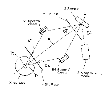
Fig. 1 is an explanatory plan view of one embodiment of the
present invention;
Figs. 2(A)-2(C) are front views of slit plates used in the
embodiment shown in Fig. 1; and
Fig. 3 shows X-ray spectra showing that two kinds of elements
can be analyzed by primary X-rays of two kinds of wavelengths.
Detailed Description of Preferred Embodiments
:LO One of the embodiments of the invention is shown in Fig. 1.
In this embodiment, it is structured that three kinds of primary X-
rays are taken and used. In the drawing, numeral 1 is an X-ray
tube; numeral 2 is a sample; and numeral 3 is X-ray detection
means, w~hich detects fluorescent X-rays ejected or radiated from
the samp:Le.
A point P of a target lt of the X-ray tube l is a focal point
of an electron beam for excitation, and is a radiation point of the
primary X-ray. A point Q is a sample analyzing position or an
analyzing point for the sample 2. Six spectral crystals 51-56 are
~'0 arranged around a linear :Line A linking between the points P and Q
(four spectral crystals 52, 53, 55, 56 are not seen in the
drawing)
Each spectral crystal is a curved crystal, and is located on
each Row]and circle passing through the points P and Q, to thereby
form an X-ray spectroscope wherein the point P is an X-ray incident
point and the point Q is an X-ray convergent point. In these
spectrosc:opes, two spectroscopes form one pair to take or obtain
one X-ray wavelength. Incidentally, numeral 4 is an X-ray incident
CA 02234316 1998-04-08
slit plate, and numeral 6 is an X-ray ejection slit plate.
Fig. 2(A) is a front view of the X-ray incident slit plate 4.
The slit plate 4 is movable in a vertical direction in Fig. 1, and
two groups of slit holes 41-46 and 47 are formed. The slit holes
41-46 are arranged at 60 degrees different from each other relative
to a point C as a center. The distances from the center to the
respective slit holes are set to values determined by the kind of
the spectral crystals corresponding to the respective slit holes
and the lwavelength of the X-ray to be ejected. The slit plate 4
can take two positions such that the line A can pass through the
point C and the center of the slit hole 47.
Fig. 2(B) is a front view of the slit plate 6. The slit plate
6 is also movable in the vertical direction in Fig. 1. In the slit
plate 6, five groups of s:Lit holes are formed. The first group is
~5 formed of slit holes 61-66 corresponding to all the spectral
crystals 51-56; the second group includes slit holes 63a, 64a
correspollding to the slit: holes 63, 64 for the first group; the
third group includes slit holes 65a, 66a; fourth group includes two
slit holes 61a, 62a; and t:he fifth group includes only one central
~!0 hole 67. The slits of the slit plate 6 for the respective groups
are arranged relative to points Dl-D5 as centers, respectively. In
regard to the slit holes 61a-66a in the respective groups, the slit
holes in the same numbers are located at the same distances away
from the centers in the re,pective slit holes 61-66. The distances
are set to form spectroscopes for obtaining the X-rays with the
desired wavelengths by the slit holes 41-46 of the slit plate 4 and
the corresponding spectral crystals 51-56.
The slit plate 4 is set such that the point C is located on
CA 02234316 1998-04-08
the center line A, and the center of the point D1 of the slit holes
in the first group of the slit plate 6 is located on the center
line A i.n Fig. 1. When the X-ray tube is turned on, X-rays with
three ki.nds of wavelengths irradiate the point Q on the sample at
the same time. Also, when the location of the slip plate 4 remains
as stated above, and in case, for example, the point D3 of the slit
plate 6 is located on the center line A in Fig. 1, the X-ray with
one wavelength selected by the spectral crystals 55, 56 is only
irradiat.ed on the sample. In case the slit hole 47 of the slit
plate 4 and the slit hole 67 in the fifth group on the slit plate
6 are located on the center line A as shown in Fig. 1, it is
possible to radiate the sample by the X-rays with all the
wavelengths ejected from the X-ray tube 1. Therefore, there is no
structural and operational complications, such as by turning on and
off of plural X-ray tubes to select or change the X-rays to be
irradiat.ed.
In the present invention, a desired type of X-ray detection
means may be used, such as a device to be able to scan wavelengths
by using~ spectral crysta.ls, a device having an ability to select
wavelengths like a proportional counter tube, or a counter tube to
be able to detect X-rays with all the wavelengths. In case the
primary X-rays with plur~l kinds of wavelengths as stated in the
above example is irradiated, a plurality of proportional counter
tubes is designed to have windows corresponding to the X-ray
wavelengths, which are desired to be detected, respectively, so
that it :is possible to detect and meter plural elements at the same
time.
The target lt of the X-ray tube 1 radiates continuous X-rays
CA 02234316 1998-04-08
and, in addition, characteristic X-rays with the elements contained
therein. The respective X-ray spectroscopes formed of the slit
plate 4, the spectral crystals 51-56 and the slit plate 6 are set
to match the wavelengths of these characteristic X-rays according
to their locations and the kinds of the spectral crystals. The
sample is being irradiated while the X-rays in the characteristic
X-rays are selected.
For example, the target is Rh, and RhK~-rays and RhLa-rays are
radiated; (200) phase of LiF is used for the spectral crystals 51-
53 corresponding to RhKa-rays, and TAP is used for the spectral
crystals 54-56 for RhLa-rays. In this selection, a sample is
irradiated with both RhLa--rays and RhK~-rays. In this combination,
three spectral crystals correspond to one X-ray wavelength, so that
two kind:, of X-ray wavelengths can be taken out or obtained. It is
possible to design such that the six spectral crystals provide all
different X-ray wavelengths, respectively. However, instead, it is
possible to enhance the X-ray strength of one X-ray wavelength.
Namely, in case two spectral crystals form one unit, X-rays with
double strengths can be obtained, and in case three spectral
;20 crystals form one unit, X-rays with triple strengths can be
obtained. In case the primary X-ray is made to have two
wavelenglhs, the slit plate 6 may additionally have two groups of
slit holes 64b-66b and 61b-63b, as shown in Fig. 2(C).
As stated above, in c:ase the La-ray and Ka-ray of Rh are used,
;'5 as understood from the characteristic X-ray spectra shown in Fig.
3, SKa-ray and PbL~-ray of S, Pb and so on in the sample can be
detected as fluorescent X-rays.
In t:he above examples, two or three kinds of the primary X-ray
CA 02234316 1998-04-08
wavelengths are selected, but in the invention, the kinds of the
primary X-ray wavelengths to be selected are not limited. Also,
the number of the spectral crystals need not be six, and can be
four, eight or other numbers. When the number of the spectral
crystals is increased, and the responsible angular range of the
respective X-ray spectroscopes around the center line A in Fig~ l
is reduced, utility effectiveness of the X-rays can be increased.
In the present invention, one or plural kinds of the primary
X-ray wavelengths can be selected at the same time by one X-ray
:L0 tube and irradiated to a sample. Therefore, the apparatus can be
made compact as compared to an apparatus utilizing plural X-ray
tubes an~d spectroscopes. In the invention, plural spectroscopes
are utilized, but since the spectroscopes are arranged around one
linear line linking between the X-ray tube and the sample, the
apparatu-, can be made especially compact. Also, utility
effectiveness of the X-rays is increased. Further, the selection
of the primary X-rays is not made by turning on or off of the X-ray
tube, and is made by selecting one of the groups of the slits.
Thus, it is not necessary to wait for rising of the selected X-ray
tube, so that plural kinds of elements can be analyzed quickly.
Whi:Le the invention has been explained with reference to the
specific embodiments of the invention, the explanation is
illustra1ive and the invention is limited only by the appended
claims.
