Note: Descriptions are shown in the official language in which they were submitted.
CA 022364~6 1998-04-30
W O 97/16151 PCT~US96/17228
ADAPTIVELY CONTROLLED MANDIBULAR
POSITIONING DEVICE AND ~ETHOD OF USING
TTlli'. DEV~CE
I
FIELD OF THE INVENTION
The present invention relates generally to systems and methods for
treating obstructive sleep apnea using an adaptive control system for adjusting
and positioning a mandibular positioning device.
BACKGROUND OF THE INVENTION
Obstructive sleep apnea (OSA) is a common disorder which produces
considerable morbidity and mortality. The disorder arises during sleep when the
victim undergoes repeated cessation of breathing. This cessation results from anobstruction of the throat air passage (pharynx) due to severe narrowing or a
collapse of the throat air passage. Repeated cessation of breathing reduces
blood oxygen and disturbs sleep. Reduction in blood oxygen can cause
hypertension, heart attacks and strokes. Additionally, sleep disturbances can
produce excessive daytime sleepiness, headache, depression, irritability and
cognitive hll~aillnents.
Medical research over the past decade has produced a standard approach
to obstructive sleep apnea therapy, known as nasal continuous positive airway
pressure (CPAP). In this therapeutic approach, a patient's nose is covered with
a mask that forms a pressure seal with the surrounding face. While the patient
sleeps, the mask is pressurized to a level that distends the collapsible throat air
passage, thereby preventing obstruction.
This therapeutic approach provides two significant advantages: it is
uniformly effective and it is entirely benign. A major disadvantage of this
approach is that the patient must remain overnight in a hospital sleep center toundergo a full night polysomnography study with the pressure mask in place to
determine the therapeutic level of pressure. A further disadvantage of this
approach is that the pressure delivered to the patient during the
polysomnography study is constant and fixed at the prescribed level, even
CA 022364~6 1998-04-30
W O 97/16151 PCT~US96/17228
though the patient's requirements may vary throughout the night and from night-
to-night.
The overnight study presents a potential bottleneck to treating a high
volume of patients with obstructive sleep apnea because it typically requires two
5 full night polysomnographic studies for each new patient: one to establish the diagnosis (diagnostic-polysomnogram) and another to establish the
aforementioned therapeutically optimal pressure (therapeutic-polysomnogram).
The ~herapeutic polysoIEmogra~hic study is necessa~ to determIne the minimnn~
level of pressure required to produce a patent pharyngeal airway (i.e., to
10 determine the necessary therapeutic pressure required for properly treating the
patient). These studies, performed in a specialized hospital sleep center, allow a
specialist to specify the pressure to be used when prescribing nasal CPAP
therapy. For this reason, the therapy cannot be prescribed by an internist or
general practitioner.
Due to the requirement of two night polysomnographic studies, hospital
sleep centers are crowded even though only a small percentage of obstructive
sleep apnea victims are presently being treated. Further, the significant cost of
the overnight polysomnographic study by a hospital sleep center represents a
significant obstacle to diagnosing and treating the large population of sleep
20 apneics. The backlog of undiagnosed and untreated obstructive sleep apnea
patients thus represents a substantial public health problem.
To address the foregoing drawbacks of existing approaches to diagnosis
and treatment of obstructive sleep apnea, recent commercial technology provides
overnight, unattended monitoring of breathing in the patient's home. Such
25 unattended monitoring generally permits the physician to diagnose obstructivesleep apnea without requiring a diagnostic overnight study in the hospital sleepcenter. However, a hospital sleep center is still required for establishing the
therapeutically optimal pressure of nasal CPAP in each patient. Accordingly,
medical practitioners have been slow to use the new monitoring technology for
30 diagnostic purposes since the patient must, in any case, be referred to a sleep
center for a full night therapeutic polysomnographic study.
CA 022364~6 1998-04-30
W O 97/16151 PCT~US96/17228
- 3 -
While there is a continuing need for CPAP technologies, clinical studies
and general clinical experience indicate that nasal CPAP is not always an
effective treatment for many patients with obstructive sleep apnea, particularlythose with symptoms of mild to moderate severity.
Various surgical approaches have been employed to correct the strLIctural
abnormality of the pharyngeal airway. Excluding massive reconstruction of the
mandibular, maxilla and/or tongue, the only widely employed surgery has been
uvlllopalatophary~A1goplasty (UPPP). However, results with UPPP are
disappointing unless patients are selected by pharyngeal endoscopy during sleep
10 and, even then, the long term benefits are questionable. Laser-assisted
uvulopalatoplasty (LAUP) is a new approach which has been recommended for
obstructive sleep apnea. No studies have reported the effectiveness of LAUP in
the treatment of obstructive sleep apnea, but there is little reason to anticipate
that it will be more effective than UPPP although it may be more convenient,
15 less expensive and may prove to be a useful adjunct therapy to be used in
combination with mandibular positioner (MP) therapy for patients in which MP
therapy does not elimin~te apneas and hypopneas.
Stationary oral appliances which draw the tongue forward have been
used in the treatment of snoring. In addition, some recent studies suggest that a
20 fixed oral appliance (i.e., mandibular positioner) which holds the lower jaw (i.e.,
mandible) of the patient forward as the patient sleeps is effective in treating
obstructive sleep apnea, especially mild obstructive sleep apnea. Studies have
shown that ventral displacement of the mandible enlarges the pharyngeal airway
and acts to prevent its closure. Conventional mandibular positioners are
25 constructed by a dentist or orthodontist at a fixed position for holding the
mandible forward. The proper fixed position is determined through trial and
error by having the patient try a series of mandibular positioning devices untilthe most effective one is found. Once the mandible displacement is set for the
device, it remains stationary with no accommodation for variations in the
30 obstructive sleep apnea, such as body position, sleep state, effects of drugs, and
congestion of the patient.
~ CA 022364~6 1998-04-30
W O 97/16151 PCT~US96/17228
An adjustable mandibular positioner, developed by Dr. A. Lowe, Head,
Department of Orthodontics, University of British Columbia allows incremental
adjustment of the ventral displacement of the mandible. This device is referred
to as a screw adjustable mandibular positioner (SAMP), because its upper and
5 lower full arch orthotics are connected by a manual screw device which is
adjusted by the patient or dentist to set the m~gnitllclP. of mandibular
advancement. Thus, the patient or dentist can progressively advance the
rnandible with the .SAMP over a period of ~veeks to morlths so that mandibular
muscles and ligaments can adjust, thereby allowing greater ventral displacement
10 and minimi7ing side effects.
Accordingly, it would be desirable to render the therapy of obstructive
sleep apnea more practical and convenient. To achieve this end, a method and
system for automatically establishing the desired mandible advancement for a
patient during changing sleep conditions is needed. More particularly, a system
15 is needed with an adaptively controlled mandibular positioner that automatically
adjusts to a patient's needs throughout the night and from night to night.
SUMMARY OF INVENTION
The present invention is therefore directed to providing a practical,
convenient and cost-effective system for adaptively treating obstructive sleep
20 apnea with an automatic, self-adjusting mandibular positioner. Further, the
invention is directed to portable systems and methods for automatically and
continuously regulating the position of the patient's mandible to an optimal
position during obstructive sleep apnea treatment during long term nightly use at
home. The present invention utilizes an automatic mandibular positioning
25 system having adaptive control software which uses readily measurable, robust feedback variables to automatically adjust a mandibular positioner for
obstructive sleep apnea treatment. Obstructive sleep apnea therapy is
implemented in the present invention by automatically applying an ~lupliate
mandible advancement to a patient. The mandible position is continuously
30 reevaluated and optimized throughout the night. The optimal position varies
CA 022364~6 1998-04-30
W O 97/16151 PCTAJS96/17228
with body position, congestion, stage of sleep, and whether any deleterious
substances, such as alcohol or sleeping medicine, have been ingested.
The present invention is a portable adaptive control system which
continually searches for the optimal minimnm mandible advancement required
to adequately distend a patient's nasal pharyngeal airway. By rendering the
system portable, a large pe-ct;~ ge of obstructive sleep apnea victims can be
cost-effectively treated in their homes, thus reducing the overcrowding in
expensive hospita1 sleep centers. Optima1 minimum adv~ncement is used
because greater advancements increase the likelihood of side effects (e.g., soremuscles), and reduce the likelihood of patient compliance. A patient's
compliance in regularly using the system is a significant concern in~cmuch as
the system is a portable device used at the patient's home without the
supervision of a hospital sleep center specialist.
In one aspect of the present invention there is provided a method for
adaptively controlling mandibular displacement for the treatment of obstructive
sleep apnea in a patient by (a) monitoring the patient for evidence of obstruction
of the patient's airway, (b) displacing the patient's mandible if evidence of
obstruction is detected and (c) repeating steps (a) and (b) until evidence of
obstruction is elimin~t~ or reduced below a predetermined value.
In another aspect of the present invention there is provided a method for
adaptively controlling mandibular displacement for the treatment of obstructive
sleep apnea in a patient by detecting obstruction in a patient's upper airway
system, identifying periods of inspiration and expiration for the patient, and
incrementally adjusting a patient's mandible in response to the obstruction
information detected during the period of inspiration.
In yet another aspect of the present invention there is provided a system
for adaptively controlling mandibular displacement for the treatment of
obstructive sleep apnea in a patient having an adjustable mandibular
displacement device, a unit which detects evidence of obstruction of the
patient's airway, and a control system for adaptively controlling the adjustablemandibular displacement device in response to the detecting unit.
CA 022364~6 1998-04-30
W O 97/16151 PCTAUS96/17228
BRIEF DESCRIPTION OF THE DR~WINGS
Other objects and advantages of the present invention will become more
apparent from the following detailed description of preferred embodiments when
read in conjunction with the accompanying drawings, wherein like elements
5 have been designated by like numerals and wherein:
FIG. 1 is a diagrammatic representation of an adaptive mandibular
positioner system; and
FIG. 2 is a conceptual diagram of an operator of the adaptive control
system.
DETAILED DESCRIPTION Oli THE PREFERRED EMBODIMENTS
The present invention is an auto-mandibular positioning (i.e., auto-MP)
system for adaptively providing a mandible position effective in treating
obstructive sleep apnea. The auto-MP system is an automatic, self-adjusting
mandibular positioner and controller which performs detection, analysis, and
15 decision-making functions.
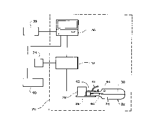
With reference to FIG. 1, there is shown an adaptive mandibular
positioner system 20 in accordance with one embodiment of the present
invention. In this embodiment, adjustable mandibular displacement device 22
comprises a lower dental appliance 24 attached to mounting bracket 26 having a
linear actuator 28 mounted thereon. Linear actuator 28 is in contact with or
attached to upper dental appliance 30. The upper and lower dental appliances
are free to slide relative to each other such that when the linear actuator 28
exerts force on the upper dental appliance (which can not move because the
patient's upper teeth are attached to the maxilla which is fixed to the skull) the
linear actuator 28, mounting bracket 26 and lower dental appliance 24 are
displaced in a direction away from the patient. As a result, the lower dental
appliance 24 draws the patient's mandible forward (i.e., ventrally) to open the
patient's upper airway. In the illustrated embodiment, the actuator 28 and
mounting bracket 26 displace the mandible in a linear manner, however it is
within the scope of the invention that the actuator and mounting bracket be
CA 022364~6 1998-04-30
W O 97/16151 PCTAUS96/17228
- 7 -
configured to displace the mandible along the patient's naturally occurring
protruding path. For example, the path may be an arcuate path forward and
downward, or forward and upward. Likewise, the patient's mandible may angle
slightly to one side or the other as it protrudes.
In one embodiment, the upper and lower dental appliances are formed by
filling an upper dental tray (which can be a partial or full arch) and a full arch
lower dental tray, which can be custom fitted to a particular patient or be in
s~andard sizes~ for e.~mple sma~l7 medium and larg~o, With a silastic impr.,ssion
material (e.g., PolyFil~ TransBite available from SciCan~) Medtech AG, Cham,
Switzerland). Inserting the upper and lower dental trays in the patient's mouth
and having the patient bite down until the molding material sets. In other
embodiments, the upper and lower dental appliances can be formed with
conventional materials such as heat deformable plastics which are placed in
heated water or other suitable heating device before being inserted in the
patient's mouth.
As illustrated in FIG. 1, the linear actuator 28 is driven by an actuator
controller 32 having an external power source 34 (or an internal power source).
Actuator controller 32 is controlled by adaptive control unit 36. The adaptive
control unit in the illustrated embodiment is a personal computer but a special
unit can be manufactured and used as well. Adaptive control unit 36 is usually
located in an area near the patient and the mandibular positioning device 22.
Attached to the adaptive control unit 36 is a recording and display device 38
(e.g., a polygraph paper chart and/or a m~gnefic recording device with a display)
which receives inputs from the adaptive control unit 36 and from patient
monitoring devices 40 (e.g., oxygen saturation, airflow, snoring sound) through
the unit 36 as will be described in more detail below. The linear actuator 28
can be any of a variety of actuators as will be recognized by one of ordinary
skill in the art and be within the scope of the present invention. Two of such
actuators are described below. The linear actuator is capable of a maximum
displacement of 25 millimt ters, but for most patient's the maximum
displacement is 16 millimeters.
CA 022364~6 1998-04-30
W O 97/16151 PCTAUS96/17228
In one such embodiment, the actuator system is comprised of a stepper
motor controller connected to a personal c~ ~utel for driving a stepper motor
connected to a micrometer which moves a first hydraulic piston. The first
hydraulic piston is in fluid communication through a 0.5 millimt-ter inside
diameter, 2 millimeter outside diameter hydraulic line with a second hydraulic
piston and cylinder mounted on the mounting bracket 26. The second hydraulic
piston has a pressure plate for contacting or ~tt~- hing to the upper dental
appliance. When the stepper motor and micromet~Pr move the first hydrqulic
piston, the pressure in the hydraulic line causes the second hydraulic piston toexert force on the patient's upper teeth through the pressure plate in contact
with the upper dental appliance and protrude the patient's mandible with the
lower dental appliance. When the pressure in the first hydraulic piston is
reduced, the natural elastic nature of the patient's muscles in the jaw cause the
patient's mandible to retrude while biasing members attached between the
second hydraulic piston and the pressure plate cause the second hydraulic pistonto retract. In this embodiment, moving the stepping motor I millimPtPr results
in 1 millimPter of displacement of the patient's mandible. Optionally, a
ples~ tr~ncdllc~r can be in fluid col~,ll-u~ication with the hydraulic line to
measure the amount of force being exerted on the patient's muscles and
ligaments to prevent excessive force that may cause patient discomfort or
arousal. Preferably, the second piston and cylinder and mounting bracket are
made of alllminnm or similar lightweight material so that the patient is not
aware of external forces applied to the patient's teeth.
In another embodiment of the actuator system, the hydraulic system just
described is replaced with a small stepper motor (e.g., model no. 20841-05
available from Haydon Switch and Instrument, Inc. in Waterbury, Connecticut)
mounted on the mounting bracket 26. The actuator controller 32 is a model
40105 Bipolar Chopper Driver available from Haydon Switch and Instrument,
Inc. in Waterbury, Connecticut. The stepper motor mounted on the mounting
bracket 26 has a screw shaft extending through the center thereof with a
pressure plate at the distal end of the shaft for contacting or attaching to the
CA 022364~6 1998-04-30
W O 97/16151 PCT~US96/17228
upper dental appliance. When the actuator controller 32 receives a protrude
signal from the adaptive control unit 36, the actuator controller sends a signal to
the stepper motor which rotates the screw shaft. The screw shaft extends
toward the upper dental appliance to exert force on the patient's upper teeth
5 through the pressure plate in contact with the upper dental appliance and
protrude the patient's mandible with the lower dental appliance. When the
actuator controller 32 receives a retract signal from the adaptive control unit, the
actuator sontroller sends a signal to the stepper motor which rot~es the screw
shaft in the opposite direction. The screw shaft retracts the pressure plate and10 the natural elastic nature of the patient's muscles in the jaw cause the patient's
mandible to retrude. In this embodiment, the adaptive control unit sends three
signals to the actuator controller. One signal tells the stepping motor to turn on
or off, another signal tells the stepping motor the direction to move (i.e.,
clockwise or counterclockwise), and another signal tells the stepping motor the
number of steps to move (e.g., I step = 15 degrees of shaft rotation - 1/40
millimf~t~r of linear displacement).
Attached to the strut 42 of the linear actuator 28 are two cannulae 41
with openings positioned to correspond to the patient's nares (not shown) (FIG.
1). These c~nn~ . are connected to a ples~ tr~ncducer (e.g., Oyster model
20 723 from Schaller) for recording an index of respiratory airflow. The kineticenergy of the expired air increases the pressure in the cannula, thereby providing
a direct index of expiratory airflow rate. Conversely, during inspiration, the
pressure in the cannula decreases providing an index of inspiratory airflow.
Snoring is sensed by a piezo-electric tr~nc-lucer applied to the neck over the
25 trachea, typically using a contact microphone. Alternatively, the piezo-electric
tr:~ncdncer can be implanted in the upper dental appliance. The signal from the
transducer is digitized and integrated. Peak snoring and duration of snoring aredetected. Snoring is deemed "~let~cte~l" when a sound of 200 milliceconds
duration is detected for 2 consecutive breaths. Arterial oxygen saturation is
30 ~letec~ ~1 by a pulse oximeter attached to the ear lobe, the finger or the lip. For
the lip, the light emitter 44 is attached to the ventral aspect of the upper dental
CA 022364~6 1998-04-30
W O 97/16151 PCT~US96/17228
-- 10 -
appllance and the sensor 46 is attached to the strut of the upper dental
appllance.
Feedback variables which provide the most useful information for the
adaptive control system include: snoring sound, oxygen saturation and nasal
5 airflow. These are selected because they are robust signals and are easily
incorporated into the auto MP nightly use.
As snoring is caused by vibration of the soft palate, it is therefore
indicative of an unstable airway and is a warning signal of the imminence of
upper airway obstruction in patients that suffer obstructive sleep apnea. Snoring
10 is itself undesirable not only as it is a disturbance to others but it is strongly
believed to be connected with hypertension. If the resultant increase in
mandibular protrusion is sufficient to completely stabilize the airway, snoring
will cease. If a further snoring sound is detected, the protruded distance is
again incrementally increased. This process is repeated until the upper airway is
15 stabilized and snoring ceases. Hence, the occurrence of obstructive apnea canbe eliminated by application of minimum mandible displacement at the time of
use.
The adaptive control unit gradually decreases the mandible displacement
if an extended period of unobstructed breathing occurs in order to ensure that
20 the degree of mandible displacement is maintained at a level as low as
practicable to prevent the onset of apnea. If, however, evidence of obstruction
is detected by the adaptive control unit, the system will again act to
incrementally increase the protruded distance of the mandible.
In use, a patient using adaptive mandibular positioner system 20 may
25 connect himself to the apparatus and go to sleep. The mandible displacement is
initially at a minimnm displacement, for example, the patient's natural mandibleposition at rest or slightly protruded so as not to cause discomfort that prevents
sleep. Not until some time after going to sleep and the patient's body relaxes
will the airway start to become unstable and the patient will begin to snore or
30 experience some obstruction of the airway. The patient inputs 40 will detect the
snore or obstruction and send a signal to adaptive control unit 36. The adaptive
CA 022364~6 1998-04-30
WO 97/16151 PCTAUS96/17228
control unit will then respond to the obstruction via the actuator controller 32 to
increase the protruded distance of the patient's mandible. The displacement can
be increased relatively rapidly, if the patient's condition so requires but care is
taken to not arouse the patient.
S If in the early stages of sleep some lesser mandible displacement will
suffice, system 20 will not increase the displacement until needed, that is, unless
the airway becomes unstable and evidence of obstruction commences, no
increase in displacement is made By continllously decreasing the displacemcnt
(unless the mandible is already in the natural position) in the absence of
evidence of obstruction, the displacement is never substantially greater than that
required to prevent apnea.
The adaptive mandibular positioner system 20 provides a system which
adjusts mandibular displacement according to variations in a patient's breathingrequirements throughout an entire sleep period. Further, system 20 will likewiseaccommodate variable displacement requirements owing to general
improvements or deteriorations in a patient's general physical condition as may
occur over an extended period of time.
Patient inputs 40 preferably comprise at least one of an oxygen saturation
monitor, a sound monitor, and an airflow monitor which continuously detects
changes in the patient's breathing patterns. Concurrently, the patient inputs unit
40 generates output signals corresponding to the continuously detected signals
and transmits these signals to adaptive control unit 36.
Depending upon the characteristics of the patient inputs signal, the
adaptive control unit may generate a command signal to either increase or
decrease the mandibular displacement. The adjustable mandibular positioner 22,
patient inputs 40 and adaptive control unit 36 thus comprise a feedback circuit
or system capable of continuously and automatically controlling the
displacement of the patient's mandible responsive to the patient's respiratory
requirements as dictated by the patient's breathing patterns.
Obstruction of the upper airway is manifested by high upper airway
resistance, hypopneas or apneas. High upper airway reSist~nce is detected when
CA 022364~6 1998-04-30
W O 97/16151 PCT~US96/17228
- 12 -
snoring is present, peak flow is reduced and/or the profile of inspiratory flow is
flat. Hypopneas are signified by snoring, reduction of peak airflow, flat
inspiratory flow trajectory and a decrease in oxygen saturation. Apneas are
manifested by absence of snoring and airflow followed by oxygen desaturation
in the range of 5 to 10 seconds.
When the patient inputs unit 40 detects breathing patterns indicative of
obstructed breathing, it transmits signals corresponding to this condition to the
adaptive control unit 36. The adaptive control unit 36 then causes the
mandibular positioner 22 to increase the protrusion of the mandible
10 incrementally (e.g., in the range of 0.25 to 2 millimeters, preferably in the range
of 0.5 to I millim~tcr) which opens the patients airway until obstructed
breathing is no longer detected. The system also includes means such as
al,plo~l;ate logic programmed into the unit 36 whereby the displacement is
gradually decreased if unobstructed breathing patterns are detected over a
15 preselected period of time (e.g., in the range of 10 seconds to 4 minutes,
preferably for 2 to 4 minutes). This feature serves to provide the patient with a
ventral displacement of the mandible minim~lly sufficient to mzlint~in airway
patency during unobstructed bre~fhing, thus enhancing patient comfort and
therapy compliance.
Several embodiments for adaptive control of the auto MP are available.
One embodiment utilizes a predetermined displacement step in position of the
mandible during the expiratory phase. Snoring (if present) and peak airflow
during a first test set (e.g., in the range of I breath to 10 breaths) after thedisplacement step are compared to the mean of the preceding breaths (e.g., in
25 the range of 2 to 10 breaths, preferably 3 to 5 breaths). In addition, measures of
the shape of the inspiratory flow profile (i.e., flatness and roundness) are
calculated and compared to preceding values.
Another embodiment utilizes a strategy of incrementing the mandibular
position by 1 millimeter when snoring and/or desaturations are present. After
30 each increment, the feedback variables will be monitored for a predetermined
period (e.g., in the range of 10 seconds to 4 minutes). Our studies indicate that
CA 022364~6 1998-04-30
W O 97/161~1 PCTAUS96/17228
often snoring will disappear shortly after the increment in mandibular position
and then reappear. Accordingly, if snoring and desaturations reappear, the
- process will be continued until snoring reaches a miniml-m value and
desaturations are elimin~t~d, or the limits of extension are reached as indicated
~ S by pressure and displacement information.
In one embodiment, airflow is used to assess the respiratory and dynamic
mechanical characteristics of a patient's pharyngeal airway (PA) during sleep
and to adjust the therapeutic mandible advancement as reqllired.
Respiratory airflow typically corresponds to patient breathing and has
two sequential, tidal components: one caused by inhalation and another caused
by exhalation. This tidal airflow is phasic and therefore allows the onset of
inspiration and the onset of expiration to be identified. Because the onset and
termination of inspiration are identifiable, parameters related to the shape of a
time profile of inspiratory flow can also be determined. In a preferred
embodiment, a degree of roundness and flatness of the inspiratory profile are
determined as will be described later.
The measurement of airflow and subsequent determination of an
inspiratory airflow profile are used to control the position of the patient's
mandible in accordance with the present invention. When the degree of
mandible displacement produces the maximal distention of the airway with the
miniml-m displacement is abruptly reduced in sleeping patients suffering from
obstructive sleep apnea, the pharynx is observed to collapse and the pharyngeal
resistance increases accordingly. This change in upper airway resistance induceschanges in peak inspiratory airflow and profile shape with little change in
airway pressure below the obstruction. Accordingly, changes in airflow
resi~tzln~e can be inferred from changes in the inspiratory airflow.
Further retrusion of the mandible leads to progressive collapse of the
pharyngeal airway which severely reduces inspiratory airflow and causes flow
limitations (i.e., increased airflow resistance). Similarly, progressive increases in
the degree of mandible protrusion leads to smaller decrements in airflow
resistance as the pharynx widens and reaches the limits of its distensibility. The
CA 022364~6 1998-04-30
W O 97/16151 PCT~US96/17228
- 14 -
collapsible behavior of the pharyngeal airway in response to progressive
reductions in the degree of mandible displacement provides a framework for
determining an optimal thelapeulic mandible displacement in accordance with
the present invention.
Accordingly, a preferred embodiment includes an adaptive control system
for displacing the patient's mandible in response to detected airflow. This
mandible displacement is adaptively adjusted to apply an optimal minimum
thera~eutic displacement.
During a testing mode of the auto-MP system, the displacement of the
10 patient's mandible is changed frequently. The position of the mandible is
changed by sending a signal from the computer 36 to controller 32 which sends
the proper signal to actuator 28.
Generally spe~king, the adaptive control system generates an optimal
desired (i.e., command) displacement by detecting airflow data over a
lS predetermined period of time, identifying periods of inspiration and expiration,
and extracting information or features from the airflow data. Using this
information, the adaptive control system identifies a critical displacement (DCrit)
at which a significant obstruction occurs during inspiration. More particularly,DCrit corresponds to a lower limit of mandibular displacement associated with a
20 significant decrease in peak inspiratory airflow and/or significant (i.e., critical)
airflow limitation. After determining DCrit7 the adaptive control system
identifies an Optilllulll (i.e., minimllm) effective mandible position (Dopt) for
elirnin~ting the obstruction during inspiration.
The adaptive control system identifies DCrit and decides upon Dopt using
25 a series of test displacements in the mandible position. Results of the tests are
evaluated by e~mining inspiratory airflow. Dopt is continuously uprlz~tecl during
testing periods which are initi~t~d throughout the night to account for changes in
the patient's sleep stages and sleeping position.
Because a testing period is used to update Dopt~ the adaptive control
30 system also decides when to test the pharyngeal airway, and when to continue
or to stop testing. Further, the adaptive control system (I) manages overall
CA 022364~6 1998-04-30
W O 97/16151 PCTAUS96/17228
operation to optimize its own performance, and (2) monitors potential airflow
measurement errors to accurately measure upper airway performance as will be
- described below.
Airflow changes and airflow profile changes in the upper airway system
S have been determined to be directly related to intra-pharyngeal pressure. Bydetermining upper and lower limits of pharyngeal resistance from changes in
airflow during a testing period, Dopt can be determined for any patient at any
tims. Accordingly, the adaptive control system searches for Dopt bet~,veen a
lower airflow limitation (DCr~t) and an upper limit (full distention of the airway).
Operating within these relative limits ensures reliable assessment of the
pharyngeal airway and an accurate determination of Dopt~ Because airflow
varies widely among patients and, for any particular patient, varies with sleep
stage, Dopt can not be determined by comparing airflow measurements with
ideal or predicted standards.
Generally speaking the adaptive control system conceptually includes
four basic components for performing the aforementioned testing and non-testing
control. As shown in FIG. 2, these four basic components are an operator, a
feature extractor, a testing protocol, and long term memory.
a. Operator
The adaptive control operator is an overseer that has access to
information of the feature extractor at all times, decides when and when not to
enter the testing protocol, controls the flow of information to and from longterm
memory, and m:~int~in~ optimal performance and reliability. Decisions are made
by the operator to ensure that the adaptive control system operates within
predetermined operating limits so that accuracy is m~int~ined.
The norrnal operating limits for the adaptive control system are based on
rules of operation. These rules of operation ensure that so called performance
indices are within predetermined physiological ranges, and that a respiratory
phase threshold detection mechanism system is functioning efficiently. Further,
CA 022364~6 1998-04-30
W O 97/16151 PCT~US96/17228
- 16
these rules are used by the adaptive control system to make decisions, such as
when to exit a testing period or when to return to a testing period.
To ensure operation within predetermined physiological limits, the rules
are designed to have the adaptive control system operate whenever there is (1) a5 low to moderate level of variation in respiratory features, (2~ no hypoventilation
and (3) no apnea.
For purposes of the present discussion of plefell~,d embodiments, a large
variation in the respiratoly features is de~ned as a variation coef~1cient value o~
0.3 or more for four or more specified features (e.g., time of inspiration (Ti),10 total time of breath (Ttot)~ mean inspiratory airflow (Vm), peak inspild~c,lyairflow (Vp), and Roundness) for a set of 2 to 40 breaths depending on whether
it is in a testing or a non-testing mode, respectively; hypoventilation is defined
as five (5) consecutive breaths with Vm less than 40 percent of the predicted
awake supine Vm; and apnea is defined as a 10 seconds duration of no change
15 in respiratory phase.
Satisfaction of these rules is the criteria used by the adaptive controller
in deciding whether or not to enter a testing mode. If these rules are not
satisfied during a non-testing period, either a subsequent testing period is
delayed or the adjustable mandibular positioner is adjusted or both. If these
20 rules are not satisfied during a testing period, the testing ceases and there is a
return to the previous Dopt~ or to a displacement position previously set by an
outside source, whatever is higher.
As mentioned above, the operator is an overseer which decides when to
enter a testing mode. Decisions made by the adaptive control system (e.g.,
25 when to test and when to discontinue testing) are based on dynamic
characteristics, or performance indices, of the pharyngeal airway during the non-
testing and testing periods. During non-testing and testing periods, the adaptive
control system continuously monitors breathing variations, hypoventilation, and
apnea.
CA 022364~6 1998-04-30
W O 97/16151 PCT~US96/17228
(1) Non-Testing Mode Periods
The adaptive control system operates in one of two basic modes: a non-
- testing mode (n-TM) and a testing mode (T~). Throughout the testing and non-
testing modes, characteristics of the upper airway are continuously detected andS evaluated by the feature extractor. In the non-testing mode (i.e., non-testing
period), results generated by the feature extractor are used to determine if andwhen to delay testing, to optimize rules of operation, and to identify
deteriorating changes in airflow
While in the non-testing mode, the auto-MP system monitors the
information from the feature extractor. This information is used to deterrnine
the presence of large variations in breathing frequency, hypoventilation, or
apnea. Testing under these conditions could lead to erroneous results.
Therefore entering into the testing mode may be delayed.
(2) Testing Mode Periods
When the adaptive control operator decides to redetermine DCrit and
Dopt7 then the testing mode is executed in accordance with the testing protocol.As in a non-testing period, the operator has continuous access to the information
from the feature extractor during a testing period to determine if it should
continue to test for DCrit and Dopt
When the auto-MP system enters the testing mode, a specific testing
protocol of incremental mandible displacements is followed. Prior to identifyingDopt7 the testing protocol is only interrupted if a large breathing variation, an
apnea or hypoventilation is detected. The results from the non-testing mode and
the testing mode are retained in the longterm memory.
b. Feature Extractor
The feature extractor (FE) is the center for continuous acquisition and
analysis of data. For example, the feature extractor generates perforrnance
indices in response to respiratory airflow data. These performance indices are ameasure of the pharyngeal airway's dynamic state and are used by the operator
CA 022364~6 1998-04-30
W O 97/16151 PCT~US96/17228
- 18 -
for decision making in both the testing and non-testing modes. In alternate
embodiments, additional signals (e.g., monitoring signals related to oxygen
saturation and sound) can be input to the feature extractor to assist in the
continuous sensing of dynamic characteristics of the pharyngeal airway.
The feature extractor has two basic functional modules: a data acquisition
module and a respiratory cycle analysis (RCA) module. Data acquisition of the
input signals (e.g., airflow) occurs via the patient inputs 40 every 8 msec.
These values are then passed into an RCA module where eight consecutive
values are averaged to produce a single low pass filtered average value every 64msec. Each 64 msec average value is then continuously analyzed in the RCA
module for phase of respiration, apnea, and breath features.
Performance indices generated by the RCA module are updated
continuously as follows, where the asterisks indicate a real time occurrence of
an update for the feature listed:
During During
Inspiration Expiration
Respiratory
phase * * (continu~lly)
End of Breath * (end of expiration)
RCA Abnorm~lities * *
Apnea * *
Breath
Features:
T; * (time of inspiration)
Te (time of expiration)
Ttot (total time of breath)
V~li * (inspiratory volume)
V~le * (expiratory volume)
Vm * (mean inspiratory
airflow)
Vp * (peak inspiratory
airflow)
Flatness * (measure of inspiratory
flatness)
Roundness * (measure of inspiratory
roundness)
CA 022364~6 1998-04-30
W O 97/16151 PCTAUS96/17228
- 19 -
As mentioned previously, an o~lhllulll mandibular position is determined by
evaluating the effects of incremental protruded steps on inspiratory airflow.
~ Accordingly, the RCA module is designed to continuously report breath changes
in upper airway state (i.e., to identify respiratory phase and end of breath
5 conditions based on extracted features). A breath is defined as an inspiratoryperiod followed by an expiration period. Therefore, an end of breath condition
is updated at the end of expiration.
~ hen the ~CA module detec1:s a problem, then an RCA ~no~nalitif~s
condition is set. For example, the RCA module is designed to continuously
10 report detection of apneas based on extracted features.
The breath features listed above are the dynamic physiological
characteristics of the pharyngeal airway. Their variation, especially in
combination, are excellent measures of the pharyngeal airway behavior. Values
of Ti, Te~ Ttot~ Volj, Vole, Vm and Vp (defined in the above table) are
lS physiologically self explanatory breath features. Flatness and roundness values
are breath features which are developed as measures of inspiratory airflow. The
flatness and roundness values are used in accordance with preferred
embodiments to identify pharyngeal airway behavior.
For purposes of the present discussion, flatness is defined as the relative
20 deviation of the observed airflow from the mean airflow. In a preferred
embodiment, individual values of airflow are obtained between 40% and 80% of
the inspiratory period. The mean value is calculated and subtracted from the
individual values of inspiratory flow. These individual differences are squared
and divided by the total number of observations minus one. The square root of
25 this product is used to determine a relative variation.
The relative variation is divided by the Vm to give a relative deviation or
a coefficient of variation for that breath. This measure of airflow therefore
represents a measure of flatness over the mid-range of inspiration. A relativelylow value is used to indicate that inspiratory airflow during mid-inspiration is30 relatively constant. The common cause of this is flow-limitation secondary to
CA 022364~6 1998-04-30
W O 97/16151 PCTAUS96/17228
- 20 -
pharyngeal collapse. Thus, a low value indicates the need for greater mandible
protrusion.
For purposes of the present ~licc~ ion, the roundness feature supplies
information regarding the similarity between the norm~li7P-1 inspiratory flow
S profile and a sine wave norm~li7e~1 for observed inspiratory time and for
observed peak flow. The airflow predicted from the sine wave, Vsine, is
calculated from the following norm~li7e~1 sine wave equation:
Vsine = Vpeak * sine(F*7~)
where Vpeak is observed peak flow and F equals the fraction of inspiratory time
10 elapsed. This equation for predicting sequential airflow measurements is usedwhen the ratio of peak flow to Ti is less than 1.1 and greater than 0.45. For
values of the ratio greater than 1.1 the peak is estimated by multiplying Ti by
1.1, and for values below .45 the peak is estimated by multiplying Ti by .45.
The differences between consecutive values of observed in~h~lo,y
15 airflow and that calculated from the sine wave equation value are squared andsllmm~l, and then divided by the total number of points. The square root of
this product is then divided by the mean value of airflow for that inspiration to
give a norm~li7P~1 value for that breath.
Accordingly, the rollnlln~ index provides an estimate of the degree to
20 which the inspiratory airflow profile resembles a sine wave. As flow limitation
occurs or as the airflow signal becomes less sinusoidal, the roundness feature
becomes larger. This indicates an increase in upper airway resistance and
suggests that the protrusion of the mandible may not be adequate. Vp and
flatness are measures of flow limitation and roundness is a measure of
25 increasing upper airway resistance.
To update the performance indices and other information presented in the
above chart, the RCA module includes a respiratory phase threshold detection
mechanism (TDM). The threshold detection mechanism detects the inspiratory
and expiratory phase changes in airflow. The accuracy of the feature extraction
30 is very dependent upon accurate detection of the start of inspiration. In
CA 022364~6 1998-04-30
WO 97/16151 PCT~US96/17228
- 21 -
accordance with preferred embodiments, the start of inspiration is ascertained
solely from airflow.
~ Basic assumptions in the threshold detection mechanism are thatinspiratory and expiratory volumes are approximately equal. Two factors affect
5 the volumes causing them to be unequal. The volume of oxygen consumed per
unit time is normally greater than the volume of carbon dioxide that is producedby the body. Further, breath-to-breath variation in tidal volume and timing
d~lrin~ sleep~ as well as arousa] which alters alveolar ventilation and exact
expiration volume, can result in a variation between ins~h~loly and expiratory
10 volumes.
Normally the inspiratory tidal volume is 4% greater than the e~hdtc,ly
tidal volume. Over a 30 second period of quiet bre~thing, all variations can be
approximately averaged out of this ratio. Therefore, a resultant average
respiratory flow can be used as a basis to estimate the beginning of inspirationlS and to approximate non-respiratory flow. The actual start of inspiratory flowcan be detected when the airflow signal crosses a no-flow value. This is
because the actual zero respiratory flow corresponds to the zero flow value.
c. Testing Protocol
During testing periods, the adaptive control system first reduces the
20 protruded distance of the mandible and determines DCrit~ This conctitlltes a
characteristic lower limit for the ventral displacement of the mandible for a
given state of the patient's pharyngeal airway (e.g., sleep stage, position, and so
forth). Having established this lower limit, the o~lhl~ulll displacement value
Dopt is determined by progressively increasing the protruded distance. The
25 increases in peaK inspiratory flow and changes in the shape of the inspiratory
airflow profile are recorded and used to identify Dopt~
The determination of DCnt during a testing period is terrned the DCrit
search. The subsequent determination of Dopt during a testing period is termed
the Dopt search. Each search consists of a progressive series of incremental
CA 022364~6 1998-04-30
W O 97/16151 PCTAUS96/17228
- 22 -
changes in mandible displacement (i.e., step decreases for DCrit and step
increases for Dopt)~
A test for DCnt during a DCnt search is repeated until predetermined
decision criteria have been met (i.e., changes in peak inspiratory airflow and/or
profile shape features c~etected by the feature extractor exceed predetermined
decision criteria) or until a limit to the DCrit search set by the DCrit scan isencountered. Each DCrit test is initi~t~fl with a pre-test period which is followed
by a single breath test period and a five breath post-test period. ~lowever. when
the decision criteria for the DCrit search have been satisfied during the single10 breath test, there is no post-test period.
The Dopt search is a series of step increases in displacement (e.g., 0.5 to
2 millim~ters) which is initi~t~rl after DCnt has been determined. The search for
Dopt involves finding the mandible position at which the peak flow and the flow
profile do not improve after a predetermined step increase in displacement.
15 Thus, the minimum effective mandible protruded distance represents that
distance at which there is no improvement in the flow profile after a worsening
in the flow profile.
Each Dopt test is initiated with a pre-test similar to that of a DCnt pre-
test. A short test period and a longer post-test period follow the pre-test. A
20 Dopt search continues provided normal rules of operation are met until
predetermined decision criteria for a ,-.i,,il.l-ll.l effective mandible position have
been met.
In any test, if the decision criteria for a flow alone condition was
exceeded (DCnt) or not exceeded (Dopt)~ then the test is repeated. A flow alone
25 condition corresponds to a relatively large change in peak airflow with little or
no relative change in roundness and/or flatness. If an apnea, hypoventilation orrespiratory variation error is detected during the testing, the testing mode is
exited and the system goes directly to the mandible position of the previous
non-testing period.
The decision criteria for DCnt are considered to have been satisfied if a
relative change in extracted features exceeds the predetermined decision criteria
CA 022364~6 1998-04-30
W O 97/16151 PCT~US96/17228
(DC) in any one of four ways: (1) difference between feature values extracted
during a first breath test and currently established pre-test feature values exceed
the DC; (2) difference between feature values extracted using an average of 4th
and 5th breaths detected during the post-test (post-test average) and currently
~ 5 established pre-test feature values exceed the DC; (3) difference between
feature values extracted during subsequent single test breaths and the initial pre-
test feature values previously established during the initial pre-test exceed the
DC; or (4) difference between feature values extracted during subsequent post-
tests and feature values of the initial pre-test exceed the DC. The detection ofDCnt using the comparisons of (3) and (4) above is referred to herein as a trendtest. While comparisons similar to (1) and (2) above are used to identify Dopt7
the trend test comparisons are used only to determine DCrit~
More particularly, the trend test is used exclusively in the DCnt search to
detect a progressive decrease in the flow profile over the DCrit search that maynot show up during any one single breath test or post-test. As described above,
the trend test uses the initial pre-test features (e.g., five breath average) as the
template for subsequent comparisons during tests (3) and (4).
In an exemplary embodiment, a test is true during a DCrit search if
relative changes in the Vp feature and the flatness feature or relative changes in
the Vp feature and the roundness feature have exceeded the DC. Similarly,
during a Dopt search, if relative changes in the Vp feature and the flatness
feature or relative changes in the Vp feature and the roundness feature changes
have not exceeded the DC, the test is true.
A search for DCnt begins with the scan protocol. As mentioned above,
an exemplary scan is an incremental step decrease in mandible displacement.
This decrease is preceded by a predetermined period (e.g., 5 breaths). The
average values from the features during the pre-step decrease of a scan are usedas control values during the scan. If the comparison between the predetermined
period average and the post step decrease during a scan is significant, the
system records that the scan was significant and the post scan mandible positionbecomes the limiting position during the DCnt search.
CA 022364~6 1998-04-30
W O 97/16151 PCT~US96/17228
- 24 -
The search protocol begins with the search for DCnt at the same
mandible position as the preceding scan. The search protocol begins with a pre-
test during which, for example, S breaths prior a step decrease are averaged andused as controls for comparisons during subsequent single breath tests and post-
5 tests. Following the pre-test breaths, the protruded distance is decreased a
predeterrnined incremental step and the subsequent inspiratory breath features
are collected.
If the breath features after the decrear.e did not exceed the DC set for
this degree of displacement, then the mandible position is left unchanged and a
10 post-test period begins, for example, consisting of S breaths. The fourth andfifth breaths of this post-test period are averaged (i.e., post-test average) and the
average is tested to see if it exceeded the same DC of the single breath test. If
the DC is exceeded in either the single breath test or the post-test average, then
the mandible position is returned to the position set during the pre-test periodlS and a Dopt search is initiated. When the mandible is protruded during the Dopt
search, a slightly longer test period (e.g., in the range of lS to 60 seconds, 5 to
20 breaths) is used.
If neither the single breath test nor the post-test average exceeded the
DC, then another test is performed, in this case a DCnt test. Accordingly, during
20 a subsequent single breath test and post-test, a trend test will be used to
compare extracted features with features of the initial pre-test average. These
comparisons are performed in addition to comparisons of extracted features with
the current pre-test average as discussed above.
In an exemplary embodiment, if a second cycle of a DCnt search pre-test,
25 single breath test, and post-test does not exceed the DC, or if the previous DCnt
scan was significant but the limiting distance was not reached, then the scan
protocol is repeated at the previous search position. This basic scan-search
combined protocol is repeated until the least displacement of the mandible is
reached or until the comparisons exceed the test criteria. For example, if the
30 initial scan was not significant and DCnt has not been detected after two
incremental decreases, another scan will be performed. In this scan, an
CA 022364~6 1998-04-30
W O 97/16151 PCTnUS96/17228
- 25 -
additional decrease is introduced. The aforementioned DCnt search is then
repeated.
An exemplary search protocol for Dopt is slightly different than the
search used to identify DCnt~ A scan is not used in the testing protocol to
S identify Dopt. Further, during a preferred Dopt search, a pre-test series, for example, of S breaths, precedes an incremental increase in mandible
displacement. Further, the trend tests used to identify DCrit are not used to
identi~y l~opt. The Dopt search protocol consists o~ ~o} example~ 5 pre-test
breaths, a step increase in displacement, and an optional post-test period (e.g., in
10 the range of lS to 60 seconds) if it was the first Dopt test. This Dopt protocol is
repeated until no significant differences exist between Vp and/or profile shape
indices of the pre-test relative to the single breath test and the post-test.
d. Long Term Memory
The long term memory stores specific inforrnation for use by the
lS physician or a sleep laboratory for diagnostic or for follow-up therapeutic
applications. In addition to recording upper airway system characteristic
features during system operation, stored information can be assembled to
identify the patient's use of the auto-MP system at home or in diagnostic or
therapeutic studies. This information can be used by the physician to assess the20 integrity of results obtained during home or lab use of the system.
It will be appreciated by those of ordinary skill in the art that the present
invention can be embodied in other specific forms without departing from the
spirit or essential character thereof. The presently disclosed embodiments are
therefore considered in all respects to be illustrative and not restrictive. The25 scope of the invention is indicated by the appended claims rather than the
foregoing description, and all changes which come within the mt~:~ning and
range of equivalents thereof are intended to be embraced therein.