Note: Descriptions are shown in the official language in which they were submitted.
CA 02236616 1998-OS-04
Docket No. P-3747 PATENT
DETECTION OF NUCLEIC ACIDS
BY FLUORESCENCE QUENCHING
INVENTORS:
James G. Nadeau, J. Bruce Pitner, James L. Schram, C. Preston Linn,
Glenn P. Vonk and G. Terrance Walker
FIELD OF THE INVENTION
The invention relates to methods for detecting nucleic acid target sequences,
and in
particular to detection methods employing fluorescence quenching.
BACKGROUND OF THE INVENTION
IS
Sequence-specific hybridization of oligonucleotide probes has long been used
as a
means for detecting and identifying selected nucleotide sequences, and
labeling of such probes
with fluorescent labels has provided a relatively sensitive, nonradioactive
means for facilitating
detection of probe hybridization. Recently developed detection methods employ
the process of
fluorescence energy transfer (FET) for detection of probe hybridization rather
than direct
detection of fluorescence intensity. Fluorescence energy transfer occurs
between a donor
fluorophore and an acceptor dye (which may or may not be a fluorophore) when
the
absorption spectrum of one (the acceptor) overlaps the emission spectrum of
the other (the
donor) and the two dyes are in close proximity. The excited-state energy of
the donor
fluorophore is transferred by a resonance dipole-induced dipole interaction to
the neighboring
acceptor. This results in quenching of donor fluorescence. In some cases, if
the acceptor is
also a fluorophore, the intensity of its fluorescence may be enhanced. The
efficiency of energy
transfer is highly dependent on the distance between the donor and acceptor,
and equations
predicting these relationships have been developed by Forster (1948. Ann.
Phys. 2, 55-75).
The distance between donor and acceptor dyes at which energy transfer
efficiency is SO% is
referred to as the Forster distance (Ro). Other mechanisms of fluorescence
quenching are also
known including, for example, charge transfer and collisional quenching.
Energy transfer and other mechanisms which rely on the interaction of two dyes
in
close proximity to produce quenching are an attractive means for detecting or
identifying
nucleotide sequences, as such assays may be conducted in homogeneous formats.
Homogeneous assay formats are simpler than conventional probe hybridization
assays which
Docket No. P-3747
CA 02236616 1998-OS-04
rely on detection of the fluorescence of a single fluorophore label, as
heterogenous assays
generally require additional steps to separate hybridized label from free
label. Typically, FET
and related methods have relied upon monitoring a change in the fluorecence
properties of one
or both dye labels when they are brought together by the hybridization of two
complementary
oligonucleotides. In this format, the change in fluorescence properties may be
measured as a
change in the amount of energy transfer or as a change in the amount of
fluorescence
quenching, typically indicated as an increase in the fluorescence intensity of
one of the dyes. In
this way, the nucleotide sequence of interest may be detected without
separation of
unhybridized and hybridized oligonucleotides. The hybridization may occur
between two
separate complementary oligonucleotides, one of which is labeled with the
donor fluorophore
and one of which is labeled with the acceptor. In double-stranded form there
is decreased
donor fluorescence (increased quenching) and/or increased energy transfer as
compared to the
single-stranded oligonucleotides. Several formats for FET hybridization assays
are reviewed in
Nonisotopic DNA Probe Techniques (1992. Academic Press, Inc., pgs. 311-352).
I S Alternatively, the donor and acceptor may be linked to a single
oligonucleotide such that there
is a detectable difference in the fluorescence properties of one or both when
the
oligonucleotide is unhybridized vs. when it is hybridized to its complementary
sequence. In
this format, donor fluorescence is typically increased and energy
transfer/quenching are
decreased when the oligonucleotide is hybridized. For example, a self
complementary
oligonucleotide labeled at each end may form a hairpin which brings the two
fluorophores (i.e.,
the 5' and 3' ends) into close proximity where energy transfer and quenching
can occur.
Hybridization of the self complementary oligonucleotide to its complement on a
second
oligonucleotide disrupts the hairpin and increases the distance between the
two dyes, thus
reducing quenching. A disadvantage of the hairpin structure is that it is very
stable and
conversion to the unquenched, hybridized form is often slow and only
moderately favored,
resulting in generally poor performance. A "double imperfect hairpin" scheme
is described by
B. Bagweli, et al. (1994. Nucl. Acids Res. 22, 2424-2425; US Patent No.
5,607,834). Kramer
and Tyagi (1996. Nature Biotech. 14, 303-308) describe a hairpin with the
detector sequence
in a loop between the arms of the hairpin.
Homogeneous methods employing energy transfer or fluorescence quenching for
detection of nucleic acid amplification have also been described. R. Higuchi,
et al. ( 1992.
Biotechnology 10, 413-417) disclose methods for detecting DNA amplification in
real-time by
monitoring increased fluorescence for ethidium bromide as it binds to double-
stranded DNA.
The sensitivity of this method is limited because binding of the ethidium
bromide is not target
specific and background amplification products are also detected. L. G. Lee,
et al. (1993. Nuc.
Acids Res. 21, 3761-3766) disclose a real-time detection method in which a
doubly-labeled
2
Docket No. P-3747
CA 02236616 1998-OS-04
detector probe is cleaved in a target amplification-specific manner during
PCR. The detector
probe is hybridized downstream of the amplification primer so that the S'-3'
exonuclease
activity of Taq polymerase digests the detector probe, spearating two
fluorescent dyes which
form an energy transfer pair. Fluorescence intensity increases as the probe is
digested.
Published PCT application WO 96/21144 discloses continuous fluorometric assays
in which
enzyme-mediated cleavage of nucleic acids results in increased fluorescence.
Fluorescence
energy transfer is suggested for use in the methods, but only in the context
of a method
employing a single fluorescent label which is quenched by hybridization to the
target. There is
no specific disclosure of how a restriction endonuclease would be used in a
fluorescence
energy transfer system.
Energy transfer and fluorescence quenching detection methods have also been
applied
to detecting a target sequence by hybridization of a specific probe. Japanese
Patent No.
93015439 B discloses methods for measuring polynucleotides by hybridizing the
single-
stranded target to a single-stranded polynucleotide probe tagged with two
labels which form an
energy transfer pair. The double-stranded hybrid is cleaved by a restriction
enzyme between
the labels and fluorescence of one of the labels is measured. A shortcoming of
this method is
that the restriction site in the probe must also be present in the target
sequence being detected.
The patent does not describe adaptation of the probe for use in assays where
the target
sequence does not contain an appropriate restriction site or where cleavage of
the target is not
desired. S. S. Ghosh, et al. (1994. Nucl. Acids Res. 22, 3155-3159) describe
restriction
enzyme catalyzed cleavage reactions of fluorophore-labeled oligonucleotides
which are
analyzed using fluorescence resonance energy transfer. In these assays, the
complementary
oligonucleotides are hybridized (not amplified) to produce the double-stranded
restriction site,
and one of the fluorescent labels is linked to each of the two strands (i.e.,
they are not linked to
the same strand, see Fig. 1 of Ghosh, et al.). S. P. Lee, et al. (1994. Anal.
Biochem. 220, 377-
383) describe fluorescence "dequenching" techniques using restriction
endonucleases to cleave
double-stranded DNA. However, these methods relate to assays employing only a
single
fluorescent label which is quenched by interaction with the DNA, not by
fluorescence energy
transfer from a second fluorescent label. The observed quenching effect may
therefore be
sequence-specific and not generally applicable. Hybridization of the labeled
oligonucleotide to
its complement and cleavage of the double-stranded restriction site relieved
non-transfer
quenching of the label and quenched fluorescence was totally recovered.
Signal primers (sometimes referred to as detector probes) which hybridize to
the target
sequence downstream of the hybridization site of the amplification primers
have been described
for use in detection of nucleic acid amplification (U.S. Patent No.
5,547,861). The signal
primer is extended by the polymerase in a manner similar to extension of the
amplification
3
Docket No. P-3747
CA 02236616 1998-OS-04
primers. Extension of the amplification primer displaces the extension product
of the signal
primer in a target amplification-dependent manner, producing a double-stranded
secondary
amplification product which may be detected as an indication of target
amplification. The
secondary amplification products generated from signal primers may be detected
by means of a
variety of labels and reporter groups, restriction sites in the signal primer
which are cleaved to
produce fragments of a characteristic size, capture groups, and structural
features such as triple
helices and recognition sites for double-stranded DNA binding proteins.
Examples of
detection methods for use with signal primers are described in U.S. Patent No.
5,550,025
(incorporation of lipophilic dyes and restriction sites) and U.S. Patent No.
5,593,867
(fluorescence polarization detection).
SUMMARY OF THE INVENTION
The present invention employs hybridization and extension of a signal primer
for
detection of nucleic acid target sequences by fluorescence quenching
mechanisms. The single-
stranded signal primer is modified by linkage to two dyes which form an energy
transfer pair.
The two dyes are positioned in proximity to each other on the signal primer
such that the
fluorescence of the first dye is quenched by the second dye. The signal primer
may further
comprise a restriction endonuclease recognition site (RERS) between the two
dyes. As the
signal primer is initially single-stranded and remains single-stranded in the
absence of target,
the restriction endonuciease recognition site is not cleavable by the
restriction endonuclease.
As a result of target-dependent synthesis of a complementary strand, however,
the signal
primer and its RERS are rendered double-stranded, making the RERS cleavable or
nickable by
the restriction endonuclease. Cleavage separates the two dyes and the
fluorescence intensity of
the first dye increases (i.e., quenching is decreased) as an indication of the
presence of the
target sequence. A decrease in the fluorescence intensity of the second dye
upon cleavage or
nicking may also be detectable.
In a first embodiment, the signal primer of the invention is employed in an
amplification
reaction for detection of target sequence amplification. In an alternative
embodiment for non
amplification based detection of target sequences, the signal primer is
hybridized at the 3' end
of the target oligonucleotide such that the restriction endonuclease
recognition site forms a 5'
overhang. Extension of the target sequence on the signal primer using
polymerase produces a
fully double-stranded restriction site which is cleaved or nicked to separate
the dyes. This
results in a change in fluorescence which indicates the presence of the target
sequence.
4
CA 02236616 1998-OS-04
Docket No. P-3747
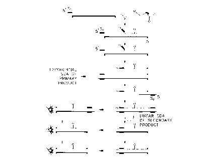
DESCRIPTION OF THE DRAWINGS
Fig. 1 illustrates the signal primer reaction' scheme for use in detection of
target
amplification according to the invention.
Fig. 2 shows the change in fluorescence intensity which occurs as a nucleic
acid target
is amplified using the signal primers of the invention.
Fig. 3 shows the change in fluorescence intensity associated with
hybridization,
extension and cleavage of a signal primer according to the invention.
DETAILED DESCRIPTION OF THE INVENTION
The present invention employs signal primers in hybridization and extension
reactions
to produce double-stranded products which contain a donor/acceptor dye pair.
Fluorescence
quenching occurs in the signal primer. Conversion of the single-stranded
signal. primer to
i 5 double-stranded form also converts a single-stranded restriction
endonuclease cleavage site in
the signal primer to double-stranded form, rendering it cleavable or nickable
by the restriction
endonuclease. Cleavage or nicking by the restriction endonuclease separates
the donor and
acceptor dyes, resulting in decreased quenching of donor fluorescence and an
increase in donor
fluorescence intensity. An associated change in a fluorescence parameter
(e.g., an increase in
donor fluorescence intensity, a decrease in acceptor fluorescence intensity or
the ratio of the
two) is monitored as a indication of target sequence amplification. Monitoring
of the change in
donor fluorescence is preferred, as this change is typically larger than the
change in acceptor
fluorescence. Other fluorescence parameters such as a change in fluorescence
lifetime may
also be monitored.
Terms relating to nucleic acid target amplification and signal primers are
defined as
follows:
An amplification primer is a primer for amplification of a target sequence by
primer
extension. For SDA, the 3' end of the amplification primer (the target binding
sequence)
hybridizes at the 3' end of the target sequence. The amplification primer
comprises a
recognition site for a restriction endonuclease near its 5' end. The
recognition site is for a
restriction endonuclease which will cleave one strand of a DNA duplex when the
recognition
site is hemimodified ("nicking"), as described in US Patent No. 5,455,166; US
Patent No.
5,270,184 and; EP 0 684 31 S. A hemimodified recognition site is a double
stranded
recognition site for a restriction endonuclease in which one strand contains
at least one
derivatized nucleotide which causes the restriction endonuclease to nick the
primer strand
rather than cleave both strands of the recognition site. Usually, the primer
strand of the
5
Docket No. P-3747
CA 02236616 1998-OS-04
hemimodified recognition site does not contain derivatized nucleotides and is
nicked by the
restriction endonuclease. Alternatively, the primer may contain derivatized
nucleotides which
cause the unmodified target strand to be protected from cleavage while the
modified primer
strand is nicked. Such restriction endonucleases can be identified in routine
screening systems
in which a derivatized dNTP is incorporated into a restriction endonuclease
recognition site for
the enzyme. Preferred hemimodified recognition sites are hemiphosphorothioated
recognition
sites for the restriction endonucleases HincII, BsoBI and BsrI. The
amplification primer also
comprises a 3'-OH group which is extendable by DNA polymerase when the target
binding
sequence of the amplification primer is hybridized to the target sequence. For
the majority of
the SDA reaction, the amplification primer is responsible for exponential
amplification of the
target sequence.
As no special sequences or structures are required to drive the amplification
reaction,
amplification primers for PCR generally consist only of target binding
sequences.
Amplification primers for 3 SR and NASBA, in contrast, comprise an RNA
polymerase
promoter near the 5' end. The promoter is appended to the target sequence and
serves to drive
the amplification reaction by directing transcription of multiple RNA copies
of the target.
Extension products are nucleic acids which comprise a primer or a portion of a
primer
and a newly synthesized strand which is the complement of the target sequence
downstream of
the primer binding site. Extension products result from hybridization of a
primer to a target
sequence and extension of the primer by polymerase using the target sequence
as a template.
A bumper primer is a primer which anneals to a target sequence upstream of the
amplification primer, such that extension of the bumper primer displaces the
downstream
amplification primer and its extension product. Extension of bumper primers is
one method for
displacing the extension products of amplification primers, but heating is
also suitable.
The terms target or target sequence refer to nucleic acid sequences to be
amplified or
detected. These include the original nucleic acid sequence to be amplified,
its complementary
second strand and either strand of a copy of the original sequence which is
produced by
replication or amplification. The target sequence may also be referred to as a
template for
extension of hybridized primers.
A signal primer comprises, at its 3' end, a target binding sequence which
hybridizes to
the target sequence and, 5' to the target binding sequence, a label;
detectable structure or
specialized sequence for detection. The signal primers of the invention
comprise a restriction
endonuclease recognition site in a tail portion S' to the target binding
sequence and a
donor/acceptor dye pair flanking the restriction endonuclease recognition site
to facilitate
detection of double-stranded products generated from the signal primer. The
signal primer
may hybridize to a target sequence downstream of an amplification primer such
that extension
6
Docket No. P-3747
CA 02236616 1998-OS-04
of the amplification primer displaces the signal primer, a portion of the
signal primer or the
signal primer extension product. It is then rendered double-stranded by
hybridization and
extension of a second amplification primer. Alternatively, for purposes of the
present
invention, the target binding sequence of the signal primer may hybridize at
the 3' end of the
target sequence forming an 5' overhang such that extension of the target on
the signal primer
renders the signal primer, including the restriction endonuclease recognition
site, double
stranded.
Amplification products, amplified products or amplicons are copies of the
target
sequence generated by hybridization and extension of an amplification primer.
This term refers
to both single stranded and double stranded amplification primer extension
products which
contain a copy of the original target sequence, including intermediates of the
amplification
reaction.
Secondary amplification products or secondary products are oligonucleotides
generated
from a signal primer in a target amplification-dependent manner. These terms
refer to single
stranded or double stranded products generated from signal primers, as well as
portions of
signal primers or signal primer extension products generated as a result of
target amplification.
Cleavage of an oligonucleotide refers to breaking the phosphodiester bonds of
both
strands of a DNA duplex or breaking the bond of single-stranded DNA. This is
in contrast to
nicking, which refers to breaking the phosphodiester bond of only one of the
two strands in a
DNA duplex.
Generation of double-stranded secondary amplification products using a signal
primer
is illustrated in Fig. 1 and may be summarized as follows. A signal primer
hybridizes to one
strand of the target sequence downstream of an amplification primer. Both the
amplification
primer and the signal primer are extended by DNA polymerase using the target
sequence as a
template. The signal primer extension product is displaced from the template
by extension of
the upstream amplification primer and in turn serves as a template for
hybridization and
extension of a second amplification primer, rendering the signal primer
extension product
double-stranded. The RERS thereby becomes a substrate for the restriction
endonuclease. A
second signal primer which hybridizes to the second, complementary strand of a
double
stranded target sequence without overlapping the the hybridization site of the
first signal
primer may optionally be included in the reaction. The second signal primer
hybridizes to the
second strand of the target sequence downstream of the second amplification
primer and is
extended and displaced by extension of the second amplification primer. The
second signal
primer extension product is rendered double stranded by hybridization and
extension of the
first amplification primer. Multiple signal primers per strand of target may
be employed if
desired, each hybridizing to the target sequence downstream of the other on
the same strand,
7
Docket No. P-3747
CA 02236616 1998-OS-04
and all signal primers being hybridized downstream of the amplification
primer. In this manner,
each signal primer is displaced by extension of the upstream signal primer and
the most 5'
signal primer is displaced by the amplification primer. Use of multiple signal
primers has the
advantage of increasing or amplifying the signal generated per target, with an
increase in
sensitivity of the assay. In SDA and other amplification reactions in which
the specialized
sequences or structures are required in the amplification primers, signal
primers do not serve as
amplification primers. Secondary amplification products are therefore either
unamplifiable or
not exponentially amplifiable and have the advantage of not contributing
significantly to
background.
The signal primers of the invention comprise a donor/acceptor dye pair linked
at
positions flanking a restriction endonuclease recognition site (RERS). In the
single-stranded
signal primer, the RERS sequence corresponds to one strand of the double-
stranded RERS.
The signal primer restriction endonuclease recognition site is positioned 5'
to the target binding
region of the signal primer so as not to interfere with hybridization of the
signal primer to the
I S target sequence or its extension by polymerase. Either the donor or
acceptor dye is linked to
the signal primer 3' to the RERS but preferably not at the 3' terminus of the
signal primer as a
3' terminal label may interfere with hybridization and/or extension of the
primer. However, if a
selected donor fluorophore or acceptor dye does not inhibit hybridization
and/or extension it
may be linked at the 3' terminus of the signal primer. The donor fluorophore
(if the acceptor is
3' to the RERS) or the acceptor (if the donor is 3' to the RERS) is linked to
the signal primer at
a position S' to the RERS. That is, the donor and acceptor dyes are linked to
the single-
stranded signal primer such that they flank the RERS. The dyes are preferably
linked on either
side of the RERS at positions sufficiently close together that fluorescence
quenching occurs
but also sufficiently far apart to allow the restriction endonuclease access
to the RERS for
cleavage or nicking.
In SDA reactions, the signal primer RERS may be a sequence which is recognized
by
the same restriction enzyme as provides the nicking function central to SDA.
That is, two
different recognition sequences for the same restriction endonuclease may be
employed - one
in the signal primer and one in the amplification primer. In this embodiment,
the sequence of
the signal primer RERS may be selected such that double-stranded cleavage is
not prevented
when the modified deoxynucleoside triphosphates (dNTPs) of SDA are
incorporated. In
contrast, the sequence of the amplification primer RERS is selected such that
nicking by the
restriction endonuclease is induced by incorporation of modified dNTPs. For
example, the
CTCGAG and CCCGAG recognition sites for BsoBI remain cleavable when
hemimodified,
whereas the CTCGGG recognition site for the same enzyme is nicked when
hemimodified.
Alternatively, a recognition site for a restriction endonuclease different
from that which
8
Docket No. P-3747
CA 02236616 1998-OS-04
provides the nicking fi~nction in the SDA reaction may be present in the
signal primer. Again,
however, the RERS in the signal primer is preferably seiected such that double-
stranded
cleavage is not compromised by incorporation of modified dNTPs. In still
another alternative
embodiment, the RERS in the signal primer is selected so as to be nicked once
by the
restriction endonuclease, regenerating an RERS which is not renickable upon
repair by the
polymerase and incorporation of the modified dNTP. Such "singly-nickable"
sites may be
recognized by either the same restriction endonuclease which provides the
nicking function in
the SDA reaction or by a different restriction endonuclease. Singly nickable
sites are generally
canonical and contain a nucleotide at the nicking site which is the same as
the modified dNTP
in the SDA reaction. For example, the CCCGGG recognition site for BsoBI is
nicked between
the first and second C's. When used as a signal primer in an SDA reaction
employing dCTPa
S, repair of the nick and displacement of the strand downstream of the nick
incorporates the
modified C nucleotide at the nicking site. Modification of the nicking site
inhibits renicking,
but the initial nick separates the donor and acceptor dyes by allowing strand
displacement of
the downstream fragment carrying one of the dyes. Singly nickable sites are
desirable in the
invention because they prevent amplification of the secondary amplification
product
independently of amplification of the target sequence, lowering background and
improving
quantitation.
The signal primer is included in a nucleic acid target amplification reaction
generally as
described in U. S. Patent No. 5,547,861. When added to the amplification
reaction, the signal
primers of the invention are converted to double-stranded form as previously
described,
converting the RERS to a double-stranded form which is cleavable by the
restriction
endonuclease. This process is illustrated in Fig. I. "Cleavage" as used herein
refers to cutting
of both strands of a nucleic acid duplex by a restriction endonuclease, in
contrast to "nicking"
which refers to cutting of only one of the two strands in a duplex nucleic
acid. Cleavage of the
RERS produces two fragments of the double-stranded secondary amplification
product.
Because the donor and acceptor dyes flank the RERS, cleavage of the RERS
results in
separation of the dyes onto the separate fragments. Nicking of the RERS with
displacement of
the single-strand downstream of the nick results in a double-stranded fragment
linked to one
dye and a separate single-stranded fragment linked to the other dye. The
distance between the
dyes increases as the two fragments diffuse in the reaction solution,
resulting in reduced
quenching. A change in a fluorescence parameter resulting from reduced
quenching, e.g., an
increase in donor fluorescence intensity or a decrease in acceptor
fluorescence intensity, may
be detected and/or monitored as an indication that target amplification is
occurring or has
occurred.
9
Docket No. P-3747
CA 02236616 1998-OS-04
Because cleavable or nickable secondary amplification products are produced
concurrently with target amplification, the change in fluorescence may be
monitored as the
amplification reaction is occurnng, i.e., in "real-time". Homogeneous assays
reduce
contamination because the reaction vessel does not have to be opened for
detection and they
allow the use of simpler instrumentation than in heterogeneous assays. In
addition, because a
change in fluorescence is monitored rather than an absolute value, the
accuracy of the assay is
not dependent on the starting point (i.e., establishing a "zero" point). The
homogeneous, real-
time assay of the invention can be used to provide semi-quantitative or
quantitative information
about the initial amount of target present. That is, the rate at which the
selected fluorescence
parameter changes during amplification is an indication of the initial target
levels. As a result,
when more initial copies of the target sequence are present, donor
fluorescence more rapidly
reaches the threshold of detection for the cleaved secondary amplification
products (i.e.,
shorter time to positivity). The decrease in acceptor fluorescence similarly
exhibits a shorter
time to positivity, detected as the time required to reach a selected minimum
value. In
1 S addition, the rate of change in the fluorescence parameter during the
course of the reaction is
more rapid in samples containing higher initial amounts of target than in
samples containing
lower initial amounts of amounts of target (i.e., increased slope of the
curve). That is, an
increased rate of change in intensity, lifetime, etc. indicates a higher
initial target level than is
present in a sample exhibiting a relatively slower rate of change.
In an alternative embodiment, the signal primer may be used in a non-
amplification
based format to detect a target oligonucleotide. In this embodiment, the
target binding
sequence of the signal primer hybridizes to the 3' end of the target
oligonucleotide such that
the RERS forms a 5' overhang. Polymerase extends the target sequence using the
S' overhang
of the signal primer, including the RERS, as a template. In this case, the
target sequence
functions as a primer in the primer extension reaction to synthesize the
complementary
sequence of the signal primer. If the target binding sequence of the signal
primer is
complementary to the entire length of the target sequence there are no other
single-stranded
overhangs and only the target is extended. However, if the target binding
sequence of the
signal primer hybridizes to only a portion of the target sequence, the target
sequence forms a
second 5' overhang. In this embodiment, the signal primer is also extended
using the 5'
overhang of the target as a template. In either case, the RERS of the signal
primer is thus
rendered double-stranded and cleavable or nickable. Extension to produce the
double-stranded
RERS and the resulting change in fluorescence can take place only in the
presence of target,
and the method is independent of the presence or absence of a restriction site
in the target
sequence itself. As this method does not require SDA or any other
amplification reaction,
modified nucleotides are not necessary. Any restriction site may be employed
in the signal
Docket No. P-3747
CA 02236616 1998-OS-04
primer. However, if the RERS is to be nicked rather than cleaved, modified
nucleotides may
be employed as described above to produce a singly-nickable site.
Many donor/acceptor dye pairs known in the art are usefial in the present
invention.
These include, for example, fluorescein isothiocyanate
(FITC)/tetramethylrhodamine
isothiocyanate (TRITC), FITC/Texas RedTM (Molecular Probes), FITC/N-
hydroxysuccinimidyl
1-pyrenebutyrate (PYB), FITC/eosin isothiocyanate (EITC), N-
hydroxysuccinimidyl 1-
pyrenesulfonate (PYS)/FITC, FITC/Rhodamine X, FITC/tetramethylrhodamine
(TANiR.A), N-
(4-aminobutyl)-N-ethylisoluminol (ABEI)/TAMRA., and others. Near-IR dyes such
as Cy5 (N,
N-modified tetramethyl indodicarbocyanine) may also be employed, e.g., paired
with ROX.
The selection of an appropriate quenching donor/acceptor pair is routine in
the art. For energy
transfer quenching it is only necessary that the emission wavelengths of the
donor fluorophore
overlap the excitation wavelengths of the acceptor fluorophore, i.e., there
must be sufficient
spectral overlap between the two dyes to allow efficient energy transfer,
charge transfer or
fluorescence quenching. p-(Dimethyl aminophenylazo) benzoic acid (DABCYL) is a
non-
I S fluorescent acceptor dye which effectively quenches fluorescence from a
neighboring
fluorophore, e.g., fluorescein or 5-((2'-aminoethyl) amino) naphthalenel-
sulfonic acid
(EDANS). Certain donor/acceptor pairs are exemplified above and in the
following Examples,
however, others will be apparent to those skilled in the art and are also
usefi~l in the invention.
Any dye pair which produces fluorescence quenching in the signal primers of
the invention is
suitable for use in the methods of the invention, regardless of the mechanism
by which
quenching occurs.
Terminal and internal labeling methods are also known in the art and may be
used to
link the donor and acceptor dyes at their respective sites in the signal
primer. Examples of 5'-
terminal labeling methods include a) periodate oxidation of a S'-to-5'-coupled
ribonucleotide
followed by reaction with an amine-containing label, b) condensation of
ethylenediamine with a
5'-phosphorylated polynucleotide followed by reaction with an amine-reactive
label, and c)
introduction of an aliphatic amine substituent using an aminohexyl phosphite
reagent in solid-
phase DNA synthesis followed by reaction with an amine-reactive label. Labels
may also be
linked to synthetic DNA oligonucleotides at specific locations using special
aliphatic amine-
containing nucleotide phosphoramidite reagents. Selection of an appropriate
method for
linking the selected labels to the signal primer and performing the linking
reactions are routine
in the art.
The signal primers of the invention have a donor and an acceptor linked to the
single-
stranded signal primer such that donor fluorescence is totally or partially
quenched. Between
the two dyes, the signal primer comprises a RERS (in single-stranded form).
The two dyes
must be in sufficiently close spatial proximity for quenching to occur,
however, the distance
II
Docket No. P-3747
CA 02236616 1998-OS-04
between them must also allow the restriction endonuclease access to its
recognition site for
binding and cleavage or nicking when the signal primer is rendered double-
stranded. To study
the relationship of these two parameters, signal primers and their complements
were chemically
synthesized. The signal primer sequence selected was SEQ ID NO:1:
5 ~-TAGCC'i'~CGAGT 11 AGAGT 16CTTCAAAT24ATCAGAGCTTTACCTAACAA-3'
6 = nucleotide position 6 16 = nucleotide position 16
11 = nucleotide position 11 24 = nucleotide position 24
The BsoBI site for cleavage is shown bolded, with additional tail sequence 5'
to it to
accommodate the "footprint" of the restriction enzyme when it binds. Double-
stranded
cleavage of this BsoBI recognition sequence is not inhibited by incorporation
of the modified
deoxynucleoside triphosphates during SDA, in contrast to the CTCGGG
recognition sequence
for BsoBI which is rendered nickable by incorporation of modified dNTPs during
SDA. The
sequence 3' to the BsoBI site is the target binding sequence, which is
complementary to the
target sequence to be amplified. The assay was performed at 52-53°C in
200 pL KPDG buffer
(40 mM KPi, 3% DMSO, 5% glycerol) with 5 mM Mg(OAc)2 added prior to the
experiment.
Measurements were obtained with an SLM 8100 research grade fluorometer
equipped with a
circulating bath for maintaining sample compartment temperature, a xenon arc
lamp and
grating monochromators for controlling excitation and emission wavelengths.
Experiments
with fluorescein (FAM) as the donor used 488 nm for the excitation wavelength
and 525 nm
for emission. Experiments in which Rhodamine X (ROX) was the donor used an
excitation at
580 nm and emission at 604 nm. Experiments with Cy-5 used 640 nm and 665 nm
respectively. Samples were prepared with 20 nM of the labeled signal primer
for initial
measurements of the emission spectrum using the optimal donor excitation
wavelength.
The selected donor fluorophore was conjugated to the S' phosphate. The
selected
acceptor dye was conjugated to either T6, T11, T16 or T24 to provide varying
distances
between the donor and acceptor dyes. Reactive dyes were obtained from
Molecular Probes
(Eugene, OR) or from the Applied Biosystems Division of Perkin Elmer (Foster
City, CA).
ROX-NHS (6-carboxy rhodamine X succinimidyl ester) and TAMRA-SE (5-carboxy
tetramethylrhodamine succinimidyl ester) were obtained from ABI/Perkin Elmer.
Oligonucleotides were synthesized on a 1 p,mole scale using an ABI 380B
automated DNA
synthesizer with standard reagents supplied by the manufacturer. The 6-carboxy
substituted
fluorescein (6-FAM) was incorporated at the 5' position by addition of the
phosphoramidite
reagent 6-FAM Amidite (ABI) at the final step of the synthesis. For other 5'
dye labeled
12
CA 02236616 2001-07-20
Docket No. P-3747
oligonucleotides, 5' aminohexyl phosphoramidite (ABI AMINOLINK 2) was
substituted at the
final step to provide a reactive amino group for subsequent conjugation. For
conjugating dyes
to internal positions of the oligonucleotide, a modified dT phosphoramidite
reagent, amino-
modifier C6 dT (Glen Research, Sterling, VA) was substituted in the
appropriate sequence
position in place of unmodified dT. The crude oligonucleotides were
deprotected by treatment
with ammonium hydroxide for 4 to 8 hours at 55°C, which also
deprotected the modified dT.
These were filtered and solvent was evaporated from the filtrate with a rotary
vacuum
apparatus. Oligonucleotides were purified directly following this step by
reverse phase HPLC.
Sequences with only the modified internal dT aminolinker were prepared with
the 5' terminal
dimethoxytrityl (DMT) intact and purified by RP HPLC. The resulting 5'-DMT
full length
product was deprotected using a SepPak column (Waters) with 2% trifluoroacetic
acid and
dried prior to coupling with reactive dyes.
Oligonucleotides were labeled by dissolving an aliquot (0.5 mole) in 100 ~L
NaHC03/NaZC03 buffer, pH 8Ø Th,e reactive dye was added to this as a
solution of 3 mg in
30 ltL, DMSO and the resulting mixture was allowed to stand in the dark for 12-
24 hours at 37
°C. The resulting reaction mixture was passed over a column of G-25
Sephadex resin (NAPS,
Pharmacia Biotech) eluting with 4 ml~t TAE (4 mM TRIS acetate, 0.1 mM EDTA, pH
8.0).
Typically, the first 0.5 to I.0 mL of colored material eluted contained the
highest fraction of
reactive dye-labeled oligonucleotide and was further purified by HPLC on a
Waters Deita Pak
300 ~ C18 3.9 X 150 mm reverse phase column using linear gradients over 30
minutes
followed by 20 minutes re-equilibration. Most gradients used two solvents: A-
98% 50 mM
TEAR (triethylammonium acetate)/2% acetonitrile and B - 10% 50 mM TEAA/90%
acetonitrile, typically in a gradient from 95%A to 70°,i° A over
30 minutes. The identity of the
conjugated material was confirmed by comparing peak intensities at 260 nm (for
DNA) and the
respective peak absorbances for the dyes. Concentrations of purified
oligonucleotides were
determined in TAE buffer by using the DNA absorbance at 260 nm corrected for
the respective
dye absorbance at that wavelength.
The signal primer was initially tested for the effect of the distance between
the donor
and acceptor on quenching efficiency and cleavage efficiency in a
hybridization assay. A 5-fold
excess of the complementary sequf:nce ( 100 nM) was added and the fluorescence
was
measured after hybridization was judged to be complete (typically about 20
min.). The BsoBI
enzyme was added to a concentration of 3.2 units/~tL and a final fluorescence
measurement
was recorded when no further change was observed in the emission spectrum of
the sample.
The results for the various separation distances and dye pairs are shown in
Table I.
* Trademark
13
Docket No. P-3747
CA 02236616 1998-OS-04
TABLEI
Donor Acceptor Fluorescence Ratio
ss ds cleaved ds/ss cleaved/ss
ROX @, T1 5'-Cy5 3376 3919 7605 1.16 2.25
I
5'-FAM ROX @ T6 2467 2973 3983 1.21 1.61*
5'-FAM ROX @, T11 3497 5290 18337 1.51 5.24
5'-FAM ROX @ T16 990 1325 2007 1.34 2.03
5'-FAM ROX @ T26 1900 1900 2000 1 1.1
5'-FAM Dabcyl @ 10011 25566 45167 2.55 4.51
T11
TAMItA @ 5'-Cy5 7357 8412 9744 1.14 1.32
TI I
5'-ROX ROX @ T1 18180 50080 46850 2.8 2.6
l
5'-FAM FAM @ T1 4450 6100 5150 1.37 1.16
I
5'-C~5 Cy5 @ T 11 3650 4150 4150 1.14 1.14
* Incomplete cleavage
These experiments show that the change in fluorescence intensity upon cleavage
of the
signal primer depends on the distance between the donor and acceptor
fluorophores in the
uncleaved signal primer. In general, as the distance between the dyes in the
intact
oligonucleotide increased, the change in donor emission (fluorescence
intensity) upon
conversion to double stranded form decreased in magnitude. The magnitude of
the change in
donor emission following double-stranded cleavage also generally decreased
with increasing
distance between the dyes. Dye pairs which too closely flanked the RERS
appeared to
interfere with complete cleavage, also reducing the total change in donor
fluorescence. Signal
primers with about eleven nucleotides between the donor and acceptor typically
exhibited the
greatest change in donor fluorescence upon conversion to double-stranded form
and cleavage
I S of the RERS. These results indicate, however, that about 8-20 nucleotide
separation,
preferably about 10-16 nucleotides between the donor and acceptor dyes
produces a change in
donor fluorescence of a readily detectable magnitude. These separation
distances are also
sufficient to accommodate binding of the restriction endonouclease to its
recognition site
without significant interference from the bulky dyes, while still placing the
dyes in sufficiently
close proximity to produce satisfactory quenching. Greater changes in donor
fluorescence
would be expected if the two dyes could be brought into closer proximity on
the signal primer,
14
CA 02236616 2001-07-20
Docket No. P-3747
however, placing the acceptor closer to the donor than six nucleotides
interfered with the
ability of the restriction enzyme t:o cleave the duplex, although an increase
in donor
fluorescence was still detectable. This demonstrates that even a small amount
of signal primer
conversion results in a relatively large change in fluorescence.
An increase in donor fluorescence was usually observed upon conversion to
double-
stranded form alone. This is likely to be due to a reduction in quenching
occurring by
mechanisms other than Forster transfer which may take place in the single-
stranded
oligonucleotide (e.g., charge transfer, collisional quenching). Target
amplification may
therefore be detected by monitoring only the change in fluorescence upon
conversion of the
signal primer to double-stranded form. In this case an RERS in the signal
primer is not
necessary. In most cases, however, cleavage further increased the change in
fluorescence.
Monitoring the total change in fluorescence (double-stranded conversion and
cleavage or
nicking) is preferred for this reason. Regardless of the magnitude of the
fluorescence change
at each step of the process (i.e., single-stranded to double-stranded
conversion, and conversion
of the double-stranded form to the cleaved or nicked form) a readily
detectable increase in
donor fluorescence was evidenced by a cut/ss ratio significantly greater than
1 when the dye
pair was sufficiently far apart for efficient cleavage but in sufficiently
close proximity to
optimize quenching. In an end-point assay a larger change in fluorescence may
be detectable if
end-point fluorescence is monitored at a lower temperature than initial
fluorescence. When the
change in fluorescence is monitored in real-time, its magnitude will be
affected by the
temperature of the reaction. At higher temperatures the change in fluorescence
associated with
double-stranded conversion and cleavage is generally smaller than at lower
temperatures.
It was also observed that homologous donor/acceptor dye pairs (shown in the
last three
lines of the Table) exhibited an increase in donor fluorescence intensity only
upon conversion
from single- to double-stranded form. In contrast to heterologous dye pairs,
no further
increase was obtained upon cleavage of the double-stranded oligonuceotide, and
in some cases
cleavage produced a slight reduction in donor fluorescence intensity.
Therefore, signal primers
employing these fluorophore pairs need not contain an RERS. Target may be
detected using
the ss/ds ratio or a change in fluorescence associated with the conversion to
double-stranded
form, as quenching of the fluorophores decreases (i.e., fluorescence intensity
will increase) as
the signal primer is converted to double-stranded form in the presence of
target.
It will be apparent that, in addition to SDI the methods of the invention may
be easily
adapted to other primer extension amplification methods (e.g., PCR, 3SR,
NASBA, TMA,
etc.). For example, replacing SDA amplification primers with PCR amplification
primers and
using a PCR DNA polymerase which lacks 5'-~3' exonuclease activity (e.g.,
Sequencing Grade
Taq from Promega or exo- Vent or exo- Deep Vent from New England BioLabs) in
the signal
* Trademark
IS
Docket No. P-3747
CA 02236616 1998-OS-04
primer reaction scheme also generates secondary amplification products which
contain a
cleavable, double-stranded RERS contributed by the signal primer. Of course,
in PCR any
RERS may be selected for use in the signal primer, as there are typically no
modified
deoxynucleoside triphosphates present which might induce nicking rather than
cleavage of the
RERS. The double-stranded RERS in the secondary amplification product may be
cleaved by
a restriction endonuclease to separate a donor/acceptor dye pair as described
above. As
thermocycling is a feature of amplification by PCR, the restriction
endonuclease is preferably
added at low temperature after the final cycle of primer annealing and
extension for end-point
detection of amplification. However, a thermophilic restriction endonuclease
which remains
active through the high temperature phases of the PCR reaction could be
present during
amplification to provide a real-time assay. As in SDA systems, cleavage of the
RERS and
separation of the dye pair reduces fluorescence quenching, with the increase
in fluorescence
intensity serving as an indication of target amplification.
For adaptation of the inventive methods to 3 SR, NASBA or TMA, a 5'-~3'
exonuclease deficient reverse transcriptase with strand displacing activity is
employed in the
3 SR reaction, with hybridization of the signal primer to the RNA target
downstream of an
amplification primer which contains an RNA polymerase promoter. In a reaction
scheme
similar to that previously described, the hybridized signal primer containing
the RERS is 1 )
extended, and 2) displaced by extension of the upstream amplification primer.
The displaced
extension product is then made double-stranded by hybridization and extension
of the second
amplification primer. This renders the restriction endonuclease recognition
site cleavable, and
the donor and acceptor dyes are thereby separated onto different fragments,
increasing the
distance between them and reducing fluorescence quenching of the donor dye.
The signal
primer for 3 SR or NASBA does not contain an RNA polymerase promoter sequence
and
therefore cannot function as an amplification primer, reducing nonspecific
background signal.
This is analogous to the signal primer in SDA, which does not contain a
repeatably nickable
RERS and therefore does not contribute to exponential background amplification
of non-
specific targets.
For reasons previously stated, signal primers are preferred for use in the
methods of the
invention with the signal primer extension product being separated from the
target sequence by
displacement due to extension of the upstream amplification primer. However,
it will be
apparent that the amplification primers known for use in the various nucleic
acid amplification
reactions may also be labeled and modified as described for signal primers. In
this
embodiment, the labeled amplification primer extension product may be
separated from the
target sequence by displacement due to extension of an upstream non-
amplification primer
(e.g., bumper primers as in SDA), by denaturation (e.g., heating as in PCR) or
by enzymatic
16
Docket No. P-3747
CA 02236616 1998-OS-04
digestion of the target strand (e.g., RNase H as in 3SR). Amplification
primers comprising the
RERS flanked by the donor/acceptor dye pair eliminate the need for the
additional signal
primer in the reaction, but because background may be higher in this
embodiment the
sensitivity of the assay may be decreased. For PCR, the amplification primer
is modified by
S addition of an RERS in a 5' tail and the RERS is flanked by a donor/acceptor
dye pair. This
primer is structurally identical to the PCR signal primer described above.
Functionally,
however, it is different in that there is no downstream primer to be extended
and displaced and
the amplification primer itself provides the change in fluorescence. For 3 SR,
NASBA and
TMA, the RERS may be placed 5' to the promoter of an amplification primer so
that the RERS
is cleaved in the double-stranded DNA portion of the amplification cycle.
Because the RERS
is 5' to the promoter, cleavage does not remove the promoter from the
amplification primer
and generation of RNA transcripts continues to sustain target amplification. A
second
amplification primer which does not contain a promoter sequence (e.g., as in
NASBA) may
also or alternatively contain the RERS in a 5' tail portion.
1 S Target DNA for the following experimental examples was prepared from
stocks of
Chlamydia trachomatis elementary bodies (EB's) stored at concentrations of 106
EB's/~L in
50% glycerol. EB stock solutions were diluted 1:10 in water, boiled for 1 S
minutes and
prepared as 10-fold serial dilutions in 10 ng/p.L human placental DNA. These
stock solutions
contained 1 to 100 genome copies/p.L of target. The donor fluorophore was
conjugated to the
5' phosphate. Measurements were obtained with an SLM 8100 research grade
fluorometer
equipped with a circulating bath for maintaining sample compartment
temperature, a xenon arc
lamp and grating monochromators for controlling excitation and emission
wavelengths.
Experiments with fluorescein (FAM) as the donor used 488 nm for the excitation
wavelength
and 525 nm for emission. Experiments in which ROX was the donor used an
excitation at 580
nm and emission at 604 nm.
EXAMPLE 1
SDA was performed generally as described in EP 0 684 315, with addition of the
signal
primer labeled at the 5' end with FAM and at T11 with ROX. The final
concentrations of
components in each 100 ~L reaction were 40 mM KiP04 pH 7.5, 6 mM MgOAc, 0.2 mM
each dTTP, dGTP, dATP, 1.4 mM dCTPocS, 20 ~g/mL acetylated BSA, 3% DMSO, 8%
(v/v)
glycerol, 100 ng human placental DNA, 25 units Bst polymerase (exo klenow
fragment, New
England BioLabs), 150 units AvaI (New England BioLabs, Beverly, MA), and DNA
from 0,
10, 100 or 1,000 Chlamydia trachomatis elementary bodies. Each sample further
contained 50
nM signal primer SEQ ID NO:1 (5'-FAMITI ~-ROX) and the four primers shown
below:
17
Docket No. P-3747
CA 02236616 1998-OS-04
Amplification primer S 1.1 (SEQ ID N0:2, 750 nM)
ACCGCATCGAATCGATGTCTCGGGTAGAAAA TCGCA TGCAAGA TA
Amplification primer S2.1 (SEQ ID N0:3, 188 nM)
CGATTCCGCTCCAGACTTCTCGGGAGCTGCCTCAGAATATACTCAG
S Bumper primer B 1 (SEQ ID N0:4, 75 nM)
TAAACATGAAAACTCGTTCCG
Bumper primer B2 (SEQ ID NO:S, 75 nM)
TTTTATGATGAGAACACTTAAACTCA
Each reaction was assembled to contain all reagents except Bst and AvaI, and
the
samples were then heated for 2 min. at 95°C. They were transferred to a
53.5°C water bath for
3-5 min. and the enzymes were added for a total sample volume of 100 ~.L. The
samples were
then transferred to 225 p.L cuvettes and placed into a research grade SLM 8100
spectrofluorometer (Spectronic Instruments, Rochester, NY). The temperature of
the cuvettes
was maintained at 53-54°C by a circulating water bath, and the
fluorescence emission of each
cuvette at 520 nm (,excitation - 488 nm) was recorded every 8 sec. Reactions
were typically
followed for 60-90 min.
Fig. 2 shows the results. Fluorescence remained low (quenched) in the control
reaction
containing no target (no amplification) but increased significantly in
reactions containing 100
and 1,000 targets, demonstrating specific detection of target amplification.
There was no
appreciable increase in fluorescence in the reaction containing 10 targets,
indicating a
sensitivity of detection between 10 and 100 targets. In addition, the rate of
increase in
fluorescence intensity of the donor (a measure of the rate of decrease in
donor quenching) was
more rapid in samples containing higher numbers of initial target. The rate of
increase in donor
fluorescence therefore provides not only detection of amplification in real-
time, but also a
semi-quantitative or relative measure of initial target levels. By comparing
the rate of increase
in fluorescence in a sample containing an unknown amount of target to the
increase in
fluorescence in a series of reactions containing varying known amounts of
target (producing a
standard curve as is known in the art) a quantitative measure of target levels
in the unknown
sample may be obtained. Alternatively, detection of an increase in
fluorescence intensity above
a predetermined threshold value may be used as an indication that the target
is present and
amplified in a simple positive/negative assay format.
18
Docket No. P-3747
CA 02236616 1998-OS-04
EXAMPLE 2
A signal primer according to the invention was used to detect a target
oligonucleotide
in the absence of target amplification. An unlabeled target oligonucleotide
having the
following sequence was synthesized by conventional methods:
TTGTTAGGTAAAGCTCTGATATTTGAAG (SEQ ID N0:6)
This target is complementary to the 3' target binding sequence of signal
primer SEQ ~ NO:1.
Four glass cuvettes (225 pL, Starna Cells) were each filled with 100 pL of a
solution
comprising 50 nM signal primer, 5 mM Mg(OAc)2, 0.2 mM each deoxynucleotide
triphosphate, 1.4 mM a-thio dCTP, 40 mM potassium phosphate (pH 7.5), 3% DMSO
(v/v),
and 5% glycerol. SEQ ID N0:6 was added to each cuvette to a final
concentration of 0, 2.5,
25 or 250 nM representing 0. 0.05, 0.5 and 5 molar equivalents of target per
equivalent of
signal primer. The samples were then heated briefly to 95°C and cooled
to 54°C in an SLM
8100 fluorometer. Bst polymerase (180 units) and BsoBI (240 units) were added
to each
cuvette and the fluorescence intensity was recorded at 520 nm (7~excitation -
488 nm) as
described in Example 1.
The results are shown in Fig. 3. Fluorescence did not change in the absence of
target,
but increased over the course of the hybridization, extension and cleavage
reaction in all
samples containing target. The magnitude of the change in fluorescence
intensity increased in
approximate proportion to the amount of target. Further, the rate of change in
fluorescence
intensity was greater as the amount of target increased. Either of these
parameters may be
used as a means for quantitating target levels, typically by comparison to the
results obtained
for known amounts of target used as standards.
19
Docket No. P-3747
CA 02236616 1998-OS-04
SEQUENCE LISTING
(1) GENERAL INFORMATION:
S
(i) APPLICANT: Nadeau, James G.
Pitney, James B.
Schram, James L.
Linn, Carl P.
Vonk, Glenn P.
Walker, George T.
(ii) TITLE OF INVENTION: Detection of Nucleic Acids by
Fluorescence Quenching
1S
(iii) NUMBER OF SEQUENCES: 6
(iv) CORRESPONDENCE
ADDRESS:
(A) ADDRESSEE: R. J. Rodrick, Becton Dickinson
and Company
(B) STREET: 1 Becton Drive
(C) CITY: Franklin Lakes
(D) STATE: NJ
(E) COUNTRY: US
(F) ZIP. 07417
2S
(v) COMPUTER
READABLE
FORM:
(A) MEDIUM TYPE: Floppy disk
(B) COMPUTER: IBM PC compatible
(C) OPERATING SYSTEM: PC-DOS/MS-DOS
(D) SOFTWARE: PatentIn Release #1.0, Version #1.30
(vi) CURRENT APPLICATION DATA:
(A) APPLICATION NUMBER:
(B) FILING DATE:
3S (C) CLASSIFICATION:
(viii) ATTORNEY/AGENT INFORMATION:
(A) NAME: Fugit, Donna R.
(B) REGISTRATION NUMBER: 32,135
4O (C) REFERENCE/DOCKET NUMBER: P-3747
(2) INFORMATION FOR SEQ ID NO:1:
4S (i) SEQUENCE CHARACTERISTICS:
(A) LENGTH: 44 base pairs
(B) TYPE: nucleic acid
(C) STRANDEDNESS: single
{D) TOPOLOGY: linear
SO
SS (xi) SEQUENCE DESCRIPTION: SEQ ID NO:1:
TAGCCTCGAG TAGAGTCTTC AAATATCAGA GCTTTACCTA ACAA 44
(2) INFORMATION FOR SEQ ID N0:2:
(i) SEQUENCE CHARACTERISTICS:
(A) LENGTH: 45 base pairs
(B) TYPE: nucleic acid
(C) STRANDEDNESS: single
Docket No. P-3747
CA 02236616 1998-OS-04
(D) TOPOLOGY: linear
S
(xi) SEQUENCE DESCRIPTION: SEQ ID N0:2:
ACCGCATCGA ATCGATGTCT CGGGTAGAAA ATCGCATGCA AGATA 45
(2) INFORMATION FOR SEQ ID N0:3:
(i) SEQUENCE CHARACTERISTICS:
(A) LENGTH: 46 base pairs
1S (B) TYPE: nucleic acid
(C) STRANDEDNESS: single
(D) TOPOLOGY: linear
(xi) SEQUENCE DESCRIPTION: SEQ ID N0:3:
ZS CGATTCCGCT CCAGACTTCT CGGGAGCTGC CTCAGAATAT ACTCAG 46
(2) INFORMATION FOR SEQ ID N0:4:
(i) SEQUENCE CHARACTERISTICS:
(A) LENGTH: 21 base pairs
(B) TYPE: nucleic acid
(C) STRANDEDNESS: single
(D) TOPOLOGY: linear
3S
(xi) SEQUENCE DESCRIPTION: SEQ ID N0:4:
TAAACATGAA AACTCGTTCC G 21
(2) INFORMATION FOR SEQ ID N0:5:
4S (i) SEQUENCE CHARACTERISTICS:
(A) LENGTH: 26 base pairs
(B) TYPE: nucleic acid
(C) STRANDEDNESS: single
(D) TOPOLOGY: linear
SS (xi) SEQUENCE DESCRIPTION: SEQ ID N0:5:
TTTTATGATG AGAACACTTA AACTCA 26
(2) INFORMATION FOR SEQ ID N0:6:
(i) SEQUENCE CHARACTERISTICS:
(A) LENGTH: 28 base pairs
(B) TYPE: nucleic acid
(C) STRANDEDNESS: single
21
CA 02236616 1998-OS-04
Docket No. P-3747
(D) TOPOLOGY: linear
(xi) SEQUENCE DESCRIPTION: SEQ ID N0:6:
TTGTTAGGTA AAGCTCTGAT ATTTGAAG 2 8
22