Note: Descriptions are shown in the official language in which they were submitted.
CA 02243959 1998-07-21
Wb 98/26280 PCT/LTS97/20199
METHOD AND APPARATUS FOR REDUCING
THE DISTORTION OF A SAMPLE ZONE ELUTING
FROM A CAPILLARY ELECTROPHORESIS CAPILLARY
FIELD OF THE INVENTION
This invention relates to an electrophoresis method and
apparatus for practicing the method. More specifically, this
invention relates to a capillary electrophoresis method and
apparatus wherein the distortion of a sample eluting from the
end of a capillary is reduced thereby resulting in enhanced
detectability of an eluted sample.
REFERENCES
Dovichi et al., U.S. Patent No. 5,439,578 (1995)
Grossman and Colburn, Capillary Electrophoresis Theory and
Practice, Chapter l, Academic Press (1992)
Grossman, U.S. Patent No. 5,374,527 (I994)
Holman, Heat Transfer, Fourth Edition, McGraw-Hill (1976)
Madabhushi et al., U.S. Patent No. 5,552,028 (199&)
Sambrook et al. , eds . , Molecular Cloning: A .Laboratory
Manual, Second Edition, Chapter 5, Cold Spring Harbor
Laboratory Press (1989)
Takahashi et al., U.S. Patent No. 5,529,679 (1996)
BACKGROUND
Electrophoretic separations of biopolymers and small
molecules are critically important in modern biology and
biotechnology, comprising an important component of such
techniques as DNA sequencing, protein molecular weight
determination, genetic mapping, and the like. A particularly
~ preferred electrophoresis format is capillary electrophoresis
(CE), where the electrophoresis is performed in a capillary
tube having a small internal diameter. Capillary
electrophoresis results in enhanced separation performance over
traditional slab-based formats because the superior ability of
the narrow-bore capillary to dissipate Joule heat allows for
-1-
CA 02243959 1998-07-21
WO 98/26280 PCT/US97/20199
high electrical fields to be employed thereby resulting in fast
separations in which sample diffusion is- minimized.
In traditional CE systems, detection of a sample
subsequent to separation is performed during electrophoresis
while the sample is still located inside the capillary lumen.
Thus, any excitation light required to excite the sample and
any emission light coming from the sample must be transmitted
through the wall of the capillary. A drawback of this approach
is that the fused silica capillaries typically used in CE are
poor optical elements, i_e., they cause significant scattering
of light. Problems associated with light scattering are
particularly problematic when it is desired to detect
fluorescence from samples located in a plurality of closely-
spaced capillaries by fluorescence because the scattered
excitation light form one capillary will interfere with the
detection of samples in neighboring capillaries.
One approach to solving the problem of on-capillary
detection has been to detect a sample after the sample emerges
from the capillary in a detection cell having superior optical
characteristics, e.g., a flat quartz chamber. In one class of
these systems, a "sheath flow" of liquid is used to transport
the sample from the outlet of the CE capillary to a detection
zone at which detection of the sample takes place (Takahashi;
Dovichi~. A drawback of sheath flow systems is that in order
to avoid distortion of a sample zone in the detection cell,
precise control of the flow rate of the sheath flow liquid is
required. A second drawback of sheath flow systems is that
the pressure used to drive the flow of the sheath flow liquid
can cause back flow of the separation medium in the separation
capillary thereby impacting resolution.
In another class of off-capillary detection systems, a
sample zone is transported from the outlet of a CE capillary to
a detection zone located in a detection cell by electrophoresis
under the influence of the same voltage difference used to
-2-
CA 02243959 2002-04-29
conduct the electrophoretic separation (Takahashi?. ~owevei,
because of the larger cross-sectional area of the detection
cell as compared to the lumen of the capillary, the electric
field diverges at the capillary outlet causing a distortion o'
an eluted sample zone. Such distortion results in severe loss
of spatial resolution between adjacent sample zones eluting
from a single capillary and/or between zones eluting from
adjacent capillaries. This loss of spatial resolution tends to
reduce the detectability of neighboring sample zones. As
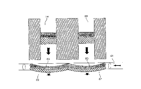
illustrated in FIG. 1, when distorted sample zones 52 and 53
eluting from capillary lumens 54 and 56 are interrogated by a
light beam 51 having dimensions larger than the distorted
sample zones in the direction of migration, it is impossible to
independently detect the adjacent sample zones 52 and 53.
SUMMARY
The present invention is directed towards the discovery of
a system for electrophoretically transporting a sample zone
from an outlet of a capillary electrophoresis capillary to a
detection zone separate from the capillary where the
distortion of the eluted sample zone is reduced, thereby
enhancing the detectability of neighboring sample zones. The
zone distortion is reduced by controlling the divergence of the
electric field at the outlet of the capillary by placing one
or more "focusing" electrodes in the vicinity of the capillary
outlet. The system finds particular application in automated
polynucleotide sequencing systems employing fluorescence
detection and a plurality of capillary electrophoresis tubes.
It is an object of an aspect of the invention to provide
a system for detecting a sample zone after separation by cE
wherein the wall of the CE capillary does not interfere with
the optical detection of the sample zone.
It is an object of an aspect of the invention to provide
a system for transporting a sample zone from an outlet of a
capillary electrophoresis capillary to a detection zone
separate from the capillary wherein a sheath flow is not
required.
-3-
CA 02243959 2002-04-29
It is yet another object of an aspect of the invention
to provide a system for electrophoretically transporting a
sample zone from an outlet of a capillary electrophoresis
capillary to a detection zone separate from the capillary
wherein distortion of the sample zone at the outlet of the
capillary caused by the divergence of an electric field at
the capillary outlet is reduced.
It is another object of an aspect of the invention to
provide a system for electrophoretically transporting a
sample zone from an outlet of a capillary electrophoresis
capillary to a detection zone separate from the capillary
wherein sample zones eluting from neighboring capillaries do
not interfere with one another.
The foregoing and other objects of the invention are
achieved by an electrophoresis apparatus including one or more
separation capillaries, each separation capillary having an
inlet end and an outlet end; a first electrode in electrical
communication with the inlet ends of the separation
capillaries; a second electrode in electrical communication
with the outlet ends of the separation capillaries; and one or
more focusing electrodes in electrical communication with the
outlet ends of the separation capillaries. In a preferred
embodiment, the outlet ends of the capillaries are located in
a detection cell. optionally, the apparatus further includes
a detector for detecting the sample after elution from the
separation capillaries, e.g., a CCD detector, and a light
source for exciting fluorescence of an eluted sample zone,
e.g., a laser.
In a second aspect, the present invention includes methods
of using-the above-described apparatus.
These and other objects, features, and advantages of the
present invention will become better understood with reference
CA 02243959 1998-07-21
Wb 98/26280 PCT/US97/20199
to the following description, drawings, and appended claims.
BRIEF DESCRIPTION OF THE DRAWINGS
FIG. 1 depicts the distortion of sample zones eluting from
a capillary caused by a diverging electrical field at a
capillary outlet.
FIG. 2 shows a schematic diagram illustrating a device of
the invention.
FIG. 3 shows a schematic diagram illustrating a
preferred multicapillary device of the invention.
FIG. 4 shows the results of a finite difference simulation
of an electric field in a detection cell not incorporating a
focusing electrode.
FIGS. 5 and 6 show the results of a finite difference
simulation of an electric field in a detection cell
incorporating a focusing electrode located at an upper surface
of the detection cell at two different values of a focusing
electrode voltage.
DETAILED DESCRIPTION OF THE PREFERRED EMBODIMENTS
Reference will now be made in detail to the preferred
embodiments of the invention, examples of which are illustrated
in the accompanying drawings. While the invention will be
described in conjunction with the preferred embodiments, it
will be understood that they are not intended to limit the
invention to those embodiments. On the contrary, the invention
is intended to cover alternatives, modifications, and
equivalents, which may be included within the invention as
defined by the appended claims.
I. DEFINITIONS
Unless stated otherwise, the following terms and phrases
as used herein are intended to have the following meanings:
-5-
CA 02243959 1998-07-21
WO 98/26280 ~.'CT/US97/20199
The term "capillary" as used herein refers to a tube or
channel or other structure capable of s~tzpporting a volume of
separation medium for carrying out electrophoresis. The
geometry of a capillary may vary widely and includes tubes with _
circular, rectangular or square cross-sections, channels,
groves, plates, and the like, and may be fabricated by a wide
range of technologies. An important feature of a capillary for
use with the invention is the surface-to-volume ratio of the
surface in contact with the volume of separation medium. High
values of this ratio permit better heat transfer from the
separation medium during electrophoresis. Preferably, values
in the range of about 0.4 to .04 are employed. These
correspond to the surface-to-volume ratios of tubular
capillaries with circular cross-sections having inside
diameters in the range of about ZO um to about 100 um.
As used herein, the term "separation medium" refers to a
medium in which an electrophoretic separation of sample
components takes place. Separation media typically comprise
several components, at least one of which is a charge-carrying
component, or electrolyte. The charge-carrying component is
usually part of a buffer system for maintaining the separation
medium at a defined pH. Media for separating polynucleotides,
proteins, or other biomolecules having different sizes but
identical charge-frictional drag ratios in free solution,
further include a sieving component. Such sieving component is
typically composed of a cross linked polymer gel, e.g., cross
linked polyacrylamide or agarose (Sambrook), or a polymer
solution, e.g., a solution of polyacrylamide, hydroxyethyl
cellulose, and the like (Grossman; Madabhushi}.
As used herein, the term "sample zone" refers to a
collection of molecules comprising a subset of sample
components having similar electrophoretic migration velocities ,
such that the molecules of a sample zone migrate as a defined
zone. In the limit, a sample zone is made up of molecules
having identical electrophoretic migration velocities.
-b-
CA 02243959 1998-07-21
WO 98/26280 PCT/US97/20199
As used herein, the term "zone distortion" refers to a
change in the size, shape, and/or veloc-ity of a sample zone
upon moving from a lumen of a separation capillary into a
detection cell having a larger cross sectional area. Such
distortion includes compression and/or expansion of the zone in
the direction of migration, and/or compression and/or expansion
of the zone in a direction normal to the direction of
electrophoretic migration.
II. THE SYSTEM
Generally, the present invention relates to a system for
reducing the distortion of a sample zone eluting from a
capillary electrophoresis separation capillary. The invention
is based on the discovery that by placing one or more focusing
IS electrodes in electrical communication with an outlet of a
separation capillary, the divergence of an electrical field at
a capillary outlet can be reduced, thereby reducing the
distortion of sample zones eluted from the capillary. With
reference to FIG. 2, the system includes one or more
separation capillaries 200, each separation capillary having an
inlet end 205 and an outlet end 210; a first electrode 2i5 in
electrical communication with the inlet ends of the separation
capillaries; a second electrode 220 in electrical communication
with the outlet ends of the separation capillaries; and a
focusing electrode 225 also in electrical communication with
the outlet ends of the separation capillaries. In operation,
the magnitude of the voltages of each of the electrodes are
adjusted such that (i) the sample zone is transported from the
inlet end to the outlet end of the separation capillaries and
(ii) the distortion of the sample upon elution from the
separation capillaries is reduced.
The electrical potential of the one or more focusing
electrodes and the positioning of the focusing electrodes will
depend on a number of factors including the shape of each of
the focusing electrodes, the geometry of the detection cell,
the electrical potential at the first and second electrodes,
CA 02243959 1998-07-21
WO 98/26280 PCT/US97/20199
and the electrical resistance of the separation capillary.
When the first electrode is the anodic electrode, the
magnitude of the voltage of the one or more focusing electrodes
is preferably set such that the voltage is larger than the
electrical potential at the outlet of the capillaries.
Conversely, when the first electrode is the cathodic electrode,
the magnitude of the voltage of the one or more focusing
electrodes is preferably set such that the voltage is smaller
IO than the electrical potential at the outlet of the capillaries.
Preferably, the voltage of the one or more focusing electrodes
is adjusted to avoid excessive Joule heating in a detection
cell containing the electrodes.
IS The focusing electrode of the invention may be a single
electrode, as shown in FIG. 2, or it may be made up multiple
electrodes. For example, it might be desirable to ring a
capillary outlet with a circular array of focusing electrodes.
20 The separation capillaries 200 used in the device of the
invention may be any capillary as defined above. Preferably,
the separation capillaries are made from an electrically
insulating material, e.g., fused silica, quartz, silicate-based
glass, such as borosilicate glass, phosphate glass, alumina-
25 containing glass, and the like, or other silica-like materials.
In addition, because the samples are detected outside of the
separation capillary, non-optically clear materials may be used
to form the capillaries, e.g., polymeric materials such as
Teflon, silicone, and the like. In practice, the separation
30 capillaries of the invention contain a separation medium to
effect the electrophoretic separation of the components of the
sample. Preferably the separation medium is a flowable ,
noncrosslinked polymer solution having a viscosity of below
about 500 cp.
FIG. 3 shows a preferred embodiment of the system of the
invention including multiple separation capillaries.
_g_
CA 02243959 1998-07-21
WO 98126280 PCT/US97/20199
Generally, the apparatus comprises one or more separation
capillaries 5, a detection cell 10 housing outlet ends 55 of
the capillaries, a first electrode 15 in electrical
communication with an inlet end 20 of the separation
capillaries, a second electrode 25 in electrical communication
with a bottom portion 30 of the detection cell, and a focusing
electrode 35 in electrical communication with a top portion 40
of the detection cell. In addition, the apparatus optionally
includes a detector (not shown) for detection of sample zones
eluting from the capillaries and a light source 50 for
stimulating emission of the sample zones.
The first electrode 15 is in electrical communication with
the inlet end 20 of the separation capillaries 5. During
operation of the device, the first electrode is maintained at
a first voltage M using a first power supply 65. Preferably,
the first electrode is physically isolated from the capillary
inlets in order to prevent bubbles formed at the surface of the
electrode from entering the capillaries or otherwise disrupting
the electrophoresis. Electrical communication between the
first electrode and the inlet ends of the separation
capillaries is established by placing both the inlet ends of
the capillaries and the first electrode in a first electrode
reservoir 70, the reservoir being filled with an electrically
conductive solution.
The second electrode 25 is in electrical communication
with the detection cell which is itself in electrical
communication with the outlet ends 55 of the separation
capillaries 5. During operation of the device, the second
electrode is maintained at a second voltage VZ also using the
first power supply 65. Preferably, electrical communication
between the second electrode and the detection cell is
established by placing the second electrode in a second
electrode reservoir 75, the reservoir being in electrical
communication with the bottom portion of the detection cell
-9-
CA 02243959 1998-07-21
WO 98/26280 PCT/US97/20199
through a first conduit 80, both the second electrode reservoir
and the first conduit being filled with an electrically
conductive solution.
The focusing electrode 35 is in electrical communication
with the detection cell which is itself in electrical
communication with the outlet ends 55 of the separation
capillaries 5. During operation of the device, the focusing
electrode is maintained at a third voltage V3 preferably using
a second power supply 85. Preferably, electrical communication
between the focusing electrode and the detection cell is
established by placing the focusing electrode in a third
electrode reservoir 90, the reservoir being in electrical
communication with the detection cell through a second conduit
95, both the third electrode reservoir and the second conduit
being filled with an electrically conductive solution.
The electrodes used in the device may be formed from any
electrically conducting materials. Preferably, the electrodes
are made from a chemically inert material, e.g., platinum,
palladium, and the like. More preferably, the electrodes are
made from a material which minimizes formation of gasses at the
electrode surface, e.g., palladium.
The electrically conductive solution used to establish
electrical continuity throughout the system may be any fluid
capable of transporting an electrical current. Preferably, the
conductive solution is an ionic solution, e.g., an aqueous
solution containing a dissolved salt. The ionic strength of
the solution is preferably chosen to be high enough to mask ion
depletion of the solution in the vicinity of the electrodes,
but not so high as to cause excessive Joule heating. Such '
Joule heating is particularly disadvantageous in the detector
cell where thermal convection may lead to mixing of neighboring
sample zones. Preferably, the conductive solution includes a
buffer for stabilizing the pH of the solution. More
preferably, the ionic composition of the conductive solution is
-10-
CA 02243959 1998-07-21
WO 98/26280 PCT/US97/20199
the same in the separation capillaries, each of the electrode
reservoirs, and the detector cell. -
Preferably, the first, second, and third electrode
reservoirs are located at the same elevation such that there is
no pressure difference established across the detection cell or
across the separation capillaries. In addition, it is desirable
to vent each of the electrode reservoirs to atmosphere to avoid
any pressure build-up in the system due to solvent degassing
and/or temperature variation. Any pressure-driven flow through
the separation capillaries can result in a severe loss of
resolution due to the resulting parabolic flow profile, while
any such flow through the flow cell can result in distortion
and/or dilution of an eluted sample zone.
The detection cell 10 may be fabricated from any suitable
electrically insulating material, e.g., glass, plastic,
ceramic, and the like. Preferably, to facilitate optical
detection of eluted sample zones, part or all of the front
face 11 of detection cell 10 is formed from a material which
efficiently transmits light, e.g., glass, quartz, and the like.
In addition, to facilitate the introduction of an excitation
light beam 51 into the detection cell to excite fluorescence of
the sample zones, part or all of the left wall 12 of the
detection cell is also formed from a material which efficiently
transmits light. Preferably, the light-transmitting material
does not significantly scatter light and has little intrinsic
fluorescence. In a particularly preferred embodiment, the
inside surfaces of the detector cell do not support
electroosmotic flow in the presence of an ionic solution and an
electric field, e.g., they are coated with an electroosmotic
suppression agent (Madabhushi).
The device shown in FIG. 3 further includes a detector
(not shown) for detecting the sample zones eluted into the
detection zone 60 of the detection cell 10. The detector may
be any type of detector for detecting emission of or absorbance
-I1-
CA 02243959 1998-07-21
WO 98126280 PCTJUS97/20199
of any type radiation, e.g., radioactivity, fluorescence, W
absorbance, and the like. Preferably the detector is capable of
detecting fluorescence from a plurality of locations
independently and simultaneously, e.g., a CCD camera, an array _
of photomultiplier tubes, a diode array, and the like. The
detector is connected to a computer to store, analyze, and ,
display data collected by the detector and/or to control the
operation of the detector.
When fluorescence is used to detect the sample zones, the
device also includes a light source 50 for exciting the
fluorescence. In a preferred embodiment of the device the
Iight source is a laser, e.g., an argon ion laser, a frequency-
doubled solid state laser, and the like.
III. EXAMPLES
The invention will be further clarified by a consideration
of the following examples, which are intended to be purely
exemplary of the invention and not to in any way limit its
scope.
EXAMPLE 1
Finite Difference Simulation of an Electrical Field
At A Capillary Outlet Located In a Detection Cell
To better understand the effect of focusing electrodes in
the present invention, the electric field at a capillary outlet
was modeled using the Gauss-Seidel finite difference method
(Holman) .
FIGS. 4-6 show the results of a finite difference
simulation of an electric field at a capillary outlet located
in a detection cell for various focusing electrode voltages. .
The figure shows in cross section the centerline 100 of a
separation capillary, the capillary lumen 105, the wall 110 of
the capillary, and the side 115, bottom 120, and top 125 walls
of a detection cell. The horizontal lines in the figure
represent lines of equal electrical potential, i.e.,
-12-
CA 02243959 1998-07-21
WO 98/26280 PCTlUS97/20199
isopotential contours. The dimensions used in the simulation
were as follows: the radius of the capillary lumen was 25 ~.un,
the capillary wall was 35 um thick, the distance between the
outer surface of the capillary and the side wall of the
detection cell was 20 um, the length of the capillary section
was 70 lun, and the distance from the outlet of the capillary to
the bottom wall of the detection cell was 180 um. The
capillary wall was assumed to be an insulator and the
electrical conductivity of the separation medium filling the
capillary lumen and the detection cell was assumed to be 0.1 X2'1
aril. The electrical potential at the bottom wall 120 of the
detection cell was set at 0 units, corresponding to VZ in FIG.
2, and the side wall of the detection cell 115 was assumed to
be an insulator. (Note that arbitrary units for electrical
potential were used in the simulation.) The electrical
potential at the capillary inlet 201 was set at 100 units,
corresponding to V1 in FIG. 2, and the electrical potential of
the top 125 wall of the detection cell, corresponding to V3 in
FIG. 2, was varied in each of the simulations shown in the
figures.
In the simulation shown in FIG. 4, the top wall I25 of the
detection cell was made to be an insulator. This situation
corresponds to a detection cell having no focusing electrode.
As can be seen in the figure, upon exiting the capillary lumen,
the isopotential contours diverge. Given that a charged
molecule will travel in a direction perpendicular to the
isopotential contours, these curved isopotential contours
indicate that a sample zone leaving the capillary under these
conditions would be substantially distorted.
FIG. 5 shows the results of a simulation essentially the
same as that shown in FIG. 4, the only difference being that
here the electrical potential at the top wall of the detection
cell 125 was set to a value of 50 units. This corresponds to
setting the third electrode in the device shown in FIG. 2 to a
potential of 50 units. Here, the divergence of the isopotential
-13-
CA 02243959 2002-04-29
counters exiting the capillary is less pronounced, ir.dicati:::,
that a sample zone exiting the capillary would be somew:~a~
less distorted than in the simulation shown in FIG. 4.
FIG. 6 shows the results of a simulation. essentially the
same as that shown in FIGS. 4 and 5, the only difference being
that here the electrical potential at the top wall 125 of the
detection cell was set a value of 130 units. This corresponds
to setting the third electrode in the device shown in FIG. 2 to
a potential of 130 units. Here, the curvature of the
isopotential contours exiting the capillary are essentially
flat, indicating that a sample zone would exit the capillary
without significant distortion.
Although only a few embodiments have been described in
detail above, those having ordinary skill in the
electrophoresis art will clearly understand that many
modifications are possible in the preferred embodiment without
departing from the teachings thereof. All such modifications
are intended to be encompassed within the following claims.
-14-