Note: Descriptions are shown in the official language in which they were submitted.
CA 02245167 1998-07-30
WO 97/28443 PCT/CA96/0085$
-1-
MULTIPLE CAPILLARY BIOCHEMICAL
ANALYZER WITH BARRIER MEMBER
FAD OF THE INVENTION
This invention relates to method and apparatus used for
biochemical analysis.
BACKGROUND OF THE INVENTION
Simultaneous analysis of a large number of biological
samples is useful in various types of analysis, for example, flow cytometry,
DNA sequencing, liquid chromatography, oligonucleotide analysis, and
various electrophoretic techniques. Rapid DNA analysis is of particular
importance in the Human Genome Project, which is an attempt to identify
the sequence of bases in human DNA.
One technique that has been applied to the sequencing of
DNA is capillary electrophoresis. In this technique, an appropriate
solution is polymerized or gelled to form a porous matrix in a fused silica
capillary tube of internal dimensions in the order of 50~,m. An electric
field is then applied across the matrix. Fragments of sampled DNA
injected into one end of the capillary tube migrate through the matrix
under the effect of the electric field at speeds that depend on the length of
the fragment. Therefore, different length fragments arrive at a detection
part of the capillary at different times. The dideoxynucleotide at one end
of the fragment may be labelled with a fluorescent marker during a
reaction step. The fluorescent marker is associated with the terminating
dideoxynucleotide. When the fragment passes through a beam of light
from a laser in a detection zone, the fluorescent marker fluoresces and the
fluorescence may be detected as an electric signal. The intensity of the
electric signal depends on the amount of fluorescent marker present in the
matrix in the detection zone. The dideoxynucleotide at the end of the
fragment may then be identified by a variety of methods. As different
length fragments migrate through the matrix under the applied field, a
CA 02245167 2003-05-28
WO 97/28443 PCT/CA96/00858
-2-
profile of the fragments may be obtained.
A multiple capillary biochemical analyzer for use in capillary
electrophoresis and for other applications is disclosed in our U.S. patent
5,439,578 issued August 8, 1995. In that patent a multiple capillary analyzer
is
disclosed which, among its other features, discloses detection of light from
multiple capillaries which terminate in a flow chamber. Sheath fluid entrains
individual sample streams from the capillaries, and collimated sample
excitation radiation is applied simultaneously across the ends of the
capillaries.
Light emitted from the excited sample is detected by an optical detection
system.
In one embodiment of the analyzer disclosed in the above-identified
patent, the rows of capillaries are offset, with the furthest back row of
capillaries furthest downstream, so that the rows of capillaries in effect
form a
staircase. 'This offset configuration allows samples migrating from a number
of
rows of multiple capillaries to be imaged simultaneously, without overlap,
onto
photo detectors. Imaging occurs through one of the walls of the cuvette.
There are several disadvantages to the staircase configuration disclosed.
First, the rows of capillaries in the back of the cuvette are imaged through a
millimeter or more of sheath fluid, while the capillaries in the front of the
cuvette are imaged through only a few micrometers of fluid. The resultant
difference in optical path lengths leads to optical aberration. While the
aberration can be largely corrected by including a prism in the optical train,
it
cannot easily be entirely corrected.
Secondly, stray laser light illuminates the capillaries, leading to
background light scatter and fluorescence. While careful adjustment of the
illumination conditions can be used to try to correct this problem, a two-
dimensional array of capillaries is inherently more sensitive to light scatter
than
a single dimensional array of capillaries. However a two-dimensional array is
preferred so that samples from a larger number of
CA 02245167 1998-07-30
WO 97/28443 PCT/CA96/00858
-3-
capillaries can simultaneously be analyzed.
Thirdly, it is desirable for the capillaries to be uniformly
spaced, to obtain good sheath flow and uniformly spaced sample streams,
and so that the position of each fluorescence spot will be known and will
not overlap a non-fluorescing spot. Achievement of this uniform spacing
is extremely difficult to obtain.
Accordingly, it is an object of the invention in one of its
aspects to produce a multiple capillary analyzer which can alleviate some
of the above disadvantages. To this end the invention provides in one of
its aspects an analyzer for analyzing an organic sample, said analyzer
comprising:
(a) a plurality of capillary tubes arranged side by side, each
capillary tube having first and second ends, the second
ends of the capillary tubes terminating adjacent each
other and the first ends being connectable to a source of
organic sample,
(b) a flow chamber having an interior cavity, the second
ends of the capillary tubes terminating inside the interior
cavity,
(c) means to force said organic sample through the capillLary
tubes from the first ends of the capillary tubes to the
second ends of the capillary tubes,
(d) means to provide sheath fluid into the interior cavity of
said flow chamber to provide a flow of sheath fluid past
the second ends of the capillary tubes and for entraining
organic sample from said capillary tubes in individual
sample streams from the second ends of the capillary
tubes,
' (e) a barrier member spaced from the second ends of said
capillary tubes, said barrier member including a plurality
of openings therein, said openings being aligned with
said second ends of said capillary tubes for the individual
CA 02245167 1998-07-30
WO 97/28443 PC~'/CA96/00858
-4-
sample streams therefrom to pass through said openings,
said barrier member having a first side facing said second
ends of said capillary tubes, and a second side opposite
said first side, ,
(f) radiation means providing electromagnetic radiation
having a wavelength that may excite said sample to emit
radiation, said radiation means being positioned to
illuminate said sample streams between said second ends
of said capillary tubes and said first side of said barrier
member, '
(g) and radiation detection means on said second side of said
barrier means for detecting radiation which is emitted
from said sample streams and which passes through said
openings to said second side of said barrier member.
Further objects and advantages of the invention will appear
from the following description, taken together with the accompanying
drawings.
'~IZIEF DESCRIPTION O~ THE DRAWINGS
In the drawings:
Fig. 1 is a diagrammatic view of an analyzer system according
to the invention;
Fig. 2 is a cross-sectional view of a portion of the analyzer of
Fig. 1;
Fig. 3 is a plan view of a plate of the analyzer portion of Fig. 2;
Fig. 4 is a cross-sectional view along Iines 4-4 of Fig. 3;
Fig. 5 is an edge view of the plate of Figs 3 and 4; ,
Fig. 6 is a plan view of a top cap of the analyzer portion of Fig.
2~
Fig. 7 is a plan view of a shim of the analyzer portion of Fig. 2;
Fig. 8 is a plan view of a washer of the analyzer portion of
Fig. 2;
CA 02245167 1998-07-30
WO 97/28443 PCT/CA96/00858
-5-
Fig. 9 is a plan view of a rubber gasket of the analyzer portion
of Fig. 2;
Fig. 10 is a plan view of a bottom plate of the analyzer portion
of Fig. 2;
Fig. 11 is an enlarged cross-sectional view of two capillaries
and other components of the analyzer system of Fig. 1; and
Fig. 12 shows a modified capillary array for the analyzer of
Fig. 1.
DETAILED DESCRII'T'ION OF PREFERRED EMBODIMENTS
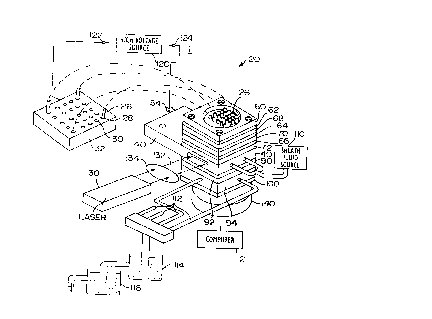
Reference is first made to Figs. 1 and 2, which show an
analyzer 20 for analyzing an organic sample such as DNA. The analyzer 20
includes a sheath flow cuvette 22 enclosing the ends 24 (shown in dotted
lines in Fig. 2) of a set of capillary tubes 26.
The capillary tubes 26 are arranged in a generally rectangular
array, which in the example shown is an array of five tubes by five tubes.
The other ends 28 of the capillary tubes 26 terminate in twenty-five wells
30 of a conventional microtiter plate 32.
The capillary tubes 26 are conventional fused silica capillaries,
having an inner diameter of about 50Nxn and an outer diameter of about
150~,m, and are available from many conventional commercial sources.
The fluid in the wells 30 contains the samples to be analyzed (a different
sample in each well).
The ends 24 of the capillaries 26 which are located in the
cuvette 22 are positioned in an interior chamber 34 (Fig. 2) in the cuvette
22. The capillary ends 24 are held in position in the chamber 34 in a leak-
proof manner by a sandwich construction for the cuvette 22. 7Che
' sandwich construction will now be described.
The cuvette 22 includes a rectangular stainless steel plate 40,
which in one example was 29mm by 59mm and 5mm thick, with a l3mm
by l3mm opening 42. The opening 42 defines the bulk of the chamber 34.
CA 02245167 1998-07-30
WO 97/28443 PCT/CA96/00858
-6-
Plate 40 is also shown in Figs. 3 to 5. Two grooves 44 are milled in one side
of the plate 40, each about 4mm thick, extending from the opening 42 to
the edges of the plate. Two glass windows 46 each l2mm by 4mm are
glued into the groove bordering each side of the opening 42. The windows
46 are for a laser beam to enter and leave chamber 34, as will be described.
Plate 40 also includes four bolt holes 48 arranged in a square
configuration, through which bolts 50 {Fig. 2) may pass, to hold the
sandwich construction together. Plate 50 also includes two openings 52 to
allow the cuvette to be mounted on a mounting fixture (not shown) and a
tab 54 for connection of a ground wire (as will be described}. Plate 40 also
includes two tubular openings 56 (e.g. 3.3mm diameter) for sheath fluid to
enter chamber 34.
Mounted above the plate 40 is a stack comprising a stainless
steel cap 60 (also shown in Fig. 6), and three identical stainless steel shims
62, 64, 66 (Fig. 7), each separated by identical plastic (e.g. TEFLONTM)
washers 68, 70 (Fig. 8). A third identical plastic (e.g. TEFLONTM) washer 72
separates shim 66 from metal plate 40. The washers 68, 70, 72 help to
prevent leaks. Each washer in the example described is 29mm by 29mm
and 1mm to 2mm thick, each with a central circular opening 74, and four
bolt holes 76 for bolts 50.
Each stainless steel shim 62, 64, 66 includes four bolt holes 78
and a five by five array of holes 80 for the capillary tubes 26. The holes 80
may be formed by any known technique, e.g. drilling, ultrasonic molding,
or electroforming, and are each of the same diameter as the outer capillary
diameter (e.g. 150~.rneter). The holes 80 are preferably normally spaced as
closely together as possible, consistent with having sufficient material
between them to provide sufficient mechanical strength to hold the ,
capillary tubes. Preferably the spacing between holes 80 does not exceed
about one outer diameter of the capillary tubes. If the spacing is too large,
it may be difficult to focus the Iaser beam (to be described) over the large
area defined by widely spaced capillaries, and collection of light from a
Iarge area may also be more difficult.
CA 02245167 2003-05-28
WO 97/28443 PCTlCA96/00858
-7-
Located in the central opening 74 of the washer 70 is a circular silicon
rubber disc or gasket 84 (FIG. 9), which is of slightly greater thickness than
that
of washer 70. The disc 84 also contains a five by five array of holes 86 for
the
capillary tubes 26. Each hole 86 may be formed by piercing the disc 84 with a
capillary when the disc is assembled in the stack, thus ensuring that holes 86
will
be of the same diameter as the outer capillary diameter. When the stack is
assembled, the rubber disc 84 is compressed between the adjacent metal shims
64, 66, thus providing a leak proof seal around the capillary tubes 26 at the
top
of the chamber 34.
Looking below the plate 40, a further thin metal shim or barrier member
90 is glued to the bottom of plate 40 (and to the windows 46). Shim or barrier
member 90 is exactly the same as shims 62, 64, 66 and has the same bolt holes
78 and the same holes 80, which are precisely aligned with holes 80 in shims
62,
64, 66.
Located below barrier member 90 is another plastic (e.g. TEFLONTM)
washer 92, and below that a second stainless steel plate 94, also shown in
FIG.
10. Plate 94 in the example shown was 29 mm by 29 mm, by 4 mm thick, and
had an interior opening 96 which was 18 mm by 18 mm. Four tubular drains 98
(e.g. 2.3 mm diameter) extend from each side of opening 96. Glued to the
bottom of plate 94 and covering opening 96 is a glass window 100. The space
between shim 90 and window 100 defines a lower chamber 102, which in the
example shown was (including the 1 mm to 2 mm thickness of washer 92)
approximately 5 mm to 6 mm thick.
A sheath fluid is supplied from source 110. The sheath fluid is chosen to
have the same or a similar index of refraction as the aqueous buffer used to
prepare the polymer mixture which fills the capillary tubes 26. The sheath
fluid
enters the chamber 34 via openings or inlets 56 in the plate 40, and is pumped
from source 110 in a non-pulsating flow, e.g. by a simple gravity feed (under
a
head, for example, of about 5 cm) or by a very low pulsation pumping means
such as a high quality syringe pump (not shown). The sheath fluid flows
through
CA 02245167 2003-05-28
WO 97/28443 PCT/CA96/00858
- 7a -
the holes 80 in the barrier member 90 and into the lower chamber 102, from
which it drains via the
CA 02245167 1998-07-30
WO 97/28443 PCT/CA96/00858
_g_
four tubular openings 98 and drain tubes 112. As described in our above
mentioned patent, droplet formation should be avoided, e.g. by draining
the sheath fluid (including the flow from the capillary tubes as will be
described) into a beaker 114 in which drain tubes 112 are submerged.
Beaker 114 in turn drains into beaker 116, which drains to waste.
A high voltage source 120 is provided, having one pole 122
connected through conductive plate 32 to the fluid in each of the wells 30.
The other pole 124 of the source 120 is connected to the tab 54 of the plate
40, which tab is grounded for safety reasons. The source 120 provides a
driving voltage of e.g. 30kV which, via the fluid in chamber 34, appears
across the length of the capillaries 26. As is well known, the electric field
created by the voltage source 120 causes fragments of sample DNA from
the wells 30 to migrate through the matrix or gel in the capillaries 26. At
the ends 24 of the capillary tubes 26, the sheath fluid entrains sample fluid
from the capillaries, in the form of individual filaments 126 of fluid, as
best shown in Fig. 11. The filaments are aligned with holes 80 in barrier
member 90 and pass through those holes 80 together with the sheath fluid.
In the lower chamber 102, the filaments 126 mingle with the sheath fluid,
and the mixed fluids are drained via openings 98.
A laser 130 or other source of collimated electromagnetic
radiation provides a collimated beam 132 of light that is aligned to pass
from a focusing lens 134 into the chamber 34, as close as possible above the
barrier member 90. Preferably the laser beam 132 is elliptically shaped, to
illuminate all of the sample streams simultaneously. Alternatively, the
beam 132 may be split into a set of parallel beams with appropriate optics,
with one parallel beam per row of capillaries. Fluorescence is excited in
the chamber 34, above the barrier member 90. The fluorescence passes ,
through the holes 80 in barrier member 90, through the glass window 100
at the bottom of lower chamber 102, and through a two element air-spaced
condenser lens 136, typically operated at unit magnification. The
condenser 136 images the fluorescence onto a photodetector 138. A
spectral filter shown diagrammatically in dotted lines at 139 may be used to
CA 02245167 1998-07-30
WO 9T/28443 PC~'/CA96/OOSSE
-9-
isolate fluorescence from specific dyes. The filter 139 can be a tunable
filter,
or a set of filters on a rotating wheel, or can be a grating or a prism. T'he
filter 139 is preferably placed in the space between the lenses of condenser
136 since that is where the light is relatively well collimated and the light
rays strike the filter roughly at right angles. If the filters were placed in
t:he
diverging portion of the beam either before or after condenser 136, then
the spectrum of the transmitted Iight would vary across the aperture of the
filter, since the transmitted spectrum depends on the incident angle,.
Preferably the photodetector 138 is a large area CCD chip of a
CCD camera 140. The area of chip 138 is as large as or larger than the area
of the capillary array, thus providing high collection efficiency. (If desired
the window 100 can be a lens or can even be a part of the CCD camera 140.)
The chip 138 is connected to a computer 142 so that the chip output can be
analyzed.
The arrangement shown has several advantages. One
advantage is that the photodetector looks straight, end-on, at the
capillaries, so optical correction elements are not needed to obtain a high
quality picture of the fluorescence. In addition, the path length through
the fluid is the same for the fluorescence from each filament or sample
stream 126, so no distortion is introduced due to differing path lengths.
The barrier member 90 ensures that the individual sample filaments 126
will remain intact above the barrier member 90 (i.e. in the region where
they are being illuminated), so that the fluorescing spots can be looked at
end-on, even though below the barrier member 90 the filaments 126 lose
their individual character. The flow in lower chamber I02 should
preferably be non-turbulent, but with the low flow rates used, turbulent
flow would be highly unlikely to occur. (For DNA sequencing, where
there is no bulk solvent flow through capillaries, but instead the anallyte
~ molecules are drawn from the tip of the capillaries and entrained in the
sheath fluid stream, the flow is essentially only the sheath fluid flow,
which may typically be about 10 microliters/minute per capillary, or e.g.
0.25 milliliters/minute for a 25 capillary design and 1 milliliters/minute
CA 02245167 1998-07-30
WO 97/28443 PCT/CA96/00858
-10-
for a 96 capillary design. In non-DNA analysis, the flow would be
augmented by a sample flow rate of typically 0.1 to 1 rnicroliters/minute
from each capillary.)
Secondly, the sandwich construction shown holds the ,
capillaries on fixed centers in a leak-proof manner, so there is no need to
worry about proper alignment of the capillaries.
Thirdly, the barrier member 90 blocks a substantial amount of
scattered laser light from reaching the photodetector, e.g. the CCD chip 138.
The reduced fluorescence background allows a higher signal to noise ratio
and improved accuracy of results.
While close spacing of the capillaries is normally preferred, if
desired they can be spaced further apart (e.g. by more than one capillary
outer diameter), and a diffraction grating (shown at 144 in Fig. 11) can be
inserted between window 100 and the camera 140, to disperse the emission
spectrum from each fluorescing spot, to help determine DNA sequences or
for other analysis.
In assembly of the cuvette 22 shown in Fig. 2, the upper part
of the cuvette is first assembled, consisting of shims 62, 64, 66 and their
associated washers. This assembly is placed on a plate such as bottom plate
94, which itself is placed on a flat, smooth, solid surface. The capillary
tubes 26 are then threaded through the holes 80 in shims 62, 64, 66, in the
process creating the holes in rubber washer or disc 84, until the capillary
tubes reach the bottom supporting surface. This ensures that the ends 24
of the capillary tubes 26 lie in a plane. When the entire chamber is
assembled, the plane in which the capillary ends 24 Iie is, in the example
shown, about lmm above the barrier member 90.
While a rectangular array of capillary tubes 26 has been
shown, if desired other forms of array can be used, e.g. a configuration as
shown in Fig. 12, where alternate rows 146 are offset so that they are
located in the spaces between adjacent rows 148. More dense packing is
advantageous for efficient illumination and detection. If the spacing is too
large, there may be poor optical excitation and collection efficiency, since
it
CA 02245167 2003-05-28
WO 97/28443 PCTlCA96/00858
-11-
is difficult to focus a laser beam over the large area defined by widely
spaced
capillaries, and it can be difficult to collect fluorescence from widely
spaced
capillaries. However the use of a large area CCD chip 138 will solve this
latter
problem, and the use of the opaque barrier member 90 blocks scattered light
which can be generated by a non-ideally focused laser beam.
In the embodiment described, the driving force created by the electric
field applied across the capillary tubes 26 is limited to the capillaries and
the
sample stream filaments are drawn from the capillaries by the sheath fluid. If
desired other driving means may be used for the sample, as described in our
above-mentioned patent. For example the sample can be forced through the
capillary tubes 26 by an appropriate pump, as in flow cytometry. In addition
the number of capillary tubes in the array used can vary. For example 96
capillaries in a 12 by 8 array may be used, to interface with a 96 well
microtiter
plate. By way of further example, 864 capillaries may be used in a 36 by 24
array to interface with an 864 well microtiter plate. Other arrays can be
designed as needed.
While preferred embodiments of the invention have been described, it
will be understood that modifications may be made within the spirit of the
invention and all such modifications are intended to be encompassed by the
appended claims.