Note: Descriptions are shown in the official language in which they were submitted.
CA 02245553 2003-05-29
English translation of German language international patent application
WO 97/29355, published August 14, 1997
Process and apparatus for contactless micro injection sorting and harvestinf;
usin;~ laser radiation of biological o~ects di~osed in a planar manner
The invention concerns a process and apparatus for micro injection, sorting
and harvesting of single biological objects. The objects are disposed side by
side on a fixed planar carrier. This process is suitable for the micro
injection
of specific substances into individual biological objects, for example cells,
and
subsequently to sort them. Further with this process single objects can be
specially separated from a very great number of objects (e.g. 105 to
10°) and
singled out. The separation of heaped up cells/cell clumps as a unit is also
possible. Also the process can be used for the separation of specific cells
from
a tissue sample. A precondition for this sorting process is the prior
recognition and selection of the objects concerned on the basis of specific
qualities (e.g. by colour, fluorescence marking or by radioactive marking).
Within the term "biological objects" in the context of the present application
is meant mainly live or trapped biological cells or parts of cells.
For injection of material into living cells usually micro capillary tubes were
used that were usually controlleei by usually a pneumatic or hydraulically
moveable micro manipulator. The desired substances are injected into the
individual cells under great mechanical stress. The manufacture of sterile
micro capillary tubes is time consuming and expensive.
Tsukakoshi et al. (1974) and Tao et al. (1987) used a focused laser beam to
drill
small self healing holes without mechanical contact in a cell membrane. The
short opening time is sufficient for the material that is dissolved in the
surrounding fluid fo penetrate the cell. A larger efficiency in laser micro
injection of genetic material was achiYVeci when the laser bl;~sts the hole
directly into the ceii.
CA 02245553 1998-08-04
2
The problem with this method is that for a precise laser micro injection in
the
submicron region the target objects have to be approached with an accuracy of
the order of nanometers in the lateral, that is in the X/Y direction, and also
in
the vertical, that is in the Z direction. For an automated micro injection the
relevant target cells have to be recoginsed via an image recognition process,
then positioned in the line of fire of the laser and most importantly then
exactly focused in the Z direction.
A further problem is to isolate or to prepare the successfully injected cells
from the other cells for further tests.
For the separation of single biological objects there exist optical methods,
such
as the optical tweezer in which the object moves in an aqueous solution (K.
Schiitze, A. Clement-Sengwald, Nature 667 (vol. 368) 1994). On account of the
very small transfer of force this method is limited to objects that are able
to
move freely in the solution. As the sorted and unsorted objects are in the
same solution a separate cultivation is only achievable with extra expenditure
of effort. For a separate cultivation of cells they have to be separated or
sucked apart by another method, for example, with micro capillary tubes.
Adherently growing cells or trapped cells on a dissected sample can be
separated with a fine needles that are moved by means of a micro
manipulator. In this situation the cells are contacted c.iirectly and thereby
can
be mechanically stressed. In addition there is the danger of contamination by
undesired cell material. Both methods are comparatively time consuming
such that they are not suitable for the manipulation ~f a large number of
objects.
CA 0_2245553 1998-08-04
3
For the separation of single cells from a large number (>106) dispersed in a
fluid there are commercially available devices for separating and sorting
biological objects. While in the fluorescence activated cell sorter (FACS)
electro static principles for the spatial separation are used the magnetic
activated cell sorter (MACS) operates with magnetic forces. In these systems
the cells are however, not disposed side by side on a planar carrier. In
addition both of these methods have the disadvantage that many of objects
can only be separated to a limited extent (FACS) or even not be separated
from each other at all ,(MACS).
The above described methods cannot release single cells from a cell
plaque/cluster such as a tissue or from a liistological tissue preparation.
An apparatus is known from JP-A-05 076342 for catching and collecting
microscopic objects such as cells. In this arrangement the object of interest
is
captured by two continuous lasers disposed opposite each other and is held in
place. Then it is guided to a collection apparatus by a third continuous laser
along its beam. From the article "Cell surgery by laser micro-dissection: a
preparative method" in Journal of Microscopy , Vol. I07 (1976) an apparatus is
known which used a quasi-continuous NZ-Laser for micro-dissection, and a
microscope is used for the subsequent observation of the selected object.
CA 02245553 1998-08-04
3a
Further there are processes known under the name of "ablative photo
decomposition" in which a directed removal of polymer material is achieved
using pulsed UV-lasers particularly Eczema lasers. This process can in the
wider sense be seen as an etching process. A similar process which however
uses a continuous UV-laser is described in US patent 5,211,805. This process
is
stated to be suitable for the industrial processing of technical polymers and
for
the biomedical treatment of biological tissue. A sorting principle is used
that
involves the destruction of undesirable biological objects on a carrier by
means of a laser radiation of a high dosage, while the selected (desired)
abject
remains behind (US 4,624,915). This procedure is relatively troublesome for
selecting a single object from a large population.
The object of the invention is among other things, is a directed manner, to
load biological objects with a selective substance by means of contactless
laser
micro-injection and subsequently to sort the successfully injected objects.
The
biological objects can be distributed side by side on a fixed planar carrier,
for
CA 02245553 1998-08-04
4
example a polymer carrier foil. In this connection the selection process
should be conductible as quickly as possible (<IOs) and without contact, e.g.
in
a separate sterile chamber. In addition the procedure must be very reliable
and therefore be able to be automated in a simple way. At the same time the
biological objects should have a high survival rate and as a rule remain
unchanged. The objects should not be damaged or injured by the micro
injection procedure and the separation process.
The Task of micro injection is conducted in accordance with the invention in
an automated manner in that an object field on the cover glass or a carrier
foil
is removed by a meander shaped scan with the motorized computer
controlled microscope carrier. In this way the single target cell (target
object)
is selected by means of an image analysis process using colour or pattern
recognition and by means of X/Y displacement is brought into the region of
the laser shot. Subsequently the cell or the desired cell structure is brought
into focus (in the Z direction), and with a directed laser shot the cell is
micro
perforated. The movement of the object in the X/Y direction can either be set
via the axis of the microscope table and the Z direction by a third axis of
the
microscope table or can be set via the focus adjustment of the objective on
the
microscope itself by computer control. Alternatively an adjustment onto the
target object can be achieved even with a fined microscope table by means of a
3-dimensional computer controlled laser focus movement. In order to sort
out the successfully micro injected cells subsequently from the remaining cell
lawn the selection process that will now be described is used.
The task of the selection and separation is achieved in accordance with the
invention in that an object field of the carrier foil on which the selected
biological object or the histological dissection is disposed, is cut out with
a
laser beam and transferred by a laser induced transport process to a
collecting
CA 02245553 1998-08-04
substrate which is directly above or below the carrier foil. The solution in
accordance with the invention is that the biological object first is either
identified visually with the eye, or by means of a colour or pattern
recognition of an image analyzing process and subsequently cut out in a
surrounding that suits the sample, for example also in a circle together with
the carrier by a laser beam and subsequent to that flung out of the carrier
foil
and onto a collector disposed in the vicinity. It was observed that the
separated out object field is always flung in the direction of the laser beam.
A
physical explanation for this laser induced transport process lies possibly in
the photokinetic impulse that is transferred from the laser beam to the cut
out object field and which is thereby responsible for the acceleration. The
spacial separation of the biological object is attributable thus in this
process to
the cutting out of the desired object field together with the previously
selected
object and its conveyance to the collecting substrate that is disposed in the
vicinity.
The cutting out of the object field can advantageously be achieved by
providing that the laser beam is guided in a closed curve around the
biological object that includes the object field by means of a relative
movement of the laser beam and the carrier foil. Alternatively, the
separation of the object field, by analogy to a stamping process can also be
conducted whereby the cutting area including the object field is
simultaneously exposed through a slit mask that is illuminated by the laser
beam and projects onto the carrier foil.
As already mentioned the collector substrate should be in the immediate
vicinity of the carrier foil so that the distances to be transported during
the
separation process are short. Good results were achieved with distances of 0.5
to IO mm, preferably I to 3 mm.
CA 02245553 1998-08-04
6
The diameter of the object field with the selected object can, on account of
the
extraordinarily precise cutting process, be selected to be in the region of 10
micro meters to 20 micro meters.
For the cutting out preferably a UV laser is used, and at the same time fhe
focus of the laser beam on the carrier foil is reproduced as an image thereon.
The carrier foil consists of a UV absorbing polymer foil with a thickness
between 5 micro meters and 15 micro meters that's absorption behavior is
matched to the wavelength of the UV laser or at least has an absorption
maximum in the region of the laser wavelength. Polymer foils have proved
to be partiv:uiariy jultabie that ~oWtaiu 5 weigh t o of au aroWatiC or yartiy
aromatic polycondensate. The geometrical form of the collector substrate is
relatively uncritical. Suitable, for example is a relatively thick foil or
plate
that is disposed at a distance of 0.5 to IO mm above or below the carrier film
and parallel thereto. The collector substrate can however be constructed in
the form of a pot shaped holder. In particular micro centrifuge containers are
recommened of the type that are used in molecular biology, for example, a
micro titration plate with 90 to 500 wells.
In accordance with a special embodiment a plate or fail is provided with an
adhesive coating. By means of such an adhesive coating the object field that
has been propelled can be retained on the collector substrate.
For the purpose of recognition and selection of the desired biological object
on
the carrier foil the method of fluorescent spectroscopy can preferably be
employed.
CA 02245553 1998-08-04
7
Alternatively the biological object can be recognised and subsequently
selected
with the help of known histochemical colour reactions or morphologically
perceptible changes, either visually or by an image analysis procedure on a
computer (digital ?).
In accordance with a further development the biological objects are coated
~Nith a fluid nutriant or buffer medium that is transparent to the laser
radiation. In these circumstances the selected object fields can in accordance
with the invention be cut out and released.
The separation process in accordance with the invention is carried out to
advantage in a closed system. To this end the carrier foil with the objects to
be
sorted and also the collector substrate are housed in a closed container that
has a UV transparent window for the laser beam.
In what now follows, the invention will be explained in more detail with
reference to the drawings and embodiments.
The drawings show:
Fig. I. schematically a carrier foil with adhered bacteria;
Fig. 2. the fundamental construction of apparatus for carrying out the
process according to the invention;
Figs. 3/4. the underlying sorting principle;
Figs. 5/6 the construction of the upright/upside down microscope; and
Fig. 7. the deposition of the object in a receptacle.
Figure 1 shows for example a population of bacteria I that is distributed in a
planar manner on a 5 micrometer thick foil of polyacrylate 2 (carrier foil).
For
CA 02245553 2003-05-29
8
the sorting process the carrier foil 2 is placed in a displaceable table 3 and
held
mechanically. This table in accordance with figure 2 is the objective table in
an inverting microscope 4 and can, for example, be pc,sitionFd by means of
computer controlled stepping motors in X/Y directions (in a horizontal
plane). In the displaceable table 3 and opposite the carrier foil 2 at a
distance
of 1.8 mm there is retained a plate shaped collector substrate 5, upon
movement of the displaceable table 3 therefore the carrier foil 2 and the
collector substrate 5 move together at right angles to the path of the
radiation
(Z direction) in the microscope. The moveable table 3 with the carrier foil 2
and the biological objects 1 that ire disposed thereon and the collector
substrate 5 are surrounded by a closed housing that is provided with a
window transparent to ultraviolet light for the laser beam (not shown ). In
this way the process can be carried out in a hermetically sealed system.
As the carrier film for the biological objects is an ultra violet at~sorbing
polymerfilm of thickness 5 micro meters to 15 micro meters that contains at
least 5 weight percent of an aromatic or pertly aromatic polycondensate, for
example polycarbonate, poUyurethane, polyacrylate, co-polyester, polyester
carbonate or a blend ~f these polycondens~tes and other thermo-pl~~tic
materials. Other types of foil are conceiv~ible within this context.
For the spacial separation, for example of single bacteria from the reroverecl
population a UV--laser beam 6 of wavelength 337 nano meters of a halsed N' -
laser 7 is used. The laser 7 delivers approximately 300 mica, joules c~f
radiation energy with a maximum pulse frequency of 20 Hz. Also s~~itable are
other pulsed or continually operating lasers for example an Eczema laser with
s;
a wave length of 193, 248 or 308 nm or a frequency quadrupled hld;YAC~-laser
with a wave length of 266 nrn or a frequency doubled AIZ-Icon laser with a
wave length of 244 nm, or 257 nm. The laser beam 6 is projected onto, the
*Trade-mark
CA 02245553 1998-08-04
9
carrier foil 2 in a dot shape via a dielectric beam splitter 8 and a reducing
microscope objective 9 (reduction 63 x, aperture NA = 0.9 or also other
objectives). This dot has a diameter of at least I micrometer.
A circular or closed cutting line with a diameter of, for example, 10
micrometers is generated around these selected bacteria by a corresponding
movement of the moveable table 3 in the horizontal plane. The area defined
by the cutting line is in this case the object area. The laser beam remains
stationary during the following described cutting process.
In the experiment the relative speed of movement of the laser beam to cut
out a closed area was 5 micro meters per second. A sharp edged narrow
bounded cutting Line was generated and as the laser beam returns to the
starting point of the cutting line a separation of the cut out area occurred.
The
area was propelled from the carrier foil 2 onto the adhesive coated collector
substrate 5 by a laser induced transport physical process of which has not yet
been explained in detail. There it remained captured. Another possibility is
to use as the collector substrate a conventional micro titration plate with,
fc~r
example 96 wells. By "wells" it is understood in the field of pharmaceutical
research, to mean the cut outs or round depressions in the micro titration
plate for receiving samples for testing and with a diameter of about ~I mm and
a depth of about 6 mm.
The sorting process will be explained again with reference to figures 3 and 4.
Figure 3 shows a bacteria IO to be separated on a carrier foil 2. On account
of
the circular movement of the moveable table 3 a cutting line 11 of width of
about 5 to 7 micro meters has already been drawn by a fixed position laser
beam 6 in a cutting process. The width of the cut depends on the absorption
properties of the foil. In this area the foil material is completely removed.
CA 02245553 1998-08-04
Immediately after the cutting line 11 has completed a closed circle the
separated piece of foil (object field) 12 with the bacteria IO on it is
propelled in
the direction of the laser beam and as shown in figure 4 catapulted onto the
adhesive tape 5 (the collector substrate). A circular hole 13 remains in the
carrier foil 2.
In place of a moveable table a stationary object table can be used and the
laser
beam can be made to draw a circle around the selected bacteria by a suitable
optical deflection element disposed in the laser beam.
A prerequisite for the laser separation is that the object field to be removed
from the carrier foil be previously recognised and selected. A method that is
frequently used in pharmacological research for the recognition and selection
of particular cell structures is fluorescent spectroscopy. For this purpose a
commercially available fluorescence microscope is used. This presupposes
that the cells or bacteria for the intended selection generate a significant
fluorescence signal that can be used as the differentiating criterion. With
the
help of a scanning program equipped with a search algorithm the moveable
table 3 can be so controlled that automatically and in turn the areas with
selected bacteria can be positioned as object fields centrally in the field of
view
of the fluorescence microscope and subsequently cut out.
For the recognition of biological objects in sections of tissue the known
histological colour reactions or morphological changes that are observable
under the microscope can be used.
The carrier foil 2 can also be coated with a laser radiation transparent
nutrient
or buffer solution, for example a PBS buffer solution.
CA 02245553 1998-08-04
11
In accordance with a further development of the process, the object of the
invention can be achieved by using the radiation pressure of the laser beam to
catapult the desired particle or biological object itself directly from the
upper
surface of the fixed planar carrier (object carrier or petri dish) and to
catch it in
a suitable container, i.e. the separation of the biological object is thus
possible,
with or without, the simultaneous release or cutting out of the area of the
carrier foil that is supporting the object. This occurs in accordance with the
further development in the following manner; that will be described with
reference to the accompanying figures 5 to 7.
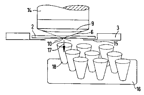
When using an upright microscope 14 in accordance with figure 5 a suitable
object carrier 2 (thickness approximately 170 micro meters) is placed upside-
down on the support 15 of a microscope table 3 especially designed for laser
micro manipulation, i.e. the biological object IO is on the under side of the
object carrier 2. The object can for example be a histological tissue section
of
only a few micro meters thick or adhering chromosome or DNA preparation.
The microscope table 3 is equipped for a displacement in 2 axes (for X/Y
movement) or 3 axes (for X/Y/Z movement) and is provided with a support
15 for, for example, the object carrier 2 or petri dishes. The table 3 is thus
motorised by means of a suitable drive that can be moved under computer
control in a known way, for example by a computer mouse (or joystick). The
hybrid stepping motors of the axis drives work with a high precision. The
smallest step is 20 nm the maximum traveling distance of the microscope
table 3 can be up to several cm, for example IO cm. The precision with which
stored points can be found again is less than 400 nm. Speeds can be chosen for
movement of the microscope table from a few nm to several mm per second.
By means of a so-called "framegrabber" actual microscope pictures, that are
taken via a video camera, are shown on a monitor and can be graphically
CA 02245553 1998-08-04
I2
overlaid with computer functions, for example command functions, control
functions, test points etc. (Video-overlay).
For the laser micro manipulation only thin object carriers (about I70 micro
meters thick) or petri dishes with thin (approximately 25 micro meters) gas
permeable foil (so-called "petri-perm" dishes) are suitable. As laser micro
manipulation in the nano meter region sets a very high requirement on a
precise holding and transport of the samples, the support 15 has been
especially configured. With a thin object carrier 2 there is the danger, for
example when an oil emersion objective is used, of slight bending and
thereby a good focus cannot be achieved. In order to prevent this the object
carrier 2 must be supported on at least three sides of the holder. The two
narrow sides of the object carrier 2 each can be held tightly with a spring
clip.
A further necessity peculiar to the laser microscopy is the exact adjustment
of
the sample holder (object carrier 2 or sample holder 15). It must be provided
that the sample is always the same distance from the tip of the objective 9
over the whole range of movement of the carrier (approximately 5 - 10 cm).
First, suitable biological objects 10 are selected optically with a lower
magnification. As soon as all objects of interest are stored in the computer a
higher magnification objective is selected. The displacement of the beam due
to the change of objective (chromatic aberration) is compensated for by a
correction function acting on the stored values that is automatically applied
to all stored points.
The computer then drives to the first object I0. A microscope image that is
viewed by a video camera is displayed on the computer monitor, which is not
shown in the figures. A marker on the monitor shows the position of the
focus of the laser beam. The microscope slide is moved either by hand
CA 02245553 1998-08-04
. I3
(controlled by a mouse or joystick), or travels automatically under the
control
of a computer program in accordance with a predetermined pattern in
essentially circular or a spiral shape around the chosen object 10. The marker
on the monitor can be regarded as a pointer with which the outline of the
desired biological object is drawn. If at the same time the laser, with a
pulse
frequency of around 15 to 20 Hz, fires, all material that is in the line of
fire in
the region of the "marker" is removed or destroyed. The extremely focused
laser beam "draws" a fine line of approximately 500 nm width around the
desired object IO and separates it thereby from its surroundings. In the case
of
cells in the histological section a desired cell can be released from the
plaque/cluster by means of this procedure and loosens from the substratum
in the shape of the object field given by the cutting line. By means of the
above mentioned spiral shaped circulation around the chosen target cell the
region around the cell that is left free can be enlarged.
The laser used in the procedure described is for example a pulsed compact
nitrogen laser with a high quality beam (wave length: 337 nm, pulse length 3
n sec, pulse frequency: from 10 to 30 Hz). Other lasers are also envisaged, as
long as the wave length of laser light used does not negatively influence the
biological material. The laser beam itself, i.e. its source remains preferably
stationary. However, the laser can also be moved in the X/Y direction with a
radius of several micrometers relative to the plane of the object, i.e. in the
final analysis it is only important that the laser beam and the plane of the
object (the microscope table) be moved relative to one another.
The object that has been isolated in this manner can then, faster and safer
than in the state of the art (for example with a needle), be automatically
catapulted into a test vessel (trajectory 17) and most importantly without any
CA 02245553 1998-08-04
14
contact, i.e. if necessary also completely sterile, with a further aimed laser
shot.
To this end it is necessary that a second sample support 16 (for example to
retain a collector vessel 18 or a microtitration plate), driven by two
motorised
and computer controlled axes, is moved in such a way beneath the first
support 15, and the object 10 that has been isolated by Ehe laser is exactly
positioned above the collecting vessel 18. F-lere is accordingly a high
precision
of the motor movement a requirement for a clean collection of the desired
object. A single aimed laser shot (possibly defocused) catapults the selected
biological object 10 and/or the cell in the direction of the beam (trajectory
I7)
into the collecting vessel I8. Afterwards a new object can be cut out and the
entire process repeated.
To accelerate Ehe collection procedure all of the desired objects can first be
released by the laser. AfEerwards Ehe collection vessel can be moved into
position beneath the microscope Eable. The microscope slide then moves to
revisit each of the stored laser micro dissected objects. Each shot of the
laser
then catapults a subsequenE one of Ehe objects into respective fresh (new)
receiving vessels 18, figure 5, Shat coordinate with Ehe movement of the
microscope slide 3. Several objects could also be collected in one container.
With an upside down microscope (figure 6) a sticky plate adhesive foil, agar
coated carrier etc. can be used, which is moved only a few micrometers
directly above the object thaE has been cut out to catch the catapulted away
object. This can, for example be a sticky member that is then thrown by
means of robot arm 20 into a suiEable container, figure 7. The robot arm 20
then picks up a new sticky member (see figure 6).
CA 02245553 1998-08-04
,~ I5
The advantage of the laser induced separation process of cells is the
selective,
and at the same time rapid, manipulation of single cells in comparison with
the prior art. On account of the simple principle the procedure is very robust
and easy to use and thereby suitable for the computerised automatic
separation of a large number of biological objects. An important advantage
considering safety aspects is that the separation process can be conducted in
a
hermetically sealed system so that the environment can be protected from
pathogenic cells. Also the cells are protected from contamination from the
environment.
The process is suitable mainly in the sub-fields of biotechnology, molecular
biology and pathology where specific cell types have to be refined. For
example transfused cells can be identified with the green fluorescent protein
(GFP) as reportergen. Marshall et al. (Neuron, Vol. 14, 211-2I5 (I995) uses
for
example the GFP method to assess the expression of ion channels. In
accordance with a further application, for example, glea cells can be
separated
from brain tissue samples for neurological experiments. For this, fluorescent
marked antibodies are used to identify the cells. For diagnosis tumor cells
can
be isolated from tissue samples that for example, show morphological
changes. In this case the tissue slice is placed on the carrier foil. The cell
to be
isolated is then cut from the tissue and the foil substrate separated so that
the
cell possibly with the substrate material is transferred to a support. The
cells
are then subsequently histologically analysed. In addition bacteria with
specific properties, for example that produce citric acid can be tested for
their
efficiency by means of suitable pf-i sensitive coloured rings of a suitable
indicator and subsequently sorted.