Note: Descriptions are shown in the official language in which they were submitted.
CA 02246961 1998-09-10
=. ~ _
PATENT
P-4012
TITLE OF THE INVENTION
SAMPLE PROCESSING METHOD USING ION EXCHANGE RESIN
BACKGROUND OF THE INVENTION
The field of the present invention broadly relates to nucleic acid
hybridization and/or
amplification. More specifically, the present invention relates to the
reduction of substances in
samples which interfere with nucleic acid hybridization and/or enzymatic
amplification events.
Such events include nucleic acid probe hybridization to determine the presence
and/or amount
of a target nucleic acid, nucleic acid primer hybridization for nucleic acid
amplification
processes and enzymatic activity including nucleic acid extension, nicking
and/or cleavage.
The present invention also relates to the removal of fluorescent compounds
from samples,
which increases sensitivity of fluorescent detection assays. Additionally, the
invention relates
to the stabilization of such samples which permits increased room temperature
storage times.
The present invention also relates to concentration of organisms.
Nucleic acid amplification processes such as strand displacement amplification
(SDA),
polymerase chain reaction (PCR), ligase chain reaction (LCR), nucleic acid
sequence based
amplification (NASBA), transcription mediated amplification (TMA) and others
are used to
create multiple copies of a particular nucleic acia sequence(s) of interest
(target sequence)
which is present in lesser copy number in a sample. However, a number of
substances
commonly found in such samples interfere with nucleic acid amplification
processes. Similarly,
such substances may interfere with or inhibit direct nucleic acid probe
hybridization reactions
used for the detection of target nucleic acids.
An example of a nucleic acid amplification inhibitory substance is porphyrin
compounds
derived from heme and hematin which are both commonly found in blood samples
and inhibit
PCR. (PCR TechnoloQV, Stockton Press, Henry A. Erlich, Ed. pp 33-34, 1989).
Protocols
CA 02246961 1998-09-10
~ ~ .-.. = ,._,.
PATENT
P-4012
using osmotic lysis and pelleting of nucleic and cell debris have been used to
reduce the
amount of these inhibitors.
Salivary samples have also been reported to contain PCR inhibitory substances.
Ochert
et al., PCR Methods and Applications 3, 365-368 (1994). Although the
inhibitory substances
were not identified, it was found that extended microwaving or boiling of the
salivary sample
totally removed PCR inhibition.
Frickhofen and Young, J. Virol. Methods 35, 65-72 (1991), report that heating
of
serum samples for 45 seconds at 70 C improves PCR amplification of viral
nucleic acid
sequences. This improvement is theorized to be due to heat inactivation of
serum enzymes
such as aprotinin, leupeptin PMSF and pepstatin which are believed to be
inhibitory to PCR
processes.
Another approach for removing PCR inhibitory substances from serum prior to
amplification of a viral nucleic acid sequence is taught by Zeldis et al., J.
Clin. Invest. 84, 1503-
1508 (1989). This approach involves adsorbing the virus to antibody coated
microparticles,
washing the microparticles, and then destroying the remaining proteins which
may be inhibitory
to PCR with proteinase K.
In attempting to detect Treponema pallidum in amniotic fluid, fetal and
neonatal sera
and cerebrospinal fluid by PCR, four different processes were attempted to
remove PCR
inhibitory compounds. Grimprel et al., J. Clin. Microbiol. 29, 1711-1718
(1991). Briefly, the
four processes for removal of PCR inhibitory compounds were: (1) a boiling
method wherein
sample in a tube was placed in a boiling water bath for 10 minutes, cooled on
ice, and then
centrifuged; (2) a low-spin separation method wherein sample was added to
sterile phosphate
buffered saline and subjected to a series of centrifugations, then the pellet
was resuspended and
boiled for 10 minutes, after which it was cooled on ice; (3) an alkaline lysis
extraction method
wherein sample was boiled for 1.5 minutes in 1 M NaCI, I N NaOH and 0.1% SDS,
then
neutralized with 0.5 M Tris-HCI (pH 8.0), and then subjected to a series of
extractions with
phenol and chloroform-isoamyl alcohol, and precipitated with isopropyl
alcohol; and (4) a spin
2
CA 02246961 1998-09-10
PATENT
P-4012
extraction method wherein sample was subjected to low-spin separation as
described in (2)
above, followed by 10 minutes of boiling and one phenol-chloroform extraction
before
precipitation in cold absolute ethanol. The authors reported varying success
of these methods
dependent on the type of samples used.
With stool samples, polyethylene glycol precipitation was found to remove a
significant
amount of small particles and soluble substances which could be inhibitory to
a reverse
transcriptase-PCR process. Jiang et al., J. Clin. Microbiol. 30, 2529-2534
(1992). Following
the precipitation, an extraction process was performed using the cationic
detergent,
cetyltrimethylammonium bromide (CTAB) in a high salt concentration in
conjunction with
phenol-chloroform extraction.
A different approach to removal of PCR inhibitory substances from stool
samples is
reported by Wilde et al., J. Clin. Microbiol. 28, 1300-1307 (1990). $efore
using PCR to
detect rotavirus nucleic acid from stool samples, the extraction process was
modified with an
added step that utilized chromatographic cellulose fiber powder (CF 11 powder)
to purify the
rotavirus RNA during a series of rapid washing and elution steps.
When performing a study to detect cytomegalovirus (CMV) in urine using PCR, it
was
found that urea is inhibitory to PCR. Khan et al., J. Clin. Pathol. 44, 360-
365 (1991). This
reference reports that the PCR inhibitory effects of urea in urine are
effectively removed by
simple dialysis or ultracentrifugation.
Another process to remove PCR inhibitory substances from urine before
detection of
CMV nucleic acid is reported by Buffone et al., Clin. Chem. 37, 1945-1949
(1991). This
process occurs subsequent to release of the nucleic acid from the CMV
organisms and uses
fine glass beads to adsorb nucleic acid such that protein and other substances
can be selectively
eluted before recovery of the nucleic acid for amplification.
As evidenced by the references described above, most of the publication
regarding
nucleic acid amplification inhibition has related to PCR. However, these same
substances
which are inhibitory to PCR, as well as a number of other substances commonly
found in
3
CA 02246961 1998-09-10
. =
PATENT
P-4012
clinical samples such as proteinaceous substances, EDTA, human DNA and iron
have been
found to be inhibitory to SDA, and other nucleic acid amplification processes
as well.
Also, most of these methods to reduce or remove inhibiting substances involve
rather
time-consuming complicated steps which must be added to the sample processing
methodology. Another problem with methods which utilize relatively severe
processing steps
or conditions, and/or require separation of target nucleic acid from other
substances is the loss
of some target nucleic acid sequence. Despite the ability of nucleic acid
amplification
processes to make multiple copies of target sequence (amplicons) from very few
original
targets, amplification efficiency and detection ability are improved if there
are greater numbers
of original targets in the sample. The greater detection ability can be very
important when
processing particularly difficult to detect samples such as acid fast Bacillus
(AFB) smear
negative Mycobacterium tuberculosis samples.
Another common problem with samples to be subjected to a molecular diagnostic
process is the stability of the sample over time. Stability of the sample
becomes more
important when samples are taken at one location, but are then transported to
another location
such as a centralized laboratory for molecular diagnostic processing.
Many clinically relevant organisms do not maintain their integrity in urine
samples and
vaginal and cervical swabs for more than about twenty-four (24) hours at room
temperature.
Thus, refrigeration of such samples during transport to centralized
laboratories and/or during
storage has become a necessity. One analyte which is commonly tested from
urine samples and
swabs and is notoriously unstable in samples stored at room temperature is
Neisseria
gonorrhoeae.
SUMMARY OF THE INVENTION
In order to address the problems associated with the presence of substances
inhibitory
to nucleic acid hybridization and/or amplification and thus, achieve the
benefits of improved
detection of target nucleic acid sequences, the present invention provides a
method for
4
CA 02246961 1998-09-10
PATENT
P-4012
reducing the amount of such substances in samples by exposing the sample to an
ion exchange
resin prior to lysis of cells in the sample.
All classes of ion exchange resins (cation, anion and mixed bed) are
potentially useful in
the present invention. Also, following exposure of the sample to the ion
exchange resin, the
ion exchange resin may be separated from the sample.
By using the ion exchange resins in dried form, an added benefit is the
concentration of
target organism. The exposure of sample to ion exchange resin also stabilizes
the sample for
storage or transport at room temperature, and permits the binding and removal
of fluorescent
substances from a sample which may interfere with subsequent fluorescence
based detection
assays. Furthermore, the ion exchange resin can be packaged in kit form for
ease of use.
BRIEF DESCRIPTION OF THE DRAWINGS
The various objects, advantages and novel features of the present invention
will be
readily understood from the following detailed description when read in
conjunction with the
appended drawings in which:
Figure 1 shows an front view of a kit or vehicle for exposure of an ion
exchange resin
to a sample; and
Figure 2 shows a cut-away side view of the kit or vehicle of Figure 1.
~
DETAILED DESCRIPTION OF THE INVENTION
As stated above, the present invention relates to a rriethod for reducing the
amount of
substances which interfere with, or are inhibitory to, nucleic acid
hybridization and/or
amplification processes from samples containing cells with nucleic acid. In
the method, the
sample is exposed to an ion exchange resin prior to lysis of cells in the
sample such that cells
containing nucleic acid will remain in the sample. Then, optionally, such
cells may be
separated from the ion exchange resin.
The results of this method were particularly unexpected because of the
complexity of
some of the processes tried by others to remove inhibitory substances as
evidenced by the
5
CA 02246961 1998-09-10
PATENT
P-4012
descriptions in the Background section above. Also, to the inventor's
knowledge, ion
exchange resins are not typically utilized prior to lysis of samples which are
to be subjected to
nucleic acid hybridization and/or amplification reactions.
Also, one of the advantages of the method of the present invention is the
ability to
increase the final concentration of target nucleic acid from the cells in a
sample. Although
nucleic acid amplification processes are capable of creating many copies of a
target sequence
(amplicons) from very few initial targets, it is beneficial to start the
amplification process with
as many initial targets as possible. Concentration occurs as a result of the
swelling of the dry
matrix. Liquid from the sample is taken up or absorbed by the dry resin while
the cells are too
large to enter the resin. The resultant decrease in available sample liquid
coupled with the
constant number of cells, yields an increase in cell concentration. In the
case of AmberliteTM
MB-150, the dry resin absorbs up to 50% of its dry weight. Other processes for
removing
nucleic acid hybridization inhibitory substances subsequent to lysis of the
cells are notoriously
inefficient, because they are based on separation of nucleic acid from other
substances in the
lysate, and thus, many initial targets are not recovered. In the present
method, where the
inhibitory substances are removed prior to cell lysis, such subsequent
separation is not
necessary, and better yields of initial target are achieved.
The samples which may be subjected to the method of the present invention
include
virtually all human and veterinary clinical samples such as sputum samples,
blood samples,
urine samples, cerebrospinal fluid ("CSF") samples, vaginal and cervical swabs
and others,
environmental samples such as water, air and soil samples, and food samples.
The samples
which may be subjected to the method of the present invention are suspected of
containing
cells with a target nucleic acid sequence to be subjected to a hybridization
process such as
direct probe hybridization or primer hybridization for initiation of an
amplification process.
Substances which are inhibitory to nucleic acid hybridization processes and
typically
found in such samples include proteinaceous materials, non-target DNA, salts,
urea, and
proteolytic enzymes. As discussed in the Background section above, these
substances are
6
CA 02246961 1998-09-10
PATENT
P-4012
known to be inhibitory of nucleic acid amplification processes such as SDA,
PCR, LCR,
NASBA, TMA and others.
The method of the present invention involves the exposure of the sample to an
ion
exchange resin. This exposure may occur at any time prior to the lysis of
cells to release target
nucleic acid.
Many ion exchange resins are useful in the method of the present invention.
Examples
of such useful ion exchange resins include resins such as those referred to as
AmberliteTM resins
available from Sigma-Aldrich, and similar resins. Typically, these ion
exchange resins are in
the form of a polymer such as polystyrene with charged functional groups
attached thereto or
incorporated therein. Thus, the polymer may bind inhibitors such as proteins
and enzymes
hydrophobically and the charged functional groups bind charged inhibitors such
as salts,
extracellular nucleic acids and protein. Other resins useful in the method of
the present
invention can be identified by one of ordinary skill in the art with a
reasonable expectation of
success by performing routine screening assays directed towards the optimal
characteristics of
such resins, e.g., removal of inhibitory substances and maintenance of the
integrity and/or
viability of the target organisms after exposure to the resin. -
Briefly, a sample containing target organisms is treated with an ion exchange
resin for a
period of time. The sample is subjected to the hybridization/amplification
test of interest. A
marked increase in hybridization and/or amplification efficiency following
exposure to the resin
would indicate efficacy of the resin.
Alternatively, or additionally, a sample is exposed to an ion exchange resin,
beyond the
amount of time which a particular sample is known to be stable at room
temperature. For
example, a N. gonorrhoeae urine sample is generally stable for no more than
about twenty-four
hours at room temperature. Thus, after such exposure, the sample is plated to
determine
whether the particular analyte, (i.e. N. gonorrhoeae) is still viable. Those
ion exchange resins
which allow the maintenance of the analyte's integrity and/or viability at
room temperature
beyond the no-resin control would be useful in the method of the present
invention.
7
CA 02246961 1998-09-10
PATENT
P-4012
The concentration and amount of the ion exchange resin used in the method of
the
present invention is dependent on the type of sample being subjected to the
method.
Generally, the amount of the ion exchange resin used in the method of the
present invention is
in a mass to mass ratio with the sample within the range of about 1:1 to about
1:20, with a
preferred range of about 1:5 to about 1:15. With most samples, a ratio of
approximately 1:10
is appropriate. By using the screening method described for selection of a
suitable resin, one
skilled in the art could vary the ratio of resin to sample volume to achieve
results similar to the
present invention.
The resin may be presented to the sample in a variety of forms. Examples of
suitable
forms of the resin for use in the methods of the present invention include a
dry granular form, a
compressed tablet of resin, a dissolvable capsule containing resin, a
permeable vehicle such as a
sack containing the resin and ion exchange paper such as Ion Exchange
Cellulose Papers Grade
P 81 available from Whatman and S&S Ion-Exchange Membranes available from
Schleicher &
Schuell. Any of these forms may be packaged by itself or with other components
in a kit for:
(a) the removal from a sample of substances which interfere with nucleic acid
hybridization
and/or amplification; (b) the stabilization of a sample in order to permit
increased room
temperature storage time; (c) the removal of fluorescent compounds from a
sample in order to
increase the sensitivity of fluorescent detection assays; and/or (d) the
concentration of
organisms in a sample.
A permeable vehicle containing resin is a preferred means for permitting
contact of
resin and sample. Such a permeable vehicle acts much like a tea bag permitting
a liquid sample
to flow over the resin, but also permitting easy separation of the resin from
the sample without
the need for an additional centrifugation or filtering step. Generally, the
permeable vehicle is a
mesh containing the ion exchange resin.
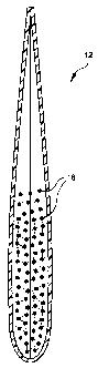
One embodiment of such a permeable vehicle is shown in Figures 1 and 2. Figure
1 is a
front view of the vehicle (12) which is constructed of a unitary sheet of mesh
(14) with a
border (16) which is either created upon heat sealing of the mesh, or may be
constructed of an
8
CA 02246961 1998-09-10
t ~^ PATENT
P-4012
adhesive or sealable material other than the mesh. As shown in Figure 1, the
unitary sheet is
folded back upon itself to create the vehicle (12).
During construction, the two sides of the unitary sheet are sealed after the
sheet is
folded back upon itself. Then, an appropriate amount of the ion exchange resin
(18) (see
Figure 2) is added from the open top of the vehicle. Following addition of the
ion exchange
resin, the top of the vehicle is crimped and sealed as well. This creates the
vehicle shown in
cut-away side view in Figure 2.
Suitable materials for the mesh (14) such vehicles include, but are not
limited to,
polypropylene and nylon. The same materials are also suitable for the adhesive
or sealable
border (16).
When the resin is contacted with the sample, nucleic acid hybridization and/or
amplification inhibitory substances adhere to the resin. The principal means
by which the ion
exchange resin binds inhibitory substances is by exchange of cations and
anions of the resin for
the inhibitory substances. Thus, innocuous cations and anions are released
into the sample, and
the inhibitory substances are bound to the resin. Then, such substances are
removed from the
sample prior to the commencement of any nucleic acid hybridization and/or
amplification
event.
The amount of time for contact of the resins and sample is dependent upon the
type of
sample, the ratio of resin to sample, and the concentration and type of
inhibitors in the sample.
Although the resin is active immediately upon contact with the sample, the
time of contact of
resin and sample for the method of the present invention is about fifteen (15)
minutes to about
four (4) days.
Optionally, following such contact, the resin and sample may be separated. As
stated
above, such separation may be conducted by a variety of means such as
centrifugation,
filtering, or if the permeable vehicle or ion exchange paper are used, then
the matrix
(permeable vehicle or ion exchange paper) can be physically removed from the
sample or the
treated sample can simply be removed by pipetting.
9
CA 02246961 1998-09-10
~ =
.. ~
PATENT
P-4012
A variety of processes are currently used to prepare target nucleic acids in
samples for
hybridization or amplification. For example, sputum samples which are
processed to amplify
mycobacterial nucleic acid sequences are typically subjected to a NALC/NaOH
process.
Similarly, other types of clinical samples are subjected to other well known
standard processes,
for example, centrifugation for large volume samples such as blood and urine.
The method of
the present invention may be used before, as part of, or after those standard
processes.
In addition to utility in the method for removing nucleic acid hybridization
and/or
amplification inhibitory substances, the contacting of ion exchange resin with
sample also
stabilizes such sample for transport at room temperature and concentrates the
sample. The
amount of resin useful for such transport stabilization is about the same as
for removing
nucleic acid hybridization andlor amplification inhibitory substances.
Transport stabilization,
sample concentration, removal of fluorescent substances, and removal of
nucleic acid inhibitory
substances all occur as a result of contacting an ion exchange resin with the
sample. Thus, the
present invention provides an extremely efficient method for one to stabilize
a sample at room
temperature for transport or other reasons, and simultaneously commence the
processing or
preparation of such sample for a molecular diagnostic assay by concentrating
the sample and
binding fluorescent compounds and nucleic acid hybridization and/or
amplification inhibitory
substances for subsequent removal from the sample.
..~
The following examples illustrate specific embodiments of the invention
described
herein. As would be apparent to skilled artisans, various changes and
modifications are
possible and are contemplated within the scope of the invention described.
r i 1 CA 02246961 2002-03-05
PATENT
P-4012
EXAMPLE 1
A Mixed Bed Ion Exchange Resin Removes a Known Amplification Inhibitor and
Background Fluorescent Compounds from Urine and Swab Samples
The purpose of this Example was to determine if a mixed bed ion exchange resin
(AmberliteTM MB-150) removes non-specific DNA from a urine sample and swab
sample. In
addition, fluorescent readings were taken of the treated and untreated samples
to determine if
fluorescent compounds, which would increase the background of a fluorescent
detection
system and potentially decrease the sensitivity of the system would be
removed.
Materials
Sample Processing Reagents:
Vaginal swab samples collected with B-D E-Z swabs
Urine samples
Sample buffer
Amberlite' MB-150 (Sigma)
BD polypropylene dispense tubes
ssDNA Assgy Reagents:
OligreenTM dye (Molecular Probes)
TE buffer
Procedure
Eight vaginal swab samples were expressed into four ml of sample buffer, the
solution
was vortexed and one ml of the sample was dispensed into two BD dispense tubes
containing
AmberliteT'` MB-150 at 0.4 gms. The solution in the BD dispense tubes was
vortexed and was
11
CA 02246961 1998-09-10
PATENT
P-4012
maintained at room temperature for 30 minutes. After 30 minutes the fluid was
transferred to
a separate tube.
One ml of urine was dispensed into two BD dispense tubes containing 0.4 gms of
AmberliteTM MB-150. The solution was vortexed and was maintained at room
temperature for
30 minutes. After 30 minutes the solution was vortexed and was then
transferred to a separate
tube.
Human placental DNA standards were prepared in TE buffer at 1000 ng/ml, 500
ng/ml,
250 ng/ml, 125 ng/ml, 62.5 ng/ml, 31.25 ng/ml and 0 ng/ml. Both AmberliteTM MB-
150 treated
samples and untreated urine and swab samples from above, were diluted 1:10 and
1:100 in TE
buffer. The samples were placed in a boiling water bath for five minutes to
denature the DNA
strands. Oligreen dye was diluted 1:200 in TE according to the manufacturer's
instructions.
An equal volume of sample and diluted Oligreen dye was combined in plastic
cuvettes and the
samples were read using a fluorescent excitation wavelength of 480 and an
emission
wavelength of 520. Background fluorescent readings on the untreated samples
were measured
using the 480 excitation and 520 emission wavelengths and the background was
subtracted
from the ssDNA measurements. Background fluorescent readings were taken on the
treated
samples as well for comparison to the untreated samples, to determine if
background
fluorescence substances were removed.
~
Results
The results are provided in the table below, using those ssDNA values which
fell on the
linear portion of the standard curve.
12
CA 02246961 1998-09-10
PATENT
P-4012
TREATMENT SAMPLE SSDNA CONC. % REDUCTION 520/480 % REDUCTION
(NG/ML ssDNA READING FLUORESCENCE
NONE SWAB 31493 ----------- 656 -------------
AMBER- SWAB 14066 55.3 320 51.2
LITE
NONE URINE 1417 ------ ---- 8514 ---------------
AMBER- URINE 546 61.5 798 93.8
LITE
Conclusions
The data of this Example indicates that a mixed bed ion exchange resin removes
background fluorescent compounds from both swab samples and urine samples.
This result is
beneficial as high background fluorescence can impact sensitivity claims in a
fluorescent assay
detection system. In addition, these data indicate that a mixed bed ion
exchange resin removes
non target DNA, a known amplification inhibitor of Strand Displacement
Amplification (SDA).
EXAMPLE 2
Direct Method for Urine Processing
The purpose of this Example was to determine if the present invention could be
used to
eliminate the traditional centrifugation and wash steps in urine processing.
13
CA 02246961 2002-03-05
1 r^ PATENT
P-4012
Materials
Sample Processing Reagents:
AmberliteTM NIB-150 (Sigma)
Antifoam
Spectra/mesh polypropylene filter, 75 um Urine samples
2X chlamydia sample buffer
1 X sample processing buffer
Chlamydia preparation
Assay Reagents:
- Oligonucleotide Devices (ODs) - microtiter plates with SDA amplification
primers, SDA
bumper primers, SDA fluorescence detector probes, dUTP and buffers dried in
each well
- Enzyme Devices (ENDs) - microtiter plates with restriction endonuclease
(BsoB 1),
polymerase (Bst), dCsTP, dATP, dGTP and buffers dried in each well
(ODs and ENDs are more completely described in co-pending U.S. Patent No.
6,077,669.
Procedure
Four urine samples were spiked at 1000 Chlamydia elementary bodies/ml.
AmberliteTM
MB-150 at 0.3 gms was dispensed into four BD dispense tubes containing ten ul
of 1.25%
anti-foam. One ml of each urine sample was added to one BD dispense tube
containing
AmberliteTM MB-150 and the samples were maintained at room temperature for 30
minutes.
The solution was squeezed through a 75 um polypropylene mesh into a tube. An
equal
14
CA 02246961 2002-03-05
PATENT
P-4012
volume of sample and 2X concentrated sample buffer were dispensed into an
tube/sample and
the tubes were placed in a 105 C heat block for 30 minutes.
Four ml of the urine sample was dispensed into tubes and the tubes were
centrifuged at
2,000 g for 30 minutes. The supernatant was decanted and three ml of Sample
buffer was
added to the tube. These tubes were placed in a 105 C heat block for 30
minutes.
Aliquots (-150 ul) of a sample from the tubes were dispensed into each well of
the
ODs. The wells of the ODs were covered, and the ODs retained at room
temperature for 20
minutes. The ODs were then uncovered, and incubated at 75 C for 10 minutes,
while the
ENDs were pre-warmed for 10 minutes to 52 C.
After the 10 minute incubation, 100 ul aliquots from each well of the ODs were
transferred (pipetted) to a corresponding well in the ENDs. The ENDs were then
sealed with
an adhesive cover, and introduced into a fluorescence reader instrument as
described in co-
pending U.S. Patent No. 6.043,880 filed September 15, 1997. (Other standard
microtiter plate
fluorescence reader instruments could also be used.)
The fluorescence signal from the wells of the ENDs were monitored for 60
minutes.
The sealed ENDs were then discarded in a sealed bag to further insure against
potential
amplicon contamination of the laboratory environment.
Results
The results are provided in the table below as units of area. Area is the
integration of
the fluorescent signal curve over time.
CA 02246961 1998-09-10
PATENT
P-4012
Sample Amberlite Method Control Method
(Area) (Area)
Fl 35774 29354
M10 15622 8138
M11 13127 2774
M12 10322 14858
Conclusions
The data of this Example indicates that, centrifiugation and associated wash
steps can
be eliminated from urine sample processing by incorporating the method of the
present
invention.
EXAMPLE 3
A Mixed Bed Ion Exchange Resin For Removal of Amplification Inhibition Using
Neisseria gonorrhoeae Spiked Negative Clinical Samples
The purpose of this Example was to determine if a mixed bed ion exchange resin
4
(Amberlite MB-150) removes amplification inhibitors and improves the recovery
of specific
target signal compared to the control method.
Materials
Sample Processing Reagents:
Urine Samples
Neisseria gonorrhoeae preparation
16
CA 02246961 1998-09-10
,
PATENT
P-4012
Sample buffer
Amberlite MB-150 (Sigma)
Amplification Reagents:
- ODs (as defined in Example 2, but containing SDA amplification primers, SDA
bumper
primers and SDA fluorescence detector probes specific for N. gonorrhoeae
rather than
Chlamydia)
- ENDs
Procedure
Thirty clinical urine samples were spiked with 1000 Neisseria gonorrhoeae
particle
forming units/ml. Six ml of each sample was transferred to a 15 ml conical
tube containing
0.12 gms Amberlite MB-150. Each tube was vortexed and the tubes were stored at
room
temperature for sixteen hours. For the control conditions, four ml of each
urine sample was
transferred directly to a centrifuge tube and the samples were stored at 4 C
for sixteen hours.
After sixteen hours at room temperature the samples containing Amberlite were
vortexed and four ml of fluid was transferred to centrifuge tubes. Both the
urine samples
treated with Amberlite and those samples stored at 4 C, without Amberlite MB-
150, were
centrifuged at 2,000 g for 30 niinutes. The supernatant was decanted from each
tube and three
ml of sample diluent was added to each tube. The tubes were placed in a heat
block at 105 C
for 30 minutes.
The samples were amplified and results detected using the same procedure as in
Example 2.
17
CA 02246961 1998-09-10
PATENT
P-4012
Results
The results are provided in a table below using the mean area of three
amplification/detection replicates. The values at the bottom of the table are
the means for all
the samples.
Sample # Amberlite Treatment (area) Control Method (area)
1 6759 4441
2 24500 1766
3 17734 1157
4 9442 4836
5 1071 791
6 2419 1477
7 2252 1664
8 6704 2105
9 5013 1072
3934 1819
11 3711 1324
12 14138 1513
13 1059 532
14 1363 4258
9598 2203
16 8466 9474
17 16810 3295
18 11105 4312
19 4098 2420
13087 2341
21 2382 1080
18
CA 02246961 1998-09-10
= / ~i
PATENT
P-4012
22 7205 8649
23 675 3430
24 6626 1091
25 19012 3147
26 4980 790
27 7968 1324
28 11034 5179
29 22049 5904
30 8910 2462
MEAN 8470 2862
Conclusions
Mixed bed ion exchange resin treatment of urine samples produces statistically
higher
areas compared to control method of processing urine samples (based on T test
results, using
equal variance with a P value = 2.36E-9), indicating that amplification
inhibitors are removed
from the system and specific amplification is performed more efficiently.
EXAMPLE 4
Comparative Example Showing Room Temperature Stability of
Neisseria gonorrhoeae in Urine
The purpose of this Example was to evaluate the stability of Neisseria
gonorrhoeae in
urine at room temperature.
19
CA 02246961 1998-09-10
. . , . ,. .
PATENT
P-4012
Materials
Sample Processing Reagents:
Sample Diluent
N. gonorrhoeae stock
Urine - Male Urine Pool
Amplification Reagents:
ODs as in Example 3
ENDs as in Example 3
Procedure
Neisseria gonorrhoeae was spiked into a normal male urine pool to yield final
reaction
concentrations of 0, 10 and 100 particles per reaction. The dilutions were
stored at 25 C.
Four ml aliquots of each dilution were transferred to sample tubes for
processing at the
following time points: 0-hr, 1 day, 2 days and 4 days. All samples were
processed as follows:
Centrifuge 2000xg for 30 minutes, decant supernatant, resuspend pellet with 2
ml of sample
diluent, heat 30 minutes at 100 C. The samples were amplified and results
detected using the
same procedure as in Example 2.
Results
Results are provided as mean areas in the table below.
CA 02246961 2002-03-05
PATENT
P-4012
Storage Time 0 pfu GC/rxn 10 pfu GC/rxn 100 yfu GC/rxn
0 hours 2376 5721 21506
1 day @ 25 C 3080 2415 12065
2 days @ 25 C 3355 4577 2797
4 days @ 25 C N/A 2599 4483
Conclusions
Neisseria gonorrhoeae is unstable when stored in urine at room temperature for
I day
or longer.
EXAMPLE 5
Screening of Additives for Stability of Neisseria gonorrhoeae and Chlamydia
trachomatis at Room Temperature
The purpose of this Example was to screen additives for stability of Neisseria
gonorrhoeae and Chlamydia trachomatis in urine at room temperature for two
days.
Materials
Sample Processing Reagents:
Urine Pools
Sample buffer
ChemStat tube (MicroSure Inc.)
Boric Acid/Formate
ProClin 300rM
AmberliteTM MB-150 (Sigma)
21
CA 02246961 1998-09-10
PATENT
P-4012
Amplification Reagents:
N. gonorrhoeae ODs as in Example 3
N. gonorrhoeae ENDs as in Example 3
C. trachomatis ODs as in Example 2
C. trachomatis ENDs as in Example 2
Procedure
Three urine pools were spiked with 750 Elementary bodies/ml of Chlamydia
strain
LGV-II. Two of the three pools were spiked with 750 particles forming units/ml
of Neisseria
gonorrhoeae. The third pool was positive for Neisseria gonorrhoeae and was not
spiked.
Four ml of each urine pool was transferred to four tubes. At 0 hr, 40 hours at
4 C, 40 hours
at 24 C and 96 hr at 4 C the tubes were centrifuged at 2,000 g for 30 minutes.
The
supernatant was decanted and three ml of sample buffer was added to each tube.
The tubes
were transferred to a heat block for 30 minutes at 105 C.
Ten ml of each pool was transferred to a ChemStat tube. The material was then
transferred to two tubes/pool at four ml/tube. At Ohr and 40 hours at 24 C the
tubes were
centrifuged at 2,000 g for 30 minutes. The supernatant was decanted and three
ml of sample
buffer was added to each tube. The tubes were transferred to a heat block for
30 minutes at
105 C.
Five ml from each pool was transferred to two Boric Acid/Formate tubes/pool.
The
material from each Boric Acid/Formate tube was transferred to an tube at four
mi. At Ohr and
40 hours at 24 C the tubes were centrifuged at 2,000 g for 30 minutes. The
supernatant was
decanted and three ml of sample buffer was added to each tube. The tubes were
transferred to
a heat block for 30 minutes at 105 C.
Nine ml from each pool was transferred to a 15 ml centrifuge tube containing
2.7 ul of
ProClin 300. Four n-d from each pool was transferred to two tubes. At Ohr and
40 hours at
22
CA 02246961 1998-09-10
PATENT
P-4012
24 C the tubes were centrifuged at 2,000 g for 30 minutes. The supernatant was
decanted and
three ml of sample buffer was added to each tube. The tubes were transferred
to a heat block
for 30 minutes at 105 C.
A 5.5 ml aliquot of each pool was transferred to two 15 ml centrifuge tubes
containing
0.72 gms of AmberliteTM MB-150. Ten minutes after addition of the sample to
Amberlite the
solution was transferred to tubes at four ml. The remaining 15 ml centrifuge
tubes containing
each pool were stored at 40 hr at 24 C. The solution was transferred to tubes
at four ml and
the tubes were centrifuged at 2,000 g for 30 minutes. The supernatant was
decanted and three
ml of sample buffer was added to each tube. The tubes were transferred to a
heat block for 30
minutes at 105 C.
The samples were amplified and the results detected using the same procedure
as in
Example 2.
Results
Results are indicated in the table below as the mean of three amplification
replicates.
Neisseria gonorrhoeae
No addition 6hemStat Boric Acid/Formate
Sample 0 hr 25 C 40 hr 4 C 40 hr 0 hr 25 C 40 hr 0 hr 25 C 40 hr
C 25 C 25 C
Pooll 19543 15970 5290 14003 2593 25843 5337
Poo12 21180 16682 403 7252 1854 12026 877
Poo13 27854 31367 30188 27486 21251 24511 24468
23
CA 02246961 1998-09-10
,. ' - , .. = ~-~
PATENT
P-4012
Amberlite ProClin (0.03%)
Sample 0 hr 25 C 40 hr, 25 C 0 hr 25 C 40 hr 25 C
Pooll 12094 27832 16925 4263
Pool2 22288 10288 14311 1117
Poo13 30564 28405 27790 30998
Chlamydia trachomatis
No addition ChemStat Boric Acid/Formate
Sample 0 hr 25 C 40 hr 4 C 40 hr 0 hr 25 C 40 hr 0 hr 25 C 40 hr
25 C 25 C 25 C
Pool1 5235 11553 12145 18339 6625 49956 16963
Pool2 10708 3631 422 16139 432 19153 5781
Pool3 14490 804 19784 18110 5378 3734 4097
Amberlite ProClin (0.03%)
Sample 0 hr 25 C 40 hr, 25 C 0 hr 25 C 40 hr 25 C
Pooll 7145 22211 637,8 21922
Pool2 43043 26650 22441 36003
Pool3 35559 34907 4097 19722
Conclusions
A mixed bed ion exchange resin (AmberliteTM M-150) at 0.13 g/ml provides
stability
of Neisseria gonorrhoeae in urine for up to 40 hours at room temperature for
Pools I and 2
which have known spike levels of Neisseria gonorrhoeae. Pool 3 which tested
initially, very
positive for Neisseria gonorrhoeae was positive for all storage conditions,
which indicates that
24
CA 02246961 1998-09-10
= .= 1 _ .
PATENT
P-4012
with very high concentrations of Neisseria gonorrhoeae, specific signal is
maintained.
ChemStat, Boric Acid Formate and ProClin additives provided little or no
stability for
Neisseria gonorrhoeae at room temperature.
AmberliteTM M-150 was effective as both inhibitor removal and for
stabilization of
Chlamydia LGV-II.
EXAMPLE 6
Mixed Bed Ion Exchange Resin Treatment of Urine Stabilizes Individual Spiked
Negative Clinical Samples for Four Days
The purpose of this Example was to determine if Neisseria gonorhhoeae and
Chlamydia trachomatis spiked into individual clinical samples could be
stabilized by a mixed
bed ion exchange resin (Amberlite) for four days at room temperature.
Materials
Sample Processing Reagents:
Negative urine samples
Sample buffer
Amberlite MB-150 (Sigma)
Neisseria gonorrhoeae preparation
Chlamydia trachomatis preparation
Amplification Reagents:
Neisseria gonorrhoeae ODs as in Example 3
CA 02246961 1998-09-10
= ' PATENT
P-4012
Neisseria gonorrhoeae ENDs as in Example 3
Chlamydia trachomatis Ods as in Example 2
Chlamydia trachomatis ENDs as in Example 2
Procedure
Thirty urine samples were spiked with 800 Neisseria gonorrhoeae/ml and 800
Chlamydia trachomatis/n-d. Additionally, seven ml of urine from each sample
was transferred
to a 15m1 conical tube containing 0.47 gms of Amberlite MB-150. Seven ml of
urine from
each sample was transferred to a 15 ml conical tube containing 1.4 gms of
Amberlite MB- 150.
The tubes were immediately vortexed. The solutions containing 0.47 gms of
Amberlite MB-
150 were transferred to separate centrifuge tubes at four ml/tube. The tubes
were placed in a
centrifuge for 30 minutes at 2,000 g. The supernatant was decanted from the
tubes and two ml
of sample diluent was added to each tube. The tubes were placed in a 110 C
heat block for 30
minutes. The tubes were stored at -20 C for four days.
The solutions containing 1.4 gms of Amberlite MB-150 were stored at room
temperature for four days. The solution was transferred to separate centrifuge
tubes at four
ml/tube. The tubes were placed in a centrifuge for 30 minutes at 2,000 g. The
supernatant
was decanted from the tubes and two ml of sample diluent was added to each
tube. The tubes
were placed in a 110 C heat block for 30 minutes. `
The samples were amplified and results detected using the same procedure as in
Example 2.
Results
The results are shown in the table below.
26
CA 02246961 1998-09-10
PATENT
P-4012
30m1 / lhr RT lOml / 4 Days, RT
Sample GC Mean CT Mean GC Mean CT Mean
M1 23268 21537 21653 20931
M2 20225 21000 19875 21541
M3 20272 27771 10136 26264
M4 12857 14305 8846 25341
M5 25794 26013 11086 34331
M6 14770 30920 16201 33743
M7 18639 34963 22402 34271
M8 15790 24579 17808 38254
M9 14108 32677 17859 37905
M10 17934 30252 19888 33420
Mil 13759 31573 7405 28978
M12 14593 28570 12464 33524
M13 12734 30523 3564 32920
M14 7877 21105 3276 24797
M15 19072 32762 ~416 22421
Fl 19098 17203 23589 33973
F2 19239 22295 12790 22651
F3 21398 23807 25668 35166
F4 24030 16982 22403 35689
F5 24875 31753 28853 34765
F6 8470 26019 20349 36289
F7 22544 27332 30676 36189
F8 19781 12821 38465 40016
27
CA 02246961 1998-09-10
PATENT
P-4012
F9 15061 32559 11625 36957
F10 31090 34744 25470 43375
F11 18029 38685 22737 36648
F12 10285 35995 16472 39225
F13 14550 30752 11455 37126
F14 22820 32537 8562 36274
F15 17515 38294 21233 36059
MEAN 18016 27678 17208 32968
Conclusions
Exposure to a mixed bed ion exchange resin (Amberlite MB-150) maintains the
integrity of target organisms in individual clinical urine samples, throughout
four days at room
temperature.
EXAMPLE 7
Comparison of Ion Exchange Resin in Permeable Vehicle to Control Free Ion
Exchange Resin
The purpose of this Example was to determine if ion exchange resin, either in
free
contact to the sample or in a permeable vehicle, would have equivalent
functional performance
in the reduction of osmolality from a sample and in the amplification of
extracted nucleic acid
from said sample.
28
CA 02246961 2002-03-05
PATENT
P-4012
Materials
Sample Processing ReaQents:
Spectra/Mesh Polypropylene Filters 75uM
American International Electric Heat Sealer
Presision Systems Inc. OsmetteTM A osmolality reader
Amberlite MB-150 (Sigma)
VWR 4oz. Specimen Collection Containers
ml FalconT" Conical tubes
10 CT/GC Sample Diluent
10 Negative Urine Pools, each consisting of unique clinical samples.
Amplification and Assay Reagents:
Chlamydia trachomatis ODs as in Example 2
Chlamydia trachomatis ENDs as in Example 2
Neisseria gonorrhoeae ODs as in Example 3
Neisseria gonorrhoeae ENDs as in Example 3
15 Procedure
Amberlite MB-150 ion exchange resin was first prepared by performing two
washes in
distilled water and vacuum drying the resin. The resin was then spread on a
flat surface and
allowed to dry overnight in a 37 C incubator. A 2.4g aliquot of the dried
resin was then placed
in an octagonal shaped heat sealed polypropylene mesh pouch. Ten of these
pouches were
constructed in such a manner that would maximize the exposure of the ion
exchange resin to
the sample. Modifications included reduction in size to accomplish the flat
placement of said
pouch at the bottom of a specimen collection container. Also, swelling of
resin by sample
absorption was accounted for. Each pouch was placed in each of 10 specimen
collection
29
CA 02246961 1998-09-10
PATENT
P-4012
containers. A 0.84 g aliquot of the dried resin was placed in each of 20
Falcon 15m1 conical
tubes.
Ten 35m1 urine pools were made. Each urine pool was then spiked with 800 GC
particles per ml and 800 CT EBs per ml. Baseline osmolality readings of
untreated urine pools
were taken. 20mis of each urine pool was then placed in a specimen collection
container with
the ion exchange resin in a permeable vehicle. 14m1s each of the remaining
urine pools was
then divided to two Falcon 15m1 conical tubes containing free ion exchange
resin. One tube
was vortexed upon addition of the 7ml urine pool. The other tube was not.
After 2 hours of treatment with the ion exchange resin, osmolality readings
were taken
on the treated urine pools. 4mls of each treated urine was then subjected to
2000xg for 30
minutes. The supernatant was decanted and CT/GC sample diluent was then added
to each
each pellet at 2ml per tube. The pellets were then vortexed for 5 seconds to
resuspend the
pellet. After heating the tubes in a heat block at 110 C for 30 minutes, all
samples were
amplified and the results detected using the same procedure as in Example 2.
Results
The results are presented in the tables below as osmolality readings and
Chlamydia
trachomatis and Neisseria gonorrhoeae areas.
Urine Pools Untreated 2 hr. Incub of Free Amberlite Free Amberlite
mOsm/kg Amberlite in Urine Vortexed Post 2 hr. Vortexed Pre & Post
Preservative Pouch Incub. (mOsm/kg) 2 hr. Incub.
(mOsm/kg) (mOsm/kg)
Prov. M Pool 1 897 606 527 520
Prov. M Pool 2 753 467 396 431
Prov. M Poo13 636 394 336 306
CA 02246961 1998-09-10
PATENT
P-4012
Prov. F Pool 1 837 597 550 555
Prov. F Pool 2 566 345 297 296
LSU F Poo1 1 738 448 401 412
LSU F Pool 2 864 582 570 532
LSU F Pool 3 852 636 568 523
LSU M Pool 1 753 566 520 frozen
LSU M Poo12 850 602 539 511
Urine Pools w/800 EBs 2 hr. Incub. of Free Amberlite Free Amberlite
per ml Amberlite in Urine Vortexed Post 2 hr. Vortexed Pre & Post 2
Preservative Pouch Incub. hr. Incub.
Mean CT Area (N=3) Mean CT Area (N=3) Mean CT Area (N=3)
Prov. Male Pool 1 28942 35687 33368
Prov. Male Pool 2 30376 28962 32419
Prov. Male Poo13 27650 25458 32562
Prov. Female Pool 1 23353 22503 37514
Prov. Female Poo12 40927 20719 44077
LSU Female Pool 1 31721 36125 25222
LSU Female Poo12 34144 25619 21939
LSU Female Pool 3 27763 29901 35388
LSU Male Poo1 1 21111 26285 36393
LSU Male Pool 2 33674 30290 32350
Negative Control 548 538 756
Positive Control 30021 29053 24737
31
CA 02246961 1998-09-10
. ~
PATENT
P-4012
Urine Pools w/800 GC 2 hr. Incub. of Free Amberlite Free Amberlite
per ml Amberlite in Urine Vortexed Post 2 hr. Vortexed Pre & Post 2
Preservative Pouch Incub. hr. Incub.
Mean GC Area (N=3) Mean GC Area (N=3) Mean GC Area (N=3)
Prov. Male Pool 1 20301 15005 20765
Prov. Male Pool 2 30946 28747 28923
Prov. Male Pool 3 23258 32763 24515
Prov. Female Pool 1 18698 17048 15900
Prov. Female Pool 2 23825 15854 20507
LSU Female Pool 1 16159 20085 9353
LSU Female Pool 2 14090 19691 11598
LSU Female Pool 3 13845 21970 22436
LSU Male Pool 1 8389 19962 14579
LSU Male Poo12 19499 21274 10361
Negative Control 400 1086 464
Positive Control 24487 9544 32709
Conclusions
Ion exchange resin, either in free contact to the sample or in a permeable
vehicle, has
equivalent functional performance in terms of osmolality reduction and
amplification.
A significant difference is observed in osmolality between the untreated pools
and the
same pools treated with the pouched resin (p value = 4.42E-05). However, no
significant
difference is observed between the pouched resin and either the free resin
with a pre-incubation
vortex step (p value = 0.06, assuming unequal variance due to frozen sample)
or free resin
without a pre-incubation vortex step (p value = 0.25). Additionally, if the
frozen sample is
32
CA 02246961 1998-09-10
t ,
PATENT
P-4012
eliminated in the comparison between the pouched resin and the free resin with
a pre-
incubation vortex step (p value = 0.19, assuming equal variance), then the
difference is even
less significant. The minimum requirement for functional removal of salts from
the sample is
simply physical contact.
No significant difference is observed in Chlamydia trachomatis area units for
the
pouched resin vs. either the free resin with a pre-incubation vortex step (p
value = 0.25) or free
resin without a pre-incubation vortex step (p value = 0.46). The same
observation can be
found for Neisseria gonorrhoeae area units. For the pouched resin vs. either
the free resin
with a pre-incubation vortex step (p value = 0.73) or free resin without a pre-
incubation vortex
step (p value = 0.39), no statistical difference could be found.
EXAMPLE 8
The Stabilization of a Sample by Ion Exchange Resin in a Permeable Vehicle for
Testing by a Molecular Diagnostic Process
The purpose of this Example was to determine if ion exchange resin in a
permeable
vehicle would provide for the stabilization of target organisms for subsequent
testing by Strand
Displacement Amplfication (SDA) of nucleic acid of such organisms.
Materials
Sample Processing Reagents:
Same as for Example 7.
Amplification and Assay Reagents:
Same as for Example 7.
33
CA 02246961 1998-09-10
PATENT
P-4012
Procedure
Same as for Example 7 except, after 5 and 7 days of treatment with the ion
exchange
resin, osmolality readings were taken on the urine pools treated with free ion
exchange resin
and urine pools treated with the ion exchange resin in a permeable vehicle.
Urine pools were
subjected to room temperature storage for the duration of the 5 and 7 day time
periods before
analysis.
Urine pools that were treated with the ion exchange resin in a permeable
vehicle were
also subjected to testing by strand displacement amplification after 5 and 7
days. At each
timepoint, a 4ml volume of each treated urine pool was subjected to 2000xg for
30 minutes.
The supernatant was decanted and CT/GC sample diluent was then added to each
each pellet
at 2ml per tube. The pellets were then vortexed for 5 seconds to resuspend the
pellet. After
heating the tubes in a heat block at I 10 C for 30 minutes, all samples were
amplified and the
results detected using the same procedure as in Example 2.
Results
The results are presented in the tables below as osmolality readings and
Chlamydia
trachomatis and Neisseria gonorrhoeae areas.
Urine Pools 2 hr. Incub of 5 day Incub of 7 day Incub of Amberlite
Amberlite in Urine Amberlite in Urine in Urine Preservative
Preservative Pouch Preservative Pouch Pouch (mOsm/kg)
(mOsm/kg) (mOsm/k )
Prov. M Pool 1 606 539 539
Prov. M Pool 2 467 402 405
Prov. M Pool 3 394 265 261
34
...r
CA 02246961 1998-09-10
ti
PATENT
P-4012
Prov. F Pool 1 597 577 565
Prov. F Pool 2 345 274 272
LSU F Pool 1 448 427 430
LSU F Pool 2 582 554 584
LSU F Pool 3 636 537 533
LSU M Pool 1 566 501 498
LSU M Poo12 602 481 483
Urine Pools w/800 2 hr. Incub. of 5 day Incub. of 7 day Incub. of
EBs per ml Amberlite in Urine Amberlite in Urine Amberlite in Urine
Preservative Pouch Preservative Pouch Preservative Pouch
Mean CT Area (N=3) Mean CT Area (N=3) Mean CT Area (N=3)
Prov. Male Pool 1 27542 26838 34194
Prov. Male Pool 2 35377 37790 37918
Prov. Male Pool 3 26533 44240 36227
Prov. Female Pool 1 30848 36650 17570
Prov. Female Pool 2 30682 34849 34969
LSU Female Pool 1 32517 18689 30677
LSU Female Pool 2 23494 32481 26919
LSU Female Pool 3 29470 39160 27402
LSU Male Pool 1 24556 31976 31595
LSU Male Pool 2 27891 32687 37414
Negative Control 515 520 573
Positive Control 18894 19081 895
CA 02246961 1998-09-10
PATENT
P-4012
Urine Pools w/800 2 hr. Incub. of 5 day Incub. of 7 day Incub. of
GC per ml Amberlite in Urine Amberlite in Urine Amberlite in Urine
Preservative Pouch Preservative Pouch Preservative Pouch
Mean GC Area (N=3) Mean GC Area (N=3) Mean GC Area (N=3 )
Prov. Male Pool 1 19020 10052 10043
Prov. Male Pool 2 25770 32026 32255
Prov. Male Pool 3 26110 21493 . 34625
Prov. Female Pool 1 22379 5929 7031
Prov. Female Pool 2 20497 15707 11580
LSU Female Pool 1 18213 9314 5382
LSU Female Pool 2 18517 16579 11179
LSU Female Pool 3 26272 17937 10682
LSU Male Pool 1 7613 21325 9332
LSU Male Pool 2 11777 21289 15567
Negative Control 834 518 775
Positive Control 21034 25312 15476
Conclusions
Osmolality readings taken after 2 hours, 5 and 7 days of treatment with free
ion
exchange resin and ion exchange resin in a permeable vehicle shows the
continuation of the
marked decrease in osmolality from untreated urine. No statistical difference
was seen within
this time frame (2hr vs. 5 day: p value = 0.17; 2hr vs. 7 day: p value =
0.18). This establishes
that ion exchange resin effects the sample throughout a time period where
stabilization of
target nucleic acid would take place.
A comparison of the amplification and detection of extracted Chlamydia
trachomatis
and Neisseria gonorrhoeae nucleic acid from all urine pools treated with ion
exchange resin in
36
dw
CA 02246961 1998-09-10
ti
PATENT
P-4012
a permeable vehicle for 2 hours, 5 and 7 days, shows no statistically
significant drop in signal
with time. It was shown in Example 4 that Neisseria gollorrhoeae is unstable
in urine samples
stored at room temperature for greater than 24 hours. It is shown here that
ion exchange resin
in a permeable vehicle provides for the stabilization of organisms for up to 7
days allowing
subsequent testing by strand displacement amplification of organism nucleic
acid.
While the invention has been described with some specificity, modifications
apparent to
those with ordinary skill in the art may be made without departing from the
scope of the
invention. Various features of the invention are set forth in the following
claims.
37